In the gray matter of the anterior horns of each spinal cord segment
there are several thousand neurons that are 50-100% larger than most other neurons. They are called anterior motor neurons. The axons of these motor neurons exit the spinal cord through the ventral roots and directly innervate skeletal muscle fibers. There are two types of these neurons: alpha motor neurons and gamma motor neurons.
Alpha motor neurons
. Alpha motor neurons give rise to large motor fibers of the A-alpha (Ace) type with an average diameter of 14 μm. After entering the skeletal muscle, these fibers branch repeatedly to innervate large muscle fibers. Stimulation of a single alpha fiber excites from three to several hundred skeletal muscle fibers, which, together with the motor neuron innervating them, constitute the so-called motor unit.
Gamma motor neurons
. Along with alpha motor neurons, the stimulation of which leads to contraction of skeletal muscle fibers, much smaller gamma motor neurons are localized in the anterior horns of the spinal cord, the number of which is approximately 2 times less. Gamma motor neurons transmit impulses along much thinner motor fibers of the A-gamma (Ay) type with an average diameter of about 5 microns.
They innervate small special fibers
skeletal muscles, called intrafusal muscle fibers. These fibers form the central part of the muscle spindles involved in the regulation of muscle tone.
Interneurons
. Interneurons are present in all areas of the gray matter of the spinal cord, in the dorsal and anterior horns, and in the space between them. These cells are approximately 30 times more numerous than anterior motor neurons. Interneurons are small in size and very excitable, often exhibit spontaneous activity and are capable of generating up to 1500 impulses/sec.
They have numerous connections
each other, and many also synapse directly with anterior motor neurons. Interconnections between interneurons and anterior motor neurons are responsible for most of the integrative functions of the spinal cord, as discussed later in this chapter.
Essentially the entire set of different types of neural circuits
, is found within a pool of spinal cord interneurons, including diverging, converging, rhythmically discharging, and other types of circuits. This chapter outlines the many ways in which these various circuits are involved in the spinal cord's execution of specific reflex actions.
Only a few sensory signals
, entering the spinal cord along the spinal nerves or descending from the brain, reach directly the anterior motor neurons. Instead, almost all signals are conducted first through interneurons, where they are processed accordingly. The corticospinal tract terminates almost entirely at the spinal interneurons, where signals from this tract combine with signals from other spinal tracts or spinal nerves before they converge on the anterior motor neurons to regulate muscle function.
A connecting neuron that lies between sensory (afferent) and motor (efferent) neurons. Located in the central nervous system. Also called an interneuron, and in older texts an association neuron.
See the meaning of Intercalary Neuron in other dictionaries
Intercalary Adj.
— 1. Intended for insertion, insertion. Explanatory Dictionary by Efremova
Neuron M.
— 1. Same as: neuron. Explanatory Dictionary by Efremova
Insertable
- (shn), insertion, insertion. Adj. to insert. Ushakov's Explanatory Dictionary
Neuron
- neuron, m. (Greek neuron - fiber, nerve) (anat.). Nerve cell. Ushakov's Explanatory Dictionary
Neuron
- -A; m. [from Greek. neuron - nerve] Special. A nerve cell with all the processes extending from it. Kuznetsov's Explanatory Dictionary
Insertion Disc
— (discus intercalatus, LNH) the general name of microscopic structures at the point of contact of adjacent muscle cells of the myocardium, ensuring their connection into muscle complexes and transmission…….. Big medical dictionary
Motor Neuron
— , a nerve cell that conducts information to EFFECTORS (usually muscles) from the CENTRAL NERVOUS SYSTEM (CNS), thus causing the appropriate response. Axons (processes,……..
Neuron
— (nerve cell), the main structural and functional unit of the NERVOUS SYSTEM, which carries out the rapid transmission of NERVE IMPULSES between various organs. Consists of…….. Scientific and technical encyclopedic dictionary
Sensory Neuron
- (sensitive neuron), a nerve cell that conducts information from RECEPTORS in any part of the body to the CENTRAL NERVOUS SYSTEM (CNS). Their nerve endings are located in…….. Scientific and technical encyclopedic dictionary
Neuron
— (neuronum, neurocytus, LNH; Greek neuron vein, nerve; synonym: nerve cell, neurocyte, neurocyte) a cell capable of perceiving irritation, becoming excited, producing…….. Big medical dictionary
Neuron Amacrine
- (n. amacrinum, LNH) N., located in the inner granular layer of the retina and providing communication between the neurons of this layer. Large medical dictionary
Neuron Associative
— see Intercalary neuron. Large medical dictionary
Neuron Afferent
- (n. afferens, n. sensorium: synonym: N. receptor, N. sensory, N. sensitive) N., which carries out the perception and transmission of excitation from receptors to other N. of the central nervous system. Large medical dictionary
Neuron Bipolar
- (n. bipolare, LNH) N., having two processes - an axon and a dendrite. Large medical dictionary
Neuron Vegetative
- the general name of N., which are part of the ganglia, plexuses and nerves of the autonomic nervous system. Large medical dictionary
Neuron Fusiform
- (n. fusiforme, LNH) multipolar intercalary N. of elongated shape, found in the molecular plate of the cerebral cortex. Large medical dictionary
Neuron Fusiform Horizontal
- (n. fusiforme horizontale, LNH) multipolar N. elongated, found mainly between the layer of piriform neurons and the granular layer of the cerebellar cortex. Large medical dictionary
Neuron Internal
— (n. internum, LNH) N. of the internal parts of the anterior horn of the spinal cord, the axon of which passes through the white commissure to the opposite half of the spinal cord. Large medical dictionary
Neuron Intercalary
- (n. intercalatum; synonym: N. associative, N. intermediate) N. involved in the transmission of excitation from afferent N. to efferent. Large medical dictionary
Neuron Input
- a formal neuron that performs an input function in a specific system of neurons (neural network), i.e., it perceives signals only from the environment external to this system. Large medical dictionary
Neuron Gigantopyramidal
- (n. gigantopyramidale, LNH; synonym: Betza cell, giant pyramidal cell) large pyramidal N. of the internal pyramidal plate of the cerebral cortex; axons of the N. g. form…….. Big medical dictionary
Neuron Horizontal
— (n. horizomale, LNH) 1) N. inner granular layer of the retina, the processes of which contact the central endings of photoreceptor cells, carrying out redistribution…….. Big medical dictionary
Neuron Piriform
- (n. piriforme, LNH; synonym Purkinje cell) efferent N. of the cerebellar cortex, located in its ganglion layer and having a pear-shaped shape. Large medical dictionary
Neuron Motor
— see Motor neuron. Large medical dictionary
Neuron Long axon
— (n. longiaxonicum, LNH; synonym for Dogel cell type I) multipolar vegetative N., the axon of which transmits impulses to smooth or cardiac muscle tissue. Large medical dictionary
Neuron Stellate
- (n. stellatum, LNH) intercalated N. star-shaped. Large medical dictionary
Neuron Stellate Long-axon
- (n. stellatum longiaxonicum, LNH) N. z., located in the granular layer of the cerebellar cortex, having an axon extending into the white matter. Large medical dictionary
Neuron Stellate Short Axon
- (n. stellatum breviaxonicum, LNH) N. z. granular layer of the cerebellar cortex, which has an axon going to the cerebellar glomeruli. Large medical dictionary
Neuron Granular
— (n. granulare, LNH) the general name of small N. round, angular and pyramidal in shape, located in the outer granular plate of the cerebral cortex, the dendrites of which rise…….. Big medical dictionary
Neuron Granular Large
— (granoneurocytus magnus, LNH) the general name of large N., located in the molecular layer of the cerebellar cortex, the dendrites of which spread in the molecular layer, and the axons go to the granular…….. Large medical dictionary
What are they needed for? Why are there so many of them? What is a sensory neuron? What function do intercalary and executive neurons perform? Let's take a closer look at these amazing cells.
Functions
Every second, many signals pass through our brain. The process does not stop even in sleep. The body needs to perceive the world around it, make movements, ensure the functioning of the heart, respiratory, digestive, genitourinary systems, etc. Two main groups of neurons are involved in organizing all this activity - sensory and motor.
When we touch cold or hot and feel the temperature of the object, this is the merit of the sensitive cells. They instantly transmit information received from the periphery of the body. This ensures reflex activity.
Neurons form our entire central nervous system. Their main tasks:
- get information;
- transmit it through the nervous system.
These unique cells are capable of instantly transmitting electrical impulses.
To ensure the process of life, the body must process a huge amount of information that comes to it from the outside world, and respond to any sign of changing environmental conditions. To make this process as efficient as possible, neurons are divided according to their functions into:
- Sensitive (afferent) are our guides to the world around us. They are the ones who perceive information from the outside, from the senses, and transmit it to the central nervous system. The peculiarity is that thanks to their contact activity, we feel temperature, pain, pressure, and have other feelings. Sensitive cells of narrow specialization transmit taste and smell.
- Motor (motor, efferent, motor neurons). Motor neurons transmit information through electrical impulses from the central nervous system to muscle groups and glands.
- Intermediate (associative, intercalary, intercalary). Now let’s take a closer look at what function interneurons perform, why they are needed, and what is their difference. They are located between sensory and motor neurons. Interneurons transmit nerve impulses from sensory fibers to motor fibers. They provide “communication” between efferent and afferent nerve cells. They should be treated as a kind of natural “extenders,” long cavities that help transmit a signal from a sensory neuron to a motor one. Without their participation this would not have been possible. This is their function.
The receptors themselves are cells of the skin, muscles, internal organs, and joints specially designated for this function. Receptors can begin in the cells of the epidermis and mucous membrane. They are able to accurately capture the smallest changes, both outside the body and inside it. Such changes may be physical or chemical. Then they are instantly transformed into special bioelectric impulses and sent directly to sensory neurons. This is how the signal travels from the periphery to the center of the body, where the brain deciphers its meaning.
Impulses from the organ to the brain are carried out by all three groups of neurons - motor, sensory and intermediate. The human nervous system consists of these groups of cells. This structure allows you to respond to signals from the outside world. They provide reflex activity of the body.
If a person ceases to feel taste, smell, hearing and vision decrease, this may indicate disorders in the central nervous system. Depending on which sense organs are affected, a neurologist can determine in which part of the brain the problems have arisen.
1) Somatic. This is conscious control of the skeletal muscles.
2) Vegetative (autonomous). This is control of internal organs uncontrolled by consciousness. The operation of this system occurs even if a person is in a state of sleep.
Sensory neurons are most often unipolar. This means that they are equipped with only one bifurcating process. It leaves the cell body (soma) and simultaneously performs the functions of both an axon and a dendrite. The axon is the input, and the dendrite of the sensory neuron is the output. After excitation of sensitive sensory cells, a bioelectric signal passes along the axon and dendrite.
There are also bipolar nerve cells that have two processes, respectively. They can be found, for example, in the retina and structures of the inner ear.
The body of the sensitive cell is shaped like a spindle. 1, and more often 2 processes (central and peripheral) extend from the body.
Peripheral in its shape is very similar to a thick long stick. It reaches the surface of the mucous membrane or skin. This process is similar to the dendrite of nerve cells.
The second, opposite process extends from the opposite part of the cell body and is shaped like a thin thread covered with swellings (they are called varicosities). This is an analogue of the nerve process of a neuron. This process is directed to a specific part of the central nervous system and branches out this way.
Sensitive cells are also called peripheral. Their peculiarity is that they are located directly behind the peripheral nervous system and the central nervous system, but without them the operation of these systems is unthinkable. For example, olfactory cells are located in the epithelium of the nasal mucosa.
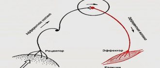
Reflex arc
The activity of the nervous system is based on reflexes (Latin reflexus - reflected). Reflex is the body's response to the action of a stimulus.
Any reflex exists on the basis of a reflex arc - a set of nerve elements connected to each other, through which a nerve impulse is sequentially transmitted during the implementation of the reflex. The simplest example is the knee reflex, which is often checked by a neurologist, which allows one to quickly draw a conclusion about the safety of the elements of the reflex arc.
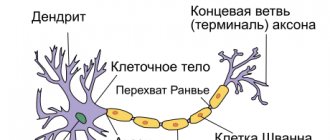
Neurons connect to each other using processes: axons and dendrites, at the end of which there are special contacts - synapses, which we studied in detail in the article about nerve tissue.
Reflex arc device
Reflex arcs can be very simple: consist of two neurons, like the reflex arc of the knee reflex (there is no interneuron), or they can include dozens of different neurons. The reflex arc can be divided into 3 parts:
- Sensitive (afferent, centripetal)
- Intercalary (associative, intermediate)
- Motor (efferent, centrifugal)
It consists of a receptor (can be located in the skin, internal organs, blood vessels) of a sensory neuron and a sensory fiber coming from this neuron, which penetrates the spinal cord through the dorsal horns.
The body of the sensory neuron is located in the dorsal roots (!) of the spinal cord. Introduced? Now imagine a dendrite running from the tip of your index finger all the way to your spinal cord. That is why it is incorrect to assume that a dendrite is always a “short” process, and an axon is always a “long” one. We discussed this issue in the article about nerve tissue.
Consists of an interneuron and its processes. The interneuron communicates between the sensory and motor parts of the reflex arc. Interneurons can communicate with other parts of the central nervous system.
The bodies of interneurons are located in the dorsal horns of the spinal cord.
Represented by a motor neuron (efferent, executive, motor neuron), from which nerve fibers go to the working organ (effector, executive organ).
Depending on what the effector is - a muscle, a gland - when nerve impulses arrive at it, its work is activated: the muscle begins to contract, the gland begins to secrete secretions.
Motor neurons lie in the anterior horns of the spinal cord, from where their processes emerge.
Let's consider the diagram of a reflex arc, on the basis of which the reflex of withdrawing a hand from a hot object is realized. Try to describe the path that a nerve impulse takes and remember the 3 parts of the reflex arc. Name the location of each neuron.
This may seem obvious, but it must be emphasized that afferent nerve fibers enter the spinal cord through the dorsal roots. Efferent nerve fibers exit the spinal cord through the ventral roots.
Types of reflex arcs
Reflex arcs are divided into somatic and autonomic. With the help of somatic reflex arcs, reflexes are carried out, providing the possibility of voluntary movements (performed by the will of a person). With the help of vegetative ones - coordination of the activity of internal organs, that is, functions that are not amenable to our conscious control (remember the autonomic nervous system).
Below you will see diagrams of somatic and autonomic reflex arcs. Under the picture there will be written a significant difference between them, which you must remember, but first try to draw a conclusion yourself by studying the picture.
The difference between the somatic and autonomic reflex arcs is that in the latter, the efferent neuron lies outside the spinal cord - in the autonomic ganglion. These ganglia can be located on the sides of the spine, near internal organs or in their wall.
Also, you most likely noticed that the interneuron of the autonomic arch is localized in a different place - in the lateral horns of the spinal cord (and not in the posterior ones, as in the somatic horn).
Nervous regulation
The reflex arc is the foundation on which the reflex is carried out. In the nervous system, not only processes of excitation occur, but also inhibition, which we will talk about in more detail in the topic devoted to higher nervous activity. Inhibition consists of weakening or delaying excitation that has already arisen.
Thus, coordination and regulation of the processes of excitation and inhibition is the basis for the coordinated work of organs and organ systems that make up a single organism.
Diseases
Paresis (Greek πάρεσις - weakening) is a neurological syndrome caused by damage to the motor (efferent) pathway and weakness in the limb, or in another organ that innervated this nerve pathway. Paresis is manifested by a decrease in muscle strength, movements are preserved in an incomplete volume.
Paralysis (Greek παράλυσις - relaxation) is the complete absence of voluntary movements, due to the same reasons as paresis.
Hypothermia may cause paresis of the facial nerve. The reason for this is tissue inflammation, as a result of which the nerve in a narrow bone canal is compressed by inflamed tissue. Nerve impulses partially or completely stop reaching the facial muscles, making it impossible for the patient to move them.
© Bellevich Yuri Sergeevich 2018-2021
This article was written by Yuri Sergeevich Bellevich and is his intellectual property. Copying, distribution (including by copying to other sites and resources on the Internet) or any other use of information and objects without the prior consent of the copyright holder is punishable by law. To obtain article materials and permission to use them, please contact Yuri Bellevich
.
How do they work
The function of a sensitive neuron is to receive a signal from special receptors located on the periphery of the body and determine its characteristics. The impulses are perceived by the peripheral processes of sensory neurons, then they are transmitted to their body, and then along the central processes they follow directly to the central nervous system.
The dendrites of sensory neurons connect to various receptors, and their axons connect to other neurons (interneurons). For a nerve impulse, the simplest path is the following - it must pass through three neurons: sensory, intercalary, motor.
The most typical example of the passage of an impulse is when a neurologist knocks on the knee joint with a hammer. In this case, a simple reflex is immediately triggered: the knee tendon, after a blow to it, sets in motion the muscle that is attached to it; Sensitive cells from the muscle transmit the signal through sensory neurons directly to the spinal cord. There, sensory neurons make contact with motor neurons, and they send impulses back to the muscle, causing it to contract, and the leg straightens.
By the way, in each section of the spinal cord (cervical, thoracic, lumbar, sacral, coccygeal) there is a pair of roots: sensory posterior, motor anterior. They form a single trunk. Each of these pairs controls its own specific part of the body and sends a centrifugal signal about what to do next, how to position a limb, torso, what to do to the gland, etc.
Sensory neurons take part in the work of the reflex arc. It consists of 5 elements:
- Receptor. Converts irritation into a nerve impulse.
- The impulse along the neuron follows from the receptor in the central nervous system.
- The interneuron, which is located in the brain, transmits a signal from the sensory neuron to the executive one.
- The motor (executive) neuron conducts the main impulse from the brain to the organ.
- An (executive) organ is a muscle, gland, etc. It reacts to the received signal by contraction, secretion, etc.
Order of interaction
The reflex regulation of body functions in an interpreted, simplified form is described in a biology textbook for the 8th grade. Interneurons, sensory and motor neurons are interconnected. The nature of the interaction depends on the type of functions of the nervous system. An approximate order of interaction in the case of the functions of sensory neurons that are localized in the skin area:
- Perception of an external stimulus by a nerve receptor located in the skin.
- Transmission of stimulus by sensory cells to areas of the brain. Typically, the signal passes through 2 synapses (in the spinal cord and thalamus), then enters the sensory zone of the cerebral cortex.
- Converting an impulse into a universal form.
- Transmission of the converted impulse to all cortical parts of the hemispheres with the help of interneurons, which are located only in the central nervous system.
Voluntary muscle movements are carried out due to the activity of motor neurons located in the cortical motor zone. Motor neurons initiate movement - the signal enters the skeletal muscles via efferent fibers. While the main signals sent by motor neurons travel to muscle tissue, the excitation spreads to other areas of the brain, for example, to the olive and cerebellum, where the planned action is fine-tuned.
Intercalary cells play the role of intermediaries, providing communication between efferent and afferent nerve cells.
Conclusion
The biology of the human body is very thought out and perfect. Thanks to the activity of many sensitive neurons, we can interact with this wonderful world and respond to it. Our body is very receptive, the development of its receptors and sensitive nerve cells has reached the highest level. Thanks to such a thoughtful organization of the central nervous system, our senses can perceive and transmit the smallest shades of taste, smell, tactile sensations, sound, and color.
We often believe that the main thing in our consciousness and the functioning of the body is the cortex and hemispheres of the brain. At the same time, we forget what enormous capabilities the spinal cord provides. It is the functioning of the spinal cord that ensures the receipt of signals from all receptors.
It is difficult to name the limit of these possibilities. Our body is very plastic. The more a person develops, the more opportunities are placed at his disposal. This simple principle allows us to quickly adapt to changes in the world around us.
A neuron is a specific, electrically excitable cell in the human nervous system and has unique characteristics. Its functions are to process, store and transmit information. Neurons are characterized by a complex structure and narrow specialization. They are also divided into three types. This article describes in detail the interneuron and its role in the action of the central nervous system.
Classification of neurons
The human brain has approximately 65 billion neurons that constantly communicate with each other. These cells are divided into several types, each of which performs its own special functions.
The sensory neuron plays the role of a transmitter of information between the sense organs and the central parts of the human nervous system. It perceives a variety of irritations, which it converts into nerve impulses, and then transmits the signal to the human brain.
Motor - sends impulses to various organs and tissues. This type is mainly involved in the control of spinal cord reflexes.
The interneuron is responsible for processing and switching impulses. The functions of this type of cell are to receive and process information from the sensory and motor neurons between which they are located. Moreover, interneurons (or intermediate) neurons occupy 90% of the human central nervous system, and are also found in large quantities in all areas of the brain and spinal cord.
Types and characteristics of neurons
Nerve cells called neurons receive, send, and conduct bioelectrical signals. There are efferent (motor) neurons - these are components of the central nervous system that redirect signals to executive organs, for example, skeletal muscles. Afferent (sensitive) neurons are cells that perceive external and internal stimuli, which ensures the body’s connection with the external environment and reactions to changes in the functional activity of internal organs.
Intercalary cells provide interconnections within the overall neural network. Neurons of all types (sensitive, efferent, associative) are functional units that support the activity of the nervous system; they are found in all tissues of the body, where they play the role of connecting links between receptor (perceiving irritating stimuli) and effector organs that respond to irritating stimuli.
Effector organs include muscles and glands, and receptor organs include sensory organs. The significance of the signals transmitted varies significantly depending on the type of cell and its role in the functioning of the central nervous system. For example, sensory neurons that perceive impulses from the external environment transmit signals from skin receptors and sensory organs in the direction of the brain; motor neurons redirect commands generated in the brain, causing contraction of skeletal muscles and initiating movement.
Despite the different meanings of bioelectric impulses, their nature is the same and consists in changing the electrical potential in the area of the plasma membrane of the nerve cell. The mechanism of propagation of nerve impulses is based on the ability of an electrical disturbance that appears in one place of the cell to be transmitted to other areas. In the absence of factors that enhance the signal, the pulses fade away as they move away from the source of excitation.
Sensory, also known as sensitive, is an afferent neuron that conducts impulses from distal parts of the body to the central parts of the central nervous system. For example, sensory fibers form fibers that extend from the light-sensitive cells of the organs of vision. Signals leave the retina, traveling along millions of axons belonging to the structures of the basal ganglia, towards a region of the visual cortex.
The sensory neuron, together with the executive (motor) neurons, forms a simple reflex arc.
For example, the knee reflex is an unconditioned stretch reflex reaction that occurs as a result of the activity of a similar reflex arc. A reaction in the form of uncontrolled extension of the lower leg occurs when there is a mechanical impact on the tendon of the femoral muscle, which lies under the patella. Reaction mechanism:
- Mechanical effects on the neuromuscular spindles located in the hip extensor muscle.
- An increase in the intensity of nerve signals in the endings entwining the neuromuscular spindles due to their stretching.
- Transmission of impulses to sensory neurons located in the spinal ganglia through dendrites extending from the femoral nerve.
- Transmission of impulses from sensory cells to alpha motor neurons located in the anterior horns within the boundaries of the spinal cord.
- Signal transmission from alpha motor neurons to contractile muscle fibers of the femoral muscle.
The knee reflex mechanism involves interneurons that transmit inhibitory impulses to motor neurons of the flexor muscles, and other interneurons, for example, Renshaw cells. The knee reflex mechanism also involves gamma motor neurons, which regulate the intensity of spindle stretch.
In the spinal cord, formed by gray matter, there are three types of neurons - motor, intercalary, and autonomic. Moreover, the vegetative ones are located in the visceral (related to the internal organs) nuclei. These cells interact with afferent (ascending pathways that transmit impulses from peripheral receptors to the central zones of the central nervous system) fibers responsible for general visceral sensitivity.
Visceral afferents conduct nerve signals (usually painful or reflex sensations) from internal organs, elements of the circulatory system, glands to the corresponding zones of the central nervous system. Visceral afferents are part of the autonomic nervous system. Reflex arcs within the autonomic department of the central nervous system differ in structure from the arcs of the somatic department.
Efferent components (descending pathways that transmit impulses from the cortical and subcortical zones of the brain to peripheral areas) are formed by two types of neurons - intercalary and effector (motor). The intercalary ones are located in the nuclei belonging to the vegetative part of the central nervous system. The name “intercalary” is due to its location between the sensory and motor neurons.
Sensitive
A sensory neuron is a component of the nervous system that transmits information to the brain about stimuli affecting a specific area of the body. Examples of irritants include factors: sunlight, mechanical impact (impact, touch), the action of a chemical substance. Sensory neurons are located in the ganglia of the brain - the spinal cord and brain.
The connection formed with a sensitive neuron can provoke excitation or inhibition, which is sent along nerve fibers to the cortical parts of the brain. As the level of sensory pathways increases, the transmitted information is processed to identify important features. Sensitive neurons belong to pseudounipolar neurons - their axon and dendrites extend from the body together, subsequently separate and are located in the spinal cord, brain (axon) and in the peripheral parts of the body (dendrites).
Insert
Interneurons transmit converted nerve impulses obtained as a result of processing sensory information received from various sources, for example, from the organs of vision and skin receptors. As a result, the processed information becomes the initial data for the formation of adequate motor commands.
Motor
Motor nerve cells are of two types - large and small. In the first case we are talking about α-motoneurons, in the second – about γ-motoneurons. Alpha motor neurons are present in the basal ganglia of lateral (closer to the lateral plane) and medial (closer to the median plane) localization. These are the largest cells present in nervous tissue.
Their axons interact with striated fibers contained in skeletal muscles. As a result, synapses (sites where nerve signals are transmitted) are formed. The axons of alpha motor neurons interconnect with intercalary analogues, also known as Renshaw cells, leading to the formation of collateral pathways and inhibitory synapses in the spinal cord.
Gamma motor neurons are part of the neuromuscular spindle, which is a complex receptor consisting of nerve endings (afferent, efferent). The main function of neuromuscular spindles is to regulate the force and rate of contraction or stretching of skeletal muscles.
The structure of intermediate neurons
An interneuron consists of a body, an axon and dendrites. Each part has its own specific functions and is responsible for a specific action. Its body contains all the components from which cellular structures are created. The important role of this part of the neuron is to generate nerve impulses and perform trophic function. The elongated process that carries the signal from the cell body is called an axon. It is divided into two types: myelinated and non-myelinated. At the end of the axon there are various synapses. The third component of neurons is dendrites. They are short processes that branch in different directions. Their function is to deliver impulses to the neuron body, which ensures communication between different types of neurons in the central nervous system.
Structure and functions
The intercalary cell consists of a body from which a single axon and dendrites extend. The dendrites of intercalary cells are often short. Their axons variably pass within the boundaries of the spinal cord from the posterior horns to the anterior ones (close the arc at the level of the spinal cord segment) or spread to other levels of brain structures - spinal, brain.
One of the functions of interneurons is inhibition of the intensity of certain signals. For example, interneurons of the neocortex (the new cortex responsible for higher mental functions - sensory perception, conscious thinking, voluntary motor activity, speech) selectively reduce the intensity of some signals coming from the thalamus to prevent the need to be distracted by extraneous, unimportant stimuli. If the impulse provoked by an external stimulus is not strong enough, it may fade before reaching the cortex of the brain.
The area of influence of intercalary cells is limited by individual structural features - the length of axonal processes, the number of collateral branches. Typically, intercalary ones are equipped with axons with terminals (the terminal section represented by the synaptic ending - the place of contact with other cells) ending within the same center, which determines integration within the group.
Interneurons close reflex arcs, they perceive excitation from afferent nerve structures, process data and transmit them to motor neurons. Associative cells play a leading role in the formation of neural networks, where the storage period of incoming and processed information is extended.
Sphere of influence
What determines the area of influence of an interneuron? First of all, his own structure. Basically, cells of this type have axons whose synapses end on neurons of the same center, which ensures their unification. Some interneurons are activated by others, from other centers, and then deliver information to their neural center. Such actions increase the impact of the signal, which is repeated in parallel paths, thereby extending the storage period of information data in the center. As a result, the location where the signal was delivered increases the reliability of the influence on the executive structure. Other interneurons can receive activation from connections of motor “brothers” from their center. Then they become transmitters of information back to their center, thereby creating feedback connections. Thus, the interneuron plays an important role in the formation of special closed networks that extend the storage period of information in the nerve center.
Excitatory type of interneurons
Interneurons are divided into two types: excitatory and inhibitory. When the former are activated, the transfer of data from one neural group to another is facilitated. This task is performed by “slow” neurons, which have the ability to activate for a long time. They transmit signals for quite a long time. In parallel with these actions, intermediate neurons activate their “fast” “colleagues”. When the activity of “slow” neurons increases, the reaction time of “fast” ones decreases. At the same time, the latter somewhat slow down the work of the “slow” ones.
The role of interneurons in the functioning of the spinal cord
In the functioning of the human spinal cord, an important role is played by the conduction pathways, which are located outside the bundles that perform the conduction function. It is along these paths that the impulses sent by the intercalary and sensory neurons move. Signals travel up and down these pathways, conveying various information to the corresponding parts of the brain. The interneurons of the spinal cord are located in the intermedial nucleus, which, in turn, is located in the dorsal horn. Interneurons are an important anterior part of the spinocerebellar tract. On the back of the spinal cord horn are fibers consisting of interneurons. They form the lateral spinothalamic tract, which performs a special function. It is a conductor, that is, it transmits signals about pain and temperature sensitivity first to the diencephalon, and then to the cerebral cortex itself.