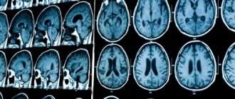
Electroencephalography is one of the ways to study biological systems using physical methods. In this case, we are talking about the study of the electrical activity of brain cells that make up both the membranes of the brain and internal structures. The uniqueness of electroencephalography is also that the method is completely non-invasive. Back in 1875, the English doctor Richard Cato and the Russian physiologist V.Ya. Danilevsky independently discovered that electrical impulses from the brain can be read from the surface of the scalp.
This fact adds another unique property to electroencephalography - the study of the functioning of the central nervous system in real time. Because The time it takes for an electrical impulse to travel from brain tissue to the surface of the skin is measured in milliseconds. It turns out that the electroencephalograph shows a picture of electromagnetic impulses that are present in the brain at a given moment. This makes it possible to study changes in electromagnetic activity in the central nervous system as accurately as possible under the influence of various factors. Positron emission tomography and magnetic resonance imaging do not have this advantage.
The essence of the method is that the patient lies down in a horizontal chair in a separate room, after which electrodes (21 pieces) are attached to his head. Electrodes are placed according to the 10-20% system. That is, for example, along the line from the bridge of the nose to the occipital protuberance: the first two electrodes are at a distance from the extreme points (the bridge of the nose and the occipital protuberance) at 10% of the line length, the next two electrodes from the first two electrodes are placed at a distance of 20% from line length, the last fifth is fixed in the parietal region. The doctor is in the next room and communicates with the patient through a speaker. The patient is then subjected to several simple testing methods:
- the patient is asked to close his eyes and relax;
- the patient is asked to imagine some visual images;
- turn off the lights in the room for a short time;
- The patient is exposed to flickering light (flashes) for a few seconds - an important experience for identifying epilepsy, etc.
Previously, the results of the encephalogram were recorded on paper tape, but now the recording occurs immediately in digital form on a computer and is saved as a file. Of course, the electrical activity of the brain takes the form of continuous electromagnetic signals. It is impossible to record them directly in such a continuous flow. We need some kind of discrete frequency. This is when the device records the value of electrical impulses at certain intervals. In the general scheme: the more often such fixation occurs, the more accurate the measurement. According to Kotelnikov's theorem, adequate restoration of the original signal is possible only if the sampling frequency is at least twice as high as the frequency of the signal being studied. In electroencephalography, a frequency of 250 Hertz was selected. This is more than enough, since all known electromagnetic impulses of the brain vary in their natural frequency from 3 to 100 Hertz.
Our mind is a colossal neural network.
When we think and experience various emotions and feelings, special cells, neurons, interact with each other through special processes called axons. This type of interaction is of an electrochemical nature. The work of the brain is accompanied by electrical activity caused by discharges of its cells (neurons). The electrical activity of the brain is small and is expressed in millionths of a volt. When large groups of neurons (hundreds of thousands) interact simultaneously, the resulting electrochemical activity generates an electric field of sufficient power to be detected from the outside of the head.
The electrical impulses that the brain constantly generates are called brain waves (brain rhythms, waves of brain activity). The frequency of these pulses is measured in hertz or cycles per second. Well, the dominant frequency of brain waves determines the general state of the brain.
Why dominant? The thing is that the brain does not work as a whole at one frequency. This means that one area of the brain may produce more beta waves while other areas of the brain emit impulses at a different frequency. In general, he may be in a state of calm relaxation, for example, but part of the subcortex will be “itching” about stress and problems at the background level.
Waves (rhythms) of human brain activity are divided by scientists into five main types: delta, theta, alpha, beta and gamma.
There are studies indicating that the rhythms of electromagnetic oscillations of our brain are directly related to electromagnetic oscillations between the Earth's surface and the ionosphere, coinciding with them in the main resonant frequencies. Probably, here is the key to the existence of large and small rhythms of the world’s existence, some of which are represented in a person in different ways, and some, resonant with them, in the surrounding space. Just as a guitar string makes a sound in unison with a tuning fork, just as a bridge begins to vibrate in resonance with the wind, and so on, so we can tune in to different cycles and frequencies in the world, entering into resonance with them.
Electrical oscillations of the brain (brain rhythms) can be recorded using a special highly sensitive device called an electroencephalograph (EEG graph), equipped with sensitive wet sensors located in certain places on the head and adjacent to the scalp, as well as an amplifier that allows recording a weak electrical signal from the brain. brain and software for a thorough mathematical analysis of detected electromagnetic oscillations.
History of the appearance of EEG.
The first person who needs to be mentioned as the person behind the creation of the modern EEG was the German physiologist Emil Heinrich Dubois-Reymond. He is considered the father of electrophysiology. He was able to establish some patterns that characterize electrical signals in nerves and muscles. In the context of describing the history of EEG, Emil Heinrich Dubois-Reymond is more interesting to us because it was he who showed in 1849 that the brain also has electrogenic properties. That is, it has electogenesis - the ability to generate weak currents.
The second significant person in the history of the emergence of modern EEG technologies is undoubtedly the Russian scientist Vladimir Vladimirovich Pravdich-Neminsky. It was he who, in 1913, while working at the Department of Physiology of Kyiv University, obtained the world's first electroencephalogram of the brain of a living creature. In his experiments he used the brain of a dog. It is also extremely important that the EEG of the dog’s brain was obtained without any damage to the animal’s scalp, that is, without the use of invasive technologies, opening the skull, and so on, from the outside of the skull. The EEG was obtained using a string galvanometer. Professor Pravdich-Neminsky introduced the term “electrocerebrogram” - a recording of the electrical activity of the brain; proposed the first classification of electroencephalogram frequencies, which formed the basis of modern classifications; discovered rhythmicity in brain activity.
At the same time, it is worth noting the English doctor Richard Cato. In 1875, he showed the very fact of the generation of electric current by the human brain. Thus, it is quite difficult to determine who exactly should recognize the priority of this discovery.
Despite all the significant achievements of the researchers described above, they failed to go down in history as the “fathers” of modern EEG technologies. In scientific historiography, the German physiologist and psychiatrist Hans Berger is considered the “father” of electroencephalography of the human brain.
Hans Berger in 1924 recorded on paper a curve obtained using a galvanometer reflecting the electrical activity of the human brain. Brain activity signals were obtained from the subject's scalp. Hans Berger also began detailed studies of the received signals, based on the signal amplitude. Until now, Alpha waves in the range of 8-12 Hz are called Berger waves. Thus, we repeat, Hans Berger is considered to be the father of EEG, and 1924 is considered to be the year the EEG of the human brain appeared.
All these first steps were the start for EEG research. In subsequent years, many discoveries were made. Research has been carried out, which continues to this day, in which it was established what rhythms, frequencies, and signal levels are responsible for this or that form of activity of the human brain.
The mechanism for obtaining an electroencephalogram of the human brain.
Electroencephalography is a method for studying the bioelectrical activity of the brain, based on determining the difference in electrical potentials generated by neurons during their life. The recording electrodes are positioned so that all major parts of the brain are represented in the recording. The resulting recording, EEG, is the total electrical activity of millions of neurons, represented primarily by the potentials of dendrites and nerve cell bodies: excitatory and inhibitory postsynaptic potentials and partly by the potentials of dendrites and nerve cell bodies. That is, EEG is a kind of visualized result of the functional activity of the brain.
Now, for those who want to learn more about the generation of an electrical signal by neurons, we will allow ourselves to quote a description of this process in a dry, scientific form. So, a little dry science:
A neuron is the main cell of the central nervous system. The shapes of neurons are extremely diverse, but the main parts are unchanged in all types of neurons: the body and numerous branched processes. Each neuron has two types of processes: an axon, along which excitation is transmitted from a neuron to another neuron, and numerous dendrites (from the Greek tree), on which axons from other neurons end in synapses (from the Greek contact). The neuron conducts excitation only from the dendrite to the axon.
The main property of a neuron is the ability to excite (generate an electrical impulse) and transmit (conduct) this excitation to other neurons, muscle, glandular and other cells.
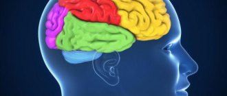
Neurons in different parts of the brain perform very diverse jobs, and accordingly, the shape of neurons from different parts of the brain is also diverse.
Many years of research in the field of neurophysiology have led to the conclusion that the following electrical events are inherent in neurons and can contribute to the total bioelectrical activity of the brain (EEG): postsynaptic excitatory and inhibitory potentials (EPSPs, IPSPs), and propagating action potentials (APs). EPSPs and IPSPs arise either in the dendrites or on the soma of the neuron. APs are generated in the area of the axon “mound” and then propagate along the axon.
Conventional spontaneous EEG, its basic rhythms arise as a result of the spatial and temporal summation of postsynaptic potentials (PSPs) of a large number of cortical neurons. The temporal characteristics of the summation process are quite slow compared to the duration of the AP.
A certain degree of synchronization is set by various subcortical structures that act as a “pacemaker” or pacemaker. Among them, the thalamus plays the most significant role in the generation of EEG rhythms.
Both postsynaptic potentials and action potentials take part in the generation of EEG. The basic rhythm of the EEG is determined by gradual changes in postsynaptic potentials due to the spatial and temporal summation of individual PSPs in large populations of neurons that are relatively synchronized and under the influence of the subcortical pacemaker.
That is, if we express the above in a simple form, we can say the following. When large groups of neurons interact, the electrical activity of this interaction becomes so powerful that it seems possible to obtain it from the surface of the human head. This received signal is called the raw signal. Many years of research have shown that certain ranges of this general spectrum are responsible for certain forms of brain activity. The science of electroencephalography, neurophysiology, psychiatry and psychology are trying to determine and identify patterns of certain combinations of levels of certain waves in the overall raw signal. This kind of information is the most valuable for the practical application of consumer-grade EEG devices, since it is precisely this that determines the scope of applications in which they can be used. Now it’s worth touching on the classical idea of ranges in the overall spectrum of the raw EEG signal. Let’s immediately make a reservation that we will be talking specifically about the five classical ranges that were identified quite a long time ago. In fact, in modern science, and even in a number of consumer devices, the total spectrum is divided into a larger number of frequency ranges.
Classic ranges in raw EEG signal.
Delta waves: range 0 to 4 Hz
Theta waves: range 4 to 8 Hz
Alpha waves: range from 8 to 12 Hz. In some programs, this range is represented as Low Alpha (8-10 Hz) and High Alpha (10-12 Hz)
Beta waves: range from 12 to 30 Hz. In some programs, this range is represented as Low Beta (12-18 Hz) and High Beta (18-30 Hz)
Gama waves: range from 30 to 70 Hz. In some programs, this range is represented as Low Gama (30-50 Hz) and High Gama (50-70 Hz)
Delta rhythm - from 0.5 to 4 oscillations per second, amplitude - 50-500 µV. This rhythm occurs both during deep natural sleep and during narcotic sleep, as well as during coma. Low-amplitude (20–30 μV) fluctuations in this range can be recorded at rest during some forms of stress and prolonged mental work. Delta waves are the slowest on the spectrum and are typically associated with deep, dreamless sleep. Normally, the level of these waves in the signal decreases when a person tries to focus. An increase in the level of delta activity is associated with a decrease in the level of awareness of the surrounding space and the level of awareness of information associated with the unconscious. At the age of 75, Delta waves, as indicators of deep sleep, may not be represented at all in the spectrum. Interestingly, these waves dominate in the EEG of children under one year of age. In a state of deep sleep, which is characterized by high Delta activity, the most effective restoration of the body occurs. There are also certain properties of Delta waves that have not yet been studied. In particular, some sources claim that Delta activity is characteristic of states of manifestation of intuition, some manifestations of the unconscious that we are not aware of.
Theta rhythm (θ rhythm) - EEG rhythm Frequency 4–8 Hz, high electrical potential 100–150 microvolts, high wave amplitude from 10 to 30 μV. The theta rhythm is most pronounced in children from two to five years old. This frequency range promotes deep relaxation of the brain, good memory, deeper and faster assimilation of information, awakening individual creativity and talents. For the most part, in children under 5 years of age, the brain functions in this wavelength range during the daytime, which allows children to phenomenally remember a huge amount of various information, which is unusual for adolescents and adults. In the natural state, this rhythm dominates in the majority of adults only during the REM sleep phase, half-asleep. Characteristic for deep meditation-dhyana. It is in this frequency range that the brain has enough energy to absorb large amounts of information and quickly transfer it to long-term memory, learning abilities are enhanced and stress is relieved. In this range, the brain is in a state of heightened sensitivity. This state is ideal for superlearning; the brain is able to maintain concentration and extroversion for a long time and is not susceptible to anxiety and neurotic manifestations.
This is the range of the upper connections of the brain, connecting both hemispheres and directly the layers of the cerebral cortex with its frontal zones. All actions that are memorized to the point of automatism are also characterized by high levels of Theta waves. For example, driving a car on an open road or taking a shower. These waves also display states of inspiration, unexpected manifestations of creative ideas, etc. Children tend to exhibit stronger Theta activity on the spectrum.
Alpha rhythm (α rhythm, alpha rhythm) is an EEG rhythm (electroencephalogram) in the frequency band from 8 to 13 Hz, the average amplitude is 30–70 μV, however, high and low amplitude α waves can be observed. It is registered in 85–95% of healthy adults. It is best expressed in the occipital regions. The α rhythm has the greatest amplitude in a state of quiet wakefulness, especially with eyes closed in a darkened room. It is blocked or weakened by increased attention (especially visual) or mental activity.
The alpha rhythm characterizes the process of a person’s internal “scanning” of mental images when focusing on some mental problem.
When we close our eyes, alpha rhythms intensify, and this property is successfully used during meditation-relaxation or a hypnosis session. For most people, alpha waves disappear when they open their eyes and a real picture appears before them. Statistical and experimental data indicate that the nature of the alpha rhythm is innate and hereditary.
Most people with a clearly defined alpha rhythm have a predominant ability for abstract thinking. A small group of people exhibit a complete absence of alpha rhythms, even with their eyes closed. These people think freely in visual images, but have difficulty solving problems of an abstract nature.
People who have learned to analyze information when their brain works in the alpha rhythm have access to much larger volumes of information, creative ideas and inspired thoughts are more likely to come to them, intuition is sharpened, which allows them to find new unexpected solutions to problems. No wonder they say: “Close your eyes, and the solution will come by itself.”
When the brain works in the alpha rhythm, a person's potential for managing his life increases. An understanding comes of how to better deal with various life problems, such as insomnia, anxiety, tension, migraines, bad habits and much more. There is an opportunity to learn how to adjust your psyche in such a way as to achieve your goals and turn your dreams into reality.
The work of the brain in the alpha rhythm allows you to quietly enter a state of shallow meditation, as during auto-training and relaxation exercises. Scientists have found that when a person engages in such practices, at the physiological level there is a decrease in the rhythm of brain functioning to the level of alpha rhythm. Taking a warm bath or shower can also increase alpha activity in the brain.
Why is the alpha rhythm so remarkable and why does the human body need it? Everything depends on the human consciousness. In a state of complete relaxation and immersion, alpha waves intensify, and healing processes begin to take place in our psyche, hidden resources awaken: intuition comes to life, concentration becomes perfectly honed, and extrasensory abilities appear. The world around begins to play with completely different colors, making a person joyful.
When you disconnect from your surroundings, close your eyes, and allow images to appear in your mind on their own, the Alpha state occurs. Alpha waves reflect the connection between consciousness and the subconscious. Training your consciousness to enter the Alpha state will be very useful, as well as training in a meditative state, as well as training in everyday stress resistance. Also, the Alpha state is very desirable for the brain to assimilate new information material, learn, and perform non-standard tasks that require action to develop them. Alpha activity can be increased if you close your eyes, relax, and begin to breathe deeply.
The decrease in the level of Alpha waves can be affected by the following: concentration on a certain task, attentiveness, opening the eyes after relaxation. It is extremely important that in the spectrum of brain waves of an ordinary person, Alpha waves are sure to show their activity. In this state, a person looks at the world positively and solves creative problems with ease. Some studies have shown that people whose EEG reveals permanently low Alpha levels under normal conditions are prone to alcoholism and drug addiction.
Low Alpha.
The waves of this part of the spectrum are more associated with relaxation, a state of alienation from the environment. We can say that this is, in a sense, a transitional state to the Theta state.
High Alpha.
The waves of this part of the spectrum are more associated with a state of anxiety and a state of focus. Reflects an increased level of composure and mental stability.
Beta rhythm (β rhythm) is a low-amplitude oscillation of the total brain potential with a frequency of 15 to 35 oscillations per second, amplitude - 5-30 μV. This rhythm is inherent in the state of active wakefulness. Refers to fast waves. This rhythm is most strongly expressed in the frontal areas, but with various types of intense activity it sharply intensifies and spreads to other areas of the brain. Thus, the severity of the beta rhythm increases when a new unexpected stimulus is presented, in a situation of attention, during mental stress, and emotional arousal. Their amplitude is 4–5 times less than the amplitude of alpha waves.
In a state of beta rhythm, our brain is immersed in the routine of life with a huge number of different problems, in an endless cycle of stressful situations, solving various problems and active concentration, a moving focus of attention. Attention is directed outward.
The beta rhythm is by no means our enemy. It was thanks to the beta rhythm that humanity reached immeasurable heights in technological progress: it built cities, went into space, created television, computers; The development of medicine is also directly related to these waves. This is the rhythm of active creation and life. Beta waves are our waking rhythm. These waves are associated with active thinking, active attention and focus on the world around us. Beta activity is especially strong when you solve problems, judge, and make difficult decisions. Beta waves are also actively emitted when a person is excited, excited or scared. With an increase in Beta activity, the efficiency of the brain, assimilation and processing of information increases. Interestingly, if the level of internal anxiety increases, then the level of Beta waves in the EEG increases, at the same time, with an increase in muscle activity, the level of Beta waves decreases.
Low Beta.
This part of the spectrum is more characteristic of states of focus, concentration, and active reflection. It is also more characteristic of a state of physical relaxation in a mental state of anxiety. Low Beta waves are also commonly associated with peak performance in training athletes. Also common in mental tasks such as reading, math, and problem solving.
High Beta.
Waves in this part of the spectrum are usually associated with vigilance, alertness, agitation, and excitement. The high level of these waves is reflected in a state of panic.
Gamma rhythm (γ rhythm) - fluctuations in EEG potentials in the range from 30 to 120–170 to fluctuations per second. The amplitude of the gamma rhythm is very low - below 10 μV and is inversely proportional to frequency. If the amplitude is higher than 15 μV, then the EEG is considered pathological. The gamma rhythm is observed when solving problems that require maximum concentrated attention. The gamma rhythm reflects oscillations that are simultaneously triggered in neurons by an incoming signal from the activating system of the reticular formation, causing a shift in the membrane potential.
The gamma rhythm is observed when solving problems that require maximum concentrated attention. This is the rhythm of composure and concentration on a problem or task, the rhythm of an active collected solution and work. There are theories connecting this rhythm with the work of consciousness. A number of publications report various disorders of gamma activity in patients with schizophrenia.
Gamma rhythm is also a state of communication between a person and “something” that is beyond the understanding of our consciousness. The brain vibration frequency of 50 Hz is called enlightenment by some researchers of meditating Buddhists. It is possible that this is simply the frequency of maximum concentration, presence here and now. That is, the gamma rhythm allows us to become someone greater and perceive the world from the point of view of this greater one. It's like a superstructure on human consciousness that we can use.
These waves reflect the cognitive processes taking place in the mind. They reflect the consolidation of information, that is, its transition from short-term memory to long-term memory. In a state of predominance of Gamma waves, insights occur. High levels of Gamma waves are associated with intellectual activity, compassion, empathy and self-control. A correlation was also identified between Gamma waves and transcendental states of consciousness. In general, these waves, being the fastest, are a reflection of the peak work of consciousness.
Low Gamma.
This part of the spectrum manifests itself during learning, activities and mental activity. Well and consistently demonstrated Gamma activity at a frequency of 40 Hz is a reflection of good memory and high efficiency in solving problems, both in children and adults. Accordingly, it is shown that a low level of waves in a given frequency range reflects a low level of ability to remember information.
High Gamma.
This part of the spectrum is associated with cognitive tasks such as reading, listening, and speaking. The low level of waves in this part of the spectrum may be associated with the cessation of cognitive activity.
It should be noted that a person’s psycho-emotional states are described not by a specific wave and its level, but by their combination.
Scientists have eavesdropped on a person's internal speech using the impulses of brain neurons
The authors of the article prepared it with the active help of 15 volunteers who suffered from epilepsy, which could only be cured by surgery. Before the operation, doctors implanted many electrodes into their brains to determine the focus of activity during seizures. Neuroscientists have used them for another purpose: monitoring the brain's electrical activity in the hearing center.
The researchers invited their subjects to a private room in the clinic, where they were asked to put on headphones and listen to a five-minute monologue or dialogue. During this time, scientists recorded signals received by electrodes in their brains.
Knight and his colleagues used data from this experiment to create an algorithm that describes the possible combinations of active and inactive nerve cells in the hearing center at the moment a person hears a particular word.
“We look at which areas of the cerebral cortex activity increases at the moment when a person hears sounds of a certain frequency, and from the combination of these areas we restore the original sound. This can be compared to how a good pianist can “hear” the music from the sequence of keys that another musician presses in a video without sound,” explained one of the team members, Brian Pasley from the University of California at Berkeley.
The scientists tested the operation of their invention, once again using the help of the same group of volunteers. This time, a one-word audio recording was played in their headphones, after which a computer program tried to reproduce it from the recorded nerve impulses.
“I didn't think it would work, but Brian managed to make it happen. “His computational model can reproduce the sounds that patients heard, and we could actually understand the overheard version of the word, although its quality left much to be desired,” Knight added.
Scientists believe that their invention can be adapted to create “neuroprostheses” that will return the gift of speech to people who have lost it for one reason or another.
“Recent experiments with neuroprosthetics have shown that the brain can control the movement of artificial limbs. Designing and building usable speech synthesizers will undoubtedly be more difficult than creating artificial limbs. Nevertheless, our experiment raised the level of research in the field of language reconstruction to a new level,” the authors of the article conclude.
Electromagnetic rhythms of the brain
Over several decades of practice, many rhythms have been identified in the electromagnetic activity of the brain, differing in frequency, amplitude and phase. It was studied in which situations which biorhythms predominate. Which factors enhance some biorhythms and which suppress them. Correlations of changes in biorhythms in certain diseases of the central nervous system were studied. Theoretically, the human brain has thousands of biorhythms. However they are very weak. So much so that it is not possible to study them with an encephalograph. A little about the basic electromagnetic rhythms of the central nervous system:
- Alpha rhythm (8-13 Hz, amplitude from 5 to 100 microvolts (µV)). It is observed in almost all healthy people. The maximum amplitude of this rhythm is observed in a state of relaxation and rest (but not drowsiness or sleep). Any increase in the functional activity of the central nervous system (intellectual/emotional/tension due to motor activity) is a depressive factor for the alpha rhythm. Chronically low alpha rhythm may indicate prolonged stress, depressive states, and neuroses.
- Beta rhythm (14-40 Hz, up to 40 µV). Normally, it is weak (amplitude no higher than 7 microvolts). Generated by the region of the anterior and central gyri, it affects the posterior frontal gyri. Increased beta rhythm is associated with intellectual processes, and depression is associated with physical motor activity (the beta rhythm fades even with imaginary motor activity).
- Gamma rhythm (one of the highest frequencies from 30 to 100 hertz, amplitude rarely exceeds 15 µV). Found in the temporal lobe, parietal cortex, as well as in the precentral and frontal zones. Its strengthening is associated with solving problems that simultaneously require great cognitive stress and maximum focus.
- Delta rhythm (1-4 Hz, but the amplitude is one of the largest from 20 to 200 μV). Delta-ri, since it is closely related to rest and recovery processes. A pronounced delta rhythm may indicate neuropsychiatric disorders and problems with focusing attention. The detection of a strong delta rhythm in atypical areas of the brain may indicate that some kind of neoplasm is located near this area.
- Theta rhythm (4-8 Hz, 20-100 μv). “Rhythm of drowsiness”, because in the physiological norm, it is most clearly expressed in a state on the verge of sleep and wakefulness (the state of drowsiness). Increased theta waves help you fall asleep completely. The theta rhythm is also observed when a person concentrates on analyzing memories. A prolonged abnormally high level of this rhythm occurs as a result of exposure to narcotic drugs (state of altered consciousness), with psychotic pathologies, and concussion.