Find out more about other diseases starting with the letter “C”: Compression of the brain; Senile chorea; Sensitive ataxia; Serous meningitis; "Rigid person" syndrome; Alien hand syndrome; Restless legs syndrome; Bogorad syndrome; West syndrome; Gaye-Wernicke syndrome; Guillain-Barre syndrome; Piriformis syndrome; Carpal tunnel syndrome; Carotid sinus syndrome; Kleine-Levin syndrome; Klippel-Feil syndrome; Cauda equina syndrome; Crumpy syndrome; Lambert-Eaton syndrome; Landau-Kleffner syndrome.
Cauda equina syndrome is a system of neurological symptoms in which the bundle of nerve fibers in the region of the spinal cord formed by the lumbosacral nerves of the conus medullaris is damaged. Clinically manifested by decreased tone of the legs, sensory disturbances in the area of innervation of damaged nerves, and dysfunction of the pelvic organs.
The diagnosis is established after consultation with a neurologist, based on the results of MRI, CT, and spinal puncture. Treatment can be conservative or surgical with further rehabilitation.
general information
The spinal cord ends at the level of the first lumbar vertebra. The nerves of the lumbar, sacral and coccygeal segments and the filum terminale form a bundle called the cauda equina. Vertebroneurologists distinguish damage to the cauda equina as a separate disease, regardless of the reasons that influenced it.
The syndrome appears in middle-aged people more often than in other patients. Children get cauda equina syndrome after injuries or pathologies of the lower parts of the spinal canal. In men, the disease is more common in women; it is associated with lifting heavy weights.
Symptoms
Symptoms of cauda equina syndrome include:
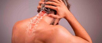
- Pain in the lumbar region
- Radicular pain in one or both legs (pain begins in the buttock and radiates down the back of the thigh and lower leg).
- Numbness in the groin or in the area of the sacrum, coccyx
- Bladder and bowel dysfunction.
- Muscle weakness in the lower extremities and decreased sensitivity
- Decreased or absent reflexes in the lower extremities.
- Pain in the lumbar region can be divided into local and radicular pain.
- Local pain is a deep, chronic pain that occurs due to irritation of the soft tissues and vertebral body.
- Radicular pain is an acute pain that occurs as a result of compression of the nerve roots. Radicular pain is projected in the dermatomes (along certain areas that are innervated by a given root).
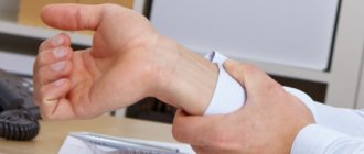
Bladder dysfunction in cauda equina syndrome is manifested by:
- Urinary retention
- Difficulty starting to urinate
- Decreased sensitivity of the urethral sphincter.
- Bowel problems may include the following symptoms:
- Incontinence
- Constipation
- Decreased tone and sensitivity of the anus.
Cauda equina syndrome is a serious surgical condition. Patients should seek medical attention if they have symptoms. Often, lower back pain and muscle weakness are associated with a disc herniation and do not require emergency surgical intervention.
Clinical studies have shown that the likelihood of regaining function and having a positive outcome is directly related to the duration of symptoms of cauda equina syndrome. Within 48 hours of the onset of symptoms of the cauda equina, surgical decompression is necessary - surgical removal of tissue causing compression of the roots (such as a herniated disc, osteophyte).
Causes of cauda equina syndrome
The main factor in damage to the terminal nerve bundle is its compression in the spinal canal. Among the most common causes, intervertebral hernia in the lumbar spine plays a key role. Sometimes inflammatory, external causes are found. The main underlying factors of the syndrome:
- Intervertebral hernia explains 15% of cases of the disease. Osteochondrosis of the spine and degenerative processes of intervertebral discs give rise to the formation of protrusion and accelerate its development. Lumbar hernias compress the cauda equina.
- Injuries. Fracture of the sacral vertebrae and spinal cord injury of the corresponding part cause defects in the nerve fibers. Compression can be generated by hemorrhages and hematomas. Cauda equina syndrome can become a complication of epidural anesthesia and surgery.
- Neoplasms. Tumors of a malignant nature grow into nerve bundles, damaging nerve tissue. Benign neoplasms arise from nerve sheaths (neurinoma), spinal ependymoma (ependymoma), fat cells (lipoma), meninges (meningioma) - all of them cause compression of the nerve roots.
- Curvature of the spine. Inherited pathologies of the spine in the lumbosacral region can significantly narrow the terminal part of the spine, creating conditions for compression damage to nerve fibers. Acquired deformities lead to age-related destruction (spondyloarthrosis deformans), dislocation of the lumbar vertebrae (spondylosis).
Causes of pathology
There are many reasons for the development of cauda equina pathology:
- the nerve endings can be compressed by the resulting intervertebral hernia;
- progressive osteochondrosis;
- congenital anomalies of the structure of the spinal column;
- tumors and metastases;
- mechanical damage to the spine;
- infectious diseases (for example, epidural abscess);
- spondylolisthesis (a pathology in which one vertebra slips off another).
When performing various types of operations in medicine, epidural anesthesia is practiced, that is, when an anesthetic substance is injected through a catheter into the epidural space of the spine. If the technology for performing such a procedure is violated and nerve endings are damaged, cauda equina syndrome may occur in humans.
Pathogenesis of the disease
The lumbar and sacral nerves, which are part of the structure of the cauda equina, provide innervation to the lower extremities, bladder, external genitalia, urethra, and rectal sphincter.
At the onset of the disease, a violation of their functionality determines irritation and increased excitation of nerve endings, with the presence of severe pain. The more the destruction or compression of nerve endings progresses, the more innervating functions are inhibited, sensitivity decreases, and muscle paresis occurs. Neoplasia, which is malignant in nature, destroys the membranes of the spinal canal and provokes metastases.
Treatment methods
The roots of the cauda equina are extremely sensitive to prolonged compression, so all therapeutic measures should be performed no later than two days after the development of the syndrome. Otherwise, even after them, a persistent neurological deficit often remains. These measures consist of emergency decompression (removal of compression) of the nerve trunks. For this purpose, Israeli clinics use a variety of surgical techniques:
- laminectomy. Through an incision of no more than 2 cm, the posterior bone structures of the vertebra are removed in portions using a high-speed diamond bur, thus increasing the space for the nerve structures;
- operations on intervertebral discs. They are most often carried out for decompression of the cauda equina, since intervertebral hernias in most cases are the cause of its compression. This is a wide range of interventions from classic open discectomy to microsurgical nucleoplasty. We perform puncture and open laser discectomy, chemonucleolysis, cold plasma coagulation of a disc herniation, electrocoagulation of a hernia, percutaneous discectomy, endoscopic discectomy;
- stabilizing operations on the lumbar spine, when the cause of compression of the roots of the cauda equina is spondylolisthesis, trauma or tumors of the vertebrae. These are all types of vertebroplasty with modern composite materials, arthrodesis, intervertebral disc replacement, installation of stationary and dynamic vertebral fixation systems.
Sometimes the disease that caused the development of cauda equina syndrome is cured radically and simultaneously with decompression. For example, a herniated disc, tumor, hematoma, or broken bone fragments are removed. In other cases, at the first stage only decompression is performed, for example, the vertebral arch is removed (laminectomy), and after some time an operation is performed for the underlying disease.
Minimally invasive operations using endoscopic and microsurgical equipment are widely practiced. In this case, the external incision is only 5-10 mm long. As a result, the paravertebral and intervertebral muscles and ligaments are practically not injured, which ensures an excellent functional result of treatment.
Israeli neurosurgeons work under conditions of excellent visibility of the surgical field. The endoscope's video camera transmits a color three-dimensional image of the smallest structures onto the screen of a large monitor, and when working without an endoscope, doctors use operating microscopes that provide 8-15x magnification. All this allows us to act extremely precisely and carefully in relation to healthy tissues.
Clinical picture
Pain appears at the very beginning of the disease. It is concentrated in the lumbar and sacral region, and is remotely felt in the leg on the side of the injury, the groin area. The patient experiences hypersensitivity and poor tactile sensations in terms of painful sensations. When coughing and sneezing, the pain increases significantly, and decreases in a semi-sitting position. A decrease in sensitivity, felt as numbness, indicates increasing hypoesthesia.
Complaints of rapid fatigue while walking and lethargy of the muscles of the lower extremities are signs of motor dysfunction. The more active the pathological process becomes, the more severe the symptoms, and the damage is distributed to the other side of the body. Over time, independent walking becomes difficult.
Disruption of the innervation of the external genitalia leads to a significant decrease in their sensitivity. This, in turn, causes anorgasmia in women and erectile disorders in men. The pelvic organs react with urinary retention and persistent constipation to sensory impairment. Patients are not aware of fullness of the bladder and rectum.
Complications of the disease
If treatment is not complete or insufficiently adequate, the patient may suffer disability associated with walking pathology. A disorder in the perception of fullness of the bladder and rectum can provoke the formation of a diverticulum, infected urethritis, cystitis, pyelonephritis along the ascending route of infection. Untimely removal of metabolic products can cause the accumulation of toxins in the intestines and general poisoning of the body. Fecal blockage and acute urinary retention are conditions that require urgent medical attention.
Prevention
Prevention of equine syndrome is aimed at identifying the symptoms of this syndrome. In most cases, patients with lower back pain, leg pain, or leg weakness do not develop cauda equina syndrome. Bowel or bladder symptoms or sensory disturbances in the groin area should be of particular concern.
Irreversible changes in nerve fibers often occur and surgical intervention does not eliminate persistent neurological deficits. Therefore, the patient will have to live with this deficiency. Therefore, in such cases, the support of a number of specialists such as a physiotherapist, social worker, and sexologist will be necessary. In addition, it will be necessary to adhere to certain rules if you have bladder and bowel dysfunction:
- Use a catheter to drain urine from the bladder (3-4 times a day).
- Drink enough fluids and practice good hygiene to prevent urinary tract infections.
- If you have constipation, do a cleansing enema or use suppositories.
- Wearing sanitary pads for possible episodes of incontinence.
In addition, it is necessary to regularly consult with your doctor regarding the presence of pain or episodic natural bowel movements. The outlook for cauda equina syndrome depends on the duration of symptoms before surgical treatment. The longer a patient has had symptoms before appropriate treatment, the less likely they are to make a full recovery.
Diagnosis of cauda equina syndrome
The initial signs of cauda equina syndrome can be mistaken for clinical symptoms of femoral nerve neuropathy, radiculitis, and lumboischialgia. It should be remembered that the manifestations of cauda equina syndrome have bilateral manifestations. It is necessary to question the patient in detail about the presence of disorders of the pelvic and genital organs.
To confirm the diagnosis, be sure to perform:
- Neurological examination. During a consultation with a neurologist, mono or paraparesis, decreased muscle tone, and degenerative disorders are discovered. Physiological reflexes (Achilles, anal, in men - bulbocavernosus) are not expressed. Sensitive disorders that occur in areas of innervation of nerve endings indicate injury to several lumbosacral nerves.
- CT scan. Spinal stenosis and pathologies of bone structures are clearly visible on CT images.
- Magnetic resonance imaging. Insufficient detection of soft tissue pathologies on CT requires MRI. The resulting images determine the presence of hematomas and neoplasia. The level of narrowing of the spinal canal is determined more accurately.
- Spinal tap. If during the procedure it is not possible to obtain cerebrospinal fluid (dry puncture), it is clear that the cerebrospinal fluid pathways are blocked by a tumor or hernia. There may be blood in the cerebrospinal fluid, which indicates hemorrhage. An increase in the amount of protein indicates neoplasia, signs of inflammation - arachnoiditis, myelitis.
- Histological analysis. It is performed in the presence of tumors to determine their structure, malignant or benign nature. The examination is carried out after the operation by examining the removed tissue.
Differential diagnosis is carried out to exclude polyneuropathies, mononeuropathies of the lower extremities, and lumbosacral plexitis. To eliminate other pathologies, the results of magnetic resonance imaging and computed tomography are important.
Diagnostics
The success of treating cauda equina entrapment syndrome in humans and eliminating the symptoms of this disease directly depend on the diagnosis. Like many other diseases of the nervous system, cauda equina syndrome is quite difficult to diagnose. The following studies can be used to identify pathology:
- Radiography
. Most often, patients with fractures in the lumbar region are referred for x-rays. X-rays help assess the degree of complexity of the fracture, the integrity of the spine, the degree of displacement of the vertebrae and the location of bone fragments. X-rays also help evaluate the condition of spinal segments in the presence of a hernia. - MRI
. This study helps to identify foci of inflammation, areas of calcification, fractures and cracks in bones, and the structure of soft tissues. - Blood tests and other routine tests.
Treatment of cauda equina syndrome is usually carried out by a neurologist
. At the initial appointment, he examines the patient, conducts special tests for sensitivity and mobility of the lower extremities, determines the exact location of the pain, and finds out what specific symptoms are bothering the patient. For diagnosis and treatment of such a complex neurological disease, you should contact experienced, qualified doctors. In particular, in St. Petersburg you can contact the Energo clinic. Our team consists of doctors of various specializations; the clinic is equipped with modern diagnostic equipment and the necessary medications for effective and quick care for patients. You can make an appointment by phone; you don’t have to wait long for an appointment in a “live queue”.
Treatment of cauda equina syndrome
Therapeutic measures aim to eliminate the main root cause, compression of nerve endings. Medications are most often unable to cope with all symptoms, so surgical treatment is required. Complex therapy of cauda equina syndrome is determined by the following methods:
- Conservative treatment. The doctor prescribes analgesics, glucocorticosteroid drugs, and muscle relaxants. In case of intense pain, therapeutic blockades are provided - injections of an anesthetic solution together with corticosteroids into the area of the nerve plexuses. Acute urinary retention requires urgent catheterization of the bladder, prolonged constipation requires a cleansing enema.
- Surgery. Depending on the MRI results, tumor removal, hematoma, and discectomy are performed for herniated intervertebral spaces. Surgical correction is necessary for native pathologies. It is possible to perform spinal stabilization. After neoplasia is diagnosed, laminectomy is performed - expansion of the spinal space. Disorders of the pelvic organs and intensive progression of lower paraparesis are indications for urgent surgical operations.
- Rehabilitation. Measures to restore lost neurological qualities consist of massages, physiotherapy, and exercise therapy complexes. If tumors are detected, all of the above measures, except physical therapy, are contraindicated.
Treatment
Treatment options depend on the underlying causes of cauda equina syndrome. Anti-inflammatory drugs such as ibuprofen (Advil, Motrin) and corticosteroids such as methylprednisolone (Solu-Medrol, Depo-Medrol) may be effective in patients with inflammatory conditions, including ankylosing spondylitis (ankylosing spondylitis). In rare cases, such as spinal metastases, it may be necessary to consider radiotherapy, especially if surgery is contraindicated.
In patients with cauda equina syndrome caused by infection, adequate antibiotic therapy must be prescribed. Patients with spinal cord tumors require radiation therapy or chemotherapy.
Treatment tactics for cauda equina syndrome should be extremely careful. If there are signs of primary cauda equina syndrome (numbness in the perineum, dysfunction of the bladder and intestines, weakness in the legs), then the patient can only undergo conservative treatment for 24 hours. Lack of effect is an indication for emergency surgery (surgical decompression minimizes the effects of compression on nerves and neurological deficits in the future)
In patients with a herniated disc (the cause of cauda equina syndrome), a laminectomy or discectomy is performed.
Many studies have been conducted to identify the dependence of various factors in determining the prognosis after surgical treatment for cauda equina syndrome.
- Patients with bilateral sciatica had a less favorable prognosis than people with unilateral sciatica.
- Patients with complete perineal anesthesia are more likely to have persistent bladder palsy.
- The degree of sensitivity impairment in the perineal area is considered the most reliable prognostic criterion.
Prognosis and prevention
The prognosis of the disease directly depends on the root cause and the level of spread of damage. Complex treatment allows you to reverse the neurological deficiency. Lack of treatment causes irreversible destructive changes. Preventive measures are aimed at preventing spinal injuries and accurately performing spinal surgeries.
A single registration center for MRI/CT provides a convenient service for choosing a clinic for diagnostics of the body. On the mrt-v-msk website you can find out the prices for the examination and sign up for a diagnostic service online at a convenient time. Telephone operators will answer all your questions and provide up to 1,000 rubles for diagnostics.
Robotic surgery
The SpineAssist robotic system has been developed and introduced into practice at leading specialized centers specifically for operations on the spinal cord and spine. With its help, the surgeon can achieve manipulation accuracy within 0.1 mm, having previously planned the operation in great detail.
The preoperative planning program in Israel is the most important design component of the robotic system. It allows:
- draw up an individual surgical plan with three-dimensional visualization based on the patient’s computer tomograms;
- carry out virtual installation of any implant from an extensive catalog and select the most suitable modification;
- plan surgery using numerous automated measurements and thin-section images of target structures.
During the operation, the robotic system compares the patient's current CT scans with the plan included in the program and provides additional advantages compared to operations performed without robotic support:
- increasing the accuracy of implant installation and other manipulations;
- adjustment of all stages in accordance with the prepared plan;
- reduction of surgical access and surgical tissue trauma;
- the possibility of performing operations percutaneously, without an incision, only with the help of a puncture;
- reducing operation time and minimizing operational risks;
- facilitating complex manipulations with small and hard-to-reach structures.
The advantage of large specialized neurosurgical centers in Israel is the ability to provide an individual approach to patients with cauda equina syndrome. The qualifications of surgeons with many years of regular experience in spinal surgery and the technical equipment of foreign clinics allow them to brilliantly perform all known open, microsurgical, endoscopic and robot-assisted operations on the spine, spinal cord, its membranes and spinal nerves. Moreover, the work is organized in such a way that the surgical team in Israeli clinics can begin emergency decompression of the cauda equina roots and other urgent interventions at any time.