Sleep paralysis is a phenomenon characterized by the inability to move or speak during sleep, despite being conscious or semi-conscious. This phenomenon occurs during the REM stage of sleep. This state lasts mainly from 30 seconds to 2 minutes.
REM (rapid eye movement) is the phase of rapid eye movement sleep or, if translated literally, the stage of Rapid Eye Movement (REM). A person goes through this stage when falling asleep, waking up and during sleep. At this stage, the brain is in an active state and the person may have dreams or hallucinations of various kinds.
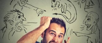
Although physical paralysis during the semi-conscious REM state does not sound as bad, in some cases it can last for more than an hour. Even worse, paralysis is often accompanied by vivid nightmares and sometimes hallucinations (such as demonic creatures). This leads to very painful experiences and makes you feel completely powerless and trapped. Those who suffer most are those who suffer from chronic attacks of sleep paralysis.
Sleep paralysis usually affects men and women equally, but first episodes most often occur during adolescence (eg, 13–19 years) or early adulthood (19–25 years). By some estimates, 36% of all cases occur in people aged 25 to 44 years. []
General information
Sleep paralysis is sometimes also called old witch syndrome .
It is expressed in the form of a special phenomenon of complete or partial stupor - paralysis of various muscles, which occurs when falling asleep or, in more rare cases, when waking up from sleep, usually lasts from several seconds to several minutes, and can be interrupted by touch or a sharp sound. Pathology refers to sleep disorders and is a secondary disorder; it can be provoked by mental, neurological and somatic diseases. For paralysis during the transition to awakening and falling asleep, the most characteristic state is immobility and it is diametrically opposed to somnambulism - the ability of a person to perform movements in a dream. In a state of sleep paralysis, people are unable to speak, walk, move their arms, or even open their eyes, although they retain the ability to control eye movements.
Being one of the most common forms of rapid eye movement sleep disorder, paralysis can occur both in completely healthy individuals and in those suffering from hypnagogia , narcolepsy and catalepsy . Sleep paralysis is usually isolated, otherwise it may be associated with other disorders.
Available statistics (source Wikipedia) indicate that approximately 7% of people surveyed have experienced an attack of sleep paralysis at least once in their lives. However, it is very important to distinguish the medical phenomenon from fictional stories, when the pathological condition is attributed to fantastic explanations such as alien abduction or demonic attacks, although this type of parasomnia has received wider publicity.
Diagnosis using the patient
How to get rid of the unpleasant “gift” of sleep paralysis? If your condition causes you insurmountable anxiety, and night attacks do not give you peace, consult a doctor.
The doctor, as a rule, will correctly diagnose your condition based on the description. Perhaps he will advise you to observe it yourself, namely, to write down everything that relates to it: the time you fall asleep and wake up, your auditory sensations, visual images, possible nuances of your life that, in your opinion, can provoke an attack. This makes it easier to find the cause or set of causes that led to regular attacks.
Perhaps, to clarify the diagnosis, you will be referred to other specialists - a somnologist, a neurologist. A polysomnography may be prescribed - a procedure for studying your sleepy state (most often, a polysomnogram does not show any oddities in connection with night paralysis - which means it does not pose a danger to the body).
If the causes of your condition seem too serious to the doctor, he may prescribe medication (usually antidepressants).
Do not try to self-medicate and take antidepressants on your own. These drugs are not harmless if they are taken according to the wrong regimen and in the wrong dosage, without the supervision of a specialist. You risk not only aggravating your condition, but also gaining additional “bonuses” in the form of side effects - and such strong remedies have a lot of them!
An interesting article about how to choose a medicine for insomnia without a prescription https://matrasium.ru/son/narusheniya/tabletki-ot-bessonnicy-bez-receptov/
Pathogenesis
This type of parasomnia, the occurrence of a motor phenomenon in a specific phase of sleep associated with the process of waking up and falling asleep, can be diagnosed by qualified neurologists even in healthy people. The condition is usually triggered by partial activation of the system responsible for REM sleep.
REM sleep is characterized by dreaming. To prevent a person’s physical ability to react to dreams and act in an emotional outburst, brain structures temporarily “paralyze” the possibility of muscle activity. Muscle dystonia occurs as a result of suppressed excitation of skeletal muscles in such parts of the brain stem as the pons and in the ventromedial region in the medulla oblongata.
Sleep structure
Inhibition occurs with the participation of glycine , γ-aminobutyric acid and other neurotransmitters that can suppress the functions of motor neurons located in the spinal cord.
Hallucinations and various strange physiological sensations are associated with the functioning of the vestibular apparatus and the need of the brain to establish the causes of disturbances and prevent the consequences of motor skills during sleep.
Night paralysis is characterized by the ability to be completely conscious while the body goes through the stages of rapid eye movement sleep. As a result, consciousness finds itself in a kind of trap: it is possible to see the world around us and everything that is happening around us, to control the breathing process, but there is no opportunity to move or speak.
Section 2. Neurophysiological basis for the occurrence of the phenomenon
2.1. Mechanisms of transition to REM sleep
The structure of our sleep includes two alternating phases: the fast REM phase (rapid eye movement phase) and the slow NREM phase (non-rapid eye movement sleep, i.e. sleep without rapid eye movements). slow sleep). In this article, we will discuss the first of these phases in more detail, since it plays a key role in the occurrence of the phenomenon of sleep paralysis. This phase is also called the paradoxical phase, as it is characterized by increased brain activity, increased cerebral blood flow and increased serotonin levels, which is accompanied by increased blood pressure and heart rate; rapid and shallow breathing is noted, as well as intense movements of the eyeballs and contractions of facial muscles. Dreams usually occur during REM sleep.
One theory suggests that REM sleep is initiated by small clusters of cells located in the pons and medulla oblongata. The main mediators of these structures are acetylcholine and glutamate [10]. Groups of these cells are not arranged into a single “center” of REM sleep, but work as individual components of a wide network (Fig. 1) [11]. For example, activation of locus coeruleus (LCα) neurons leads to the development of muscle atonia; rapid eye movements are initiated by neurons of the reticular formation (RF), located near the abducens nucleus (PAb); muscle contractions occur when neurons in the nucleus giant cell are activated [11], [12].
Activation of a certain group of brain stem cells ( red ovals ) causes a change in the corresponding indicators of the body during the REM sleep phase, which are recorded in the form of curves (Fig. 1, right column). For example, EEG signs of cortical activation during REM sleep are realized due to the simultaneous activation of midbrain RF (MRF) neurons and magnocellular nucleus (MN) neurons in the medulla oblongata; generation of the theta rhythm in the hippocampus is initiated by neurons of the pontine oral reticular nucleus (PO); an increase in brain temperature and cyclical fluctuations in the parameters of the cardiorespiratory system are provided by neurons of the parabrachial nucleus (PBN).
Figure 1. Cellular-molecular-network model of the physiological mechanisms of REM sleep formation.
Blue square: LC — neurons of the locus coeruleus, RN — neurons of the raphe nuclei; Green circle: GABA - local GABAergic neurons; Red ovals: MRF - neurons of the midbrain reticular formation, MN - neurons of the magnocellular nucleus of the medulla oblongata, LCα - neurons of the locus coeruleus alpha, PAb - neurons located near the abducens nucleus, PO - neurons of the oral reticular nucleus of the pons, PBN - neurons of the abrachial nucleus; C-PBL—neurons of the posterolateral peribrachial region, Sub C—neurons located under the locus coeruleus; Yellow rectangle: mPRF - medial pontine reticular formation; Red square: PPT - neurons of the pedunculopontine nucleus, LDT - neurons of the posterolateral part of the tegmentum; above the arrow: KR - kainate receptors; Yellow oval: Glutamate - glutamate [13]; Curves on the right (from top to bottom): EEG of a rat in the REM sleep phase, EMG of a rat in the REM sleep phase, EOG of a rat in the REM sleep phase, the theta rhythm of the hippocampus in the REM sleep phase, cyclic oscillations (BP - blood pressure, HR - heart rate, RESP - respiratory rate, BT - body temperature), PGO waves.
[11]
These groups of brainstem cells responsible for triggering REM signals are activated by increased release of cholinergic neurotransmitters, while the amount of aminergic neurotransmitters is either reduced or completely absent. Sources of acetylcholine (Fig. 1, red arrows) are cholinergic neurons of the pedunculopontine nucleus (PPT) and the posterolateral tegmentum (LDT). The sources of aminergic neurotransmitters (Fig. 1, blue arrows) are noradrenergic neurons of the locus coeruleus (LC) and serotonergic neurons of the raphe nucleus (RN). On the membrane of the former there are kainate receptors, which these neurons need to start working. A similar system is formed by both GABAergic and aminergic neurons.
The development of REM sleep begins with increased release of glutamate (Fig. 1, yellow oval); Consequently, the kainate receptors of cholinergic neurons (KR) are activated, which leads to their excitation and increases the release of acetylcholine in each of the REM sleep signal generators (Fig. 1, red arrows). Bidirectional communication of cholinergic neurons of the pedunculopontine nucleus (PPT) and a group of cells of the posterolateral tegmentum (LDT) with neurons of the medial pontine reticular formation (mPRF) is also triggered. Simultaneously with the described processes, local GABAergic cells are also activated (Fig. 1, green circle), which leads to active inhibition of aminergic neurons of the locus coeruleus and raphe nuclei and reduces or completely stops the release of aminergic neurotransmitters in the structures responsible for the generation of REM sleep.
The key role of the aminergic system is to stop its activity in time when cholinergic neurotransmitters are released. To further maintain episodes of REM sleep, increased acetylcholine release in mPRF neurons activates glutamatergic neurons, which continue to release glutamate in the PPT and LDT to maintain the activity of cholinergic neurons. Thus, cholinergic cells PPT and LDT and glutamatergic cells LC and RN form a positive feedback loop to maintain REM sleep cycles [11].
Another mechanism for the occurrence of REM sleep was proposed by Jouvet and Thompson and called it the “activation-synthesis hypothesis.” According to scientists, dreams - just as in the mechanism described above - are the result of nonspecific activation of neurons in the brainstem. But the analysis does not end there, and the signal is further transmitted to the cerebral cortex and subcortical formations for subsequent synthesis of the received information. Scientists have divided all nerve cells into two categories: cholinergic and monoaminergic REM sleep neurons. REM sleep is the result of the reciprocal interaction of these nerve cells [9].
However, the nerve centers for sleep regulation are located not only in the brainstem, but also in the diencephalon. The GABAergic “NREM sleep center” is located in the anterior hypothalamus, and the orexinergic neurons that ensure the process of turning on the “REM sleep center” are located in its middle part (Fig. 2).
Almost simultaneously, two groups of researchers from Japan and the USA discovered two amino acid sequences that were similar to each other, but named them differently [14]. It soon became clear that these were the same substance - the oligopeptides orexin A, containing 33 amino acid residues, and orexin B, consisting of 28 amino acids [15]. The protein proorexin is cleaved from preproorexin, from which orexins A and B are derived. The effect of the latter is determined by two G-protein-coupled metabotropic receptors. OX1R is a type 1 receptor that selectively binds only to orexin A, while OX2R is a type 2 receptor that is nonselective and binds to both orexins. The orexin A receptor is associated only with the Gq subclass of heterotrimeric G proteins, the orexin B receptor is associated with the Gq subclasses or Go and Gi proteins (Fig. 2) [16]. Orexinergic nerve cells interact with locus coeruleus neurons, inhibiting them [14].
Figure 2. Orexins and their receptors.
LC - locus coeruleus, VTA - ventral tegmentum, VMN - ventromedial hypothalamus, DR - raphe nuclei, LDT/PPT - dorsolateral laterodorsal/pedunculopontine tegmentum, BST - bed nucleus of the stria terminalis, PVN - paraventricular hypothalamic nucleus, Arc - semicircular nucleus, TMN - tuberomammillary nucleus, DMH - dorsomedial nucleus, PVT - paraventricular thalamic nucleus.
[16]
2.2. How can disruption of the mechanisms of transition to REM sleep lead to SP?
Research by American scientists at the University of Pennsylvania has shown that the basis of SP is the perseveration of REM activity, that is, a violation of the switch from sleep to wakefulness [2].
- Perseveration (Latin perseveratio - “persistence”, “persistence”) - a stable repetition of something, in this case - REM activity.
During the REM phase of sleep, our brain systems are actively working, as we verified above, but the body remains motionless [6]. But sometimes during this period an awakening occurs, and if the brain structures do not have time to synchronize with each other, paralysis occurs [17]. At the molecular level, awakening is mediated by the following mechanisms. It is believed that the cholinergic and glutamatergic systems are mainly associated with the bioelectrical and behavioral manifestations of the awakening process [18]. The nuclei of the brainstem, responsible for excitation and containing acetylcholine, dopamine, serotonin or norepinephrine, activate the thalamus, hypothalamus, motor neurons of the spinal cord, forebrain and have an inhibitory effect on the ventrolateral preoptic area (GABA, galanin); The hypothalamic and thalamic excitatory centers activate the cortex and arousal-related areas in the basal forebrain and brainstem [19].
One of the most notable aspects of SP is vivid hypnogogic or hypnopompic hallucinations [1]. They can occur during a sudden awakening, along with visions of demonic images, a state of panic, and a feeling of the presence of strangers [20]. A possible explanation for this phenomenon may be that during REM sleep there is a decrease in the activity of the respiratory muscles; this is due to inhibition of motor neurons. During REM sleep, breathing becomes irregular, skeletal muscle hypotension occurs, which leads to a significant decrease in lung ventilation and tidal volume, which leads to excessive accumulation of carbon dioxide in the blood - hypercapnia - and a decrease in the amount of oxygen - hypoxia [21]. In addition, oligodendrocytes, the myelin-producing cells of the central nervous system, are selectively sensitive to hypoxia and sleep fragmentation [22].
- Hypnogic hallucinations are visual illusions that appear before falling asleep.
- Hypnopompic hallucinations are visual illusions of perception that appear after awakening.
A century ago, American neurologist Weir Mitchell described the state of “night paralysis” as follows: “The subject awakens and is aware of his surroundings, but cannot move his muscles; lying there, apparently still sleeping. He is really involved in the struggle for the movement, fraught with acute mental disorder; if he had time to move, the spell would have disappeared instantly” [23].
It is assumed that in addition to the secondary (and sometimes primary) sensory cortex, dysfunction in other areas - the premotor cortex, the cingulate cortex, the subcortical and cerebellar areas - also contributes to the occurrence of hallucinations. The disorder that causes hallucinations is almost always located in areas of the brain associated with sensory systems. It is assumed that compensatory overactivation of brain regions surrounding these pathways is also the cause of hallucinations [24]. However, within seconds to minutes of awakening, the perceptual, cognitive, and motor components of the sleep cycle are synchronized, hallucinations disappear, and mobility is restored when the person is fully awake.
The mechanisms of REM-on and REM-off systems block impulses arriving along incoming sensory fibers, supplying the cortex with internal stimuli that form the content of dreams. They also block the activity of motor neurons in the cortex, thereby immobilizing the dreamer [1].
Thus, when sleep paralysis occurs during a sudden unplanned awakening during REM sleep, eye movements remain unchanged, showing the same high activity, and sensory sensations are clear.
Classification
Depending on when paralysis occurs - during the transition to falling asleep or waking up, it has its own characteristics and is divided into 2 forms:
- When going to sleep, a person’s state enters the brain-conscious phase of REM sleep, whereas normally falling asleep should be accompanied by a cessation of vigilance a couple of moments before the onset of paralysis, which explains why people practically never remember how they fell asleep. The main feature of this form is the complete or partial preservation of consciousness and the feeling of the work pattern and structure of the body, its motor skills - the ability to move the fingers, but the time for the transition of thought to the execution of the movement takes a fairly long period of time.
- The second, more common form is awakening paralysis, which is accompanied by sensations such as suffocation, irrational fear, lack of air, false perception of body movements and disorientation in space . In practice, this can be expressed in the form of a false sensation of a rapid heartbeat and a turning over of the body, although the person is actually calm and immobilized. In attempts to wake up and in anticipation of the final awakening, people groan and twitch their limbs, which is caused by strong psycho-emotional impulses. Such sensations can lead to fear of death, falling into a lethargic sleep , nightmares and auditory hallucinations of someone’s voice, creating the impression of the presence of an outside force and hostile entities (humanoids, demons, etc.), most often perceived as otherworldly or alien forces. They are usually provoked by swelling of the arms and their raised position above the head. This pathological condition is promoted by sleeping on the stomach, with the head in the pillow, and is caused by the body's wake-up signals and lack of oxygen. The position of the body while sleeping, lying on its side, rarely causes sleep paralysis.
Symptoms
What does night paralysis look like? Very unpleasant. A person feels:
- immobility - inability to move even a finger;
- a feeling of suffocation, a feeling of heaviness and pressure on the chest, throat, stomach;
- auditory and visual hallucinations, especially in the dark - hears noises of unknown origin, footsteps, voices, pulsating sounds; sees vague images perceived as threatening;
- As a result, a state of horror, panic, and doom arises—and disturbances in heart and respiratory rhythms, muscle twitching, and distortion of facial muscles are observed.
Have you had a frightening experience of night paralysis and are terrified of the possibility of it returning? Don't forget: you have nothing to fear! You will not die, you will not fall into lethargy, you will not go crazy! This state is: safe and temporary. Remember this.
The video talks about sleep paralysis, its causes and ways to combat this condition - narcologist Mikhail Tetyushkin:
Causes of sleep paralysis
As a result of surveys, it was found that students and mentally ill people are more susceptible to sleep paralysis. Approximately a third of surveyed people in these categories experience a motor phenomenon while falling asleep, and in more rare cases, upon awakening.
Therefore, presumably, the causes of sleep paralysis may lie in irregular, fragmented and restless sleep and constant stress. The incidence in women is several percent higher than the incidence in men, but the difference is too small and does not make it possible to talk about the gender predisposition of the disease. Etiogenesis has not been fully and completely studied.
Risk factors
People at risk of experiencing muscle atonia when waking up or falling asleep include those who:
- suffer from migraines and poor, disturbed sleep;
- engage in activities that stimulate the nervous system in the evening;
- often fly on an airplane with time zone changes;
- sleep on their back;
- have a neurotic type of personality development;
- experiences increased levels of anxiety;
- work in shifts;
- suffer from hyperglycemia, arterial hypertension, anxiety disorder ;
- abuse alcohol and various medications;
- suffer from hypertension , idiopathic hypersomnia , sleep deprivation, narcolepsy , obstructive sleep apnea syndrome, Wilson-Konovalov disease ;
- have an increased level of dissociation and imaginative development with a belief in paranormal or supernatural phenomena;
- have a hereditary predisposition, since the likelihood of developing sleep paralysis due to family affinity has been established (presence of genes PER1, 2, 3, ABCC9, CACNA1C, ARNTL2, CLOCK, DBP).
How to induce sleep paralysis?
Since the state of sleep paralysis causes out-of-body experiences in some people, although this state can be frightening, people try to induce it on their own of their own free will. It is believed that this technique is used by Tibetan monks and shamans of various nations of the world, who are specially trained in techniques “how to control the phases of REM sleep and get into sleep paralysis.” They do not consider this phenomenon terrible and strive to get the sensation of floating above the body.
It is believed that you can enter a state of REM sleep while maintaining consciousness through the habit of sleeping on your back, interrupted fragmented sleep, many hours of meditation, shamanic rituals accompanied by special music and dancing, taking various herbal narcotic herbs and decoctions, and smoking them. Incense, light and music can also create a special atmosphere.
Symptoms
The main manifestation of sleep paralysis is muscle atonia in the REM sleep phase, which occurs during falling asleep and waking up. In addition to immobility, most patients experience other symptoms:
- they see dreams - very vivid and quite unpleasant;
- they experience fear or, on the contrary, bliss, strange sensations of out-of-body experiences, it may seem to them as if the body is soaring, flying, accelerating, circling;
- while in a state of sleep, the brain is capable of activity and awareness of the surrounding world, control of breathing, etc.;
- chest pressure and difficulty breathing;
- pain;
- visual and auditory hallucinations, most often frightening, because atony is combined with a state of wakefulness;
- “flies” is another manifestation of illusions or hallucinations, expressed in the form of sound vibrations in the ears with increases in acoustic range and volume, smoothly turning into “white noise” and ringing in the ears, perhaps the predominance of a certain “squeak” that an ordinary person hears. a person is at rest in silence.
How else can you combat sleep paralysis?
If you are in the early stages of trying to stop sleep paralysis, it may take some time. Don't expect immediate results—it may take months before you adhere to a strict sleep schedule, your circadian rhythm changes completely, and your sympathetic nervous system becomes less sensitive. In the meantime, you should be aware of some methods to combat episodes of sleep paralysis.
Try moving your fingers/toes . Despite near-total paralysis, some people have found that consciously trying to move their fingers/toes helps them stop sleep paralysis. You may not be able to lift your arm, move your leg, or scream for help, but consciously using your awareness to “move” your fingers can help you “wake up.”
Accept this state . When you experience sleep paralysis, it can be scary and uncomfortable. You may feel the presence of another being, and in extreme situations you may even experience hallucinations. Although the natural tendency will be to resist, accepting what is happening and realizing that you have nothing to fear can reduce your REM activity and bring you out of this state.
Keep a sleep journal . For some people, it might be helpful to consider using a sleep diary. When you wake up, write about what you experienced during sleep paralysis. Document how long it lasted and keep track of how well you slept. Make a note of whether you maintain a sleep schedule, whether you avoid stimulation before bed, etc. Keeping sleep diaries will help you keep track of all the factors that may contribute to sleep paralysis.
Tests and diagnostics
Old witch syndrome is most often identified during a conversation with a patient or a questionnaire. In this case, the patient has the ability to control eye movements and breathing, hallucinates and experiences suffocation.
At the moment, no types of tests similar to polysomnography have been developed to diagnose this type of parasomnia, but it has been assigned an ICD-10 code: G47.0 and the name “Disturbances in falling asleep and maintaining sleep, expressed in the form of complaints of unwanted and disturbing events and sensations during sleep." To diagnose pathology, a few repeated episodes that cause anxiety and fear are enough.
Prevention
It is believed that repeated episodes of muscle atony during falling asleep and waking up can be eliminated by following the rules of sleep hygiene:
- fall asleep and wake up at the same appointed time;
- take water procedures in preparation for bed;
- practice meditation and breathing exercises to relax in the evening;
- adhere to the normal amount of sleep typical for your age;
- choose a comfortable mattress and bed linen;
- ventilate the room before going to bed;
- worry about the room temperature – 18-20°C;
- Avoid drinking alcoholic and caffeinated drinks several hours before bedtime;
- Avoid falling asleep lying on your back or stomach.
Diet for sleep paralysis
Diet for the nervous system
- Efficacy: therapeutic effect after 2 months
- Timing: constantly
- Cost of food: 1700-1800 rubles per week
Reducing stress, avoiding drugs, alcohol, excessive physical activity, and most importantly, proper diet can help reduce the risk of attacks of paralysis. It is important to strictly arrange meal times, choose a balanced menu, make breakfasts more filling and dinners lighter, and 3-4 hours before going to bed. It is recommended to enrich the diet with the following products that improve sleep quality:
- bananas;
- boiled eggs (1-2 per day);
- a handful of almonds for an afternoon snack or salad;
- seafood (at least 2 times a week);
- light fermented milk drinks (a glass of yogurt in the evening will improve the general condition of the body and give a blooming appearance);
- different types of cabbage and leafy greens should be included in both soups and salads on your plate every day;
- fresh berries are good if they complement morning oatmeal;
- honey, molasses and grapes are the best sweet substitutes.
Shackled by fear
A dark room, your eyelids lift heavily, your breathing quickens, your heart beats like crazy. You lie in your bed with a feeling of vague anxiety, which will soon turn into real horror. The eyes frantically search for the source of danger. But the darkness has already shackled you. Your arms and legs don't move, you feel completely helpless. And at that moment an incomprehensible black shadow hovers over you, as if out of curiosity it stopped to look at you. After a few seconds the shadow disappears. And you still can't move. Only the eyes look around in horror. A whole swarm of shadows gathers around, each wanting to look at the one chained by sleep. You try to scream, call for help, but your mouth feels like it’s been sealed with tape. You feel like a dry crust has covered your lips. At some point, one of the shadows hovers very close above you, and you notice its small red glowing eyes. You are again trying to break out of captivity, but it is useless. The shadow opens its huge tarry mouth. There is a pounding sound at the temple, like a drum roll. Suddenly silence. The shadows disappeared. Someone persistently pats you on the shoulder, it’s your husband, or maybe your brother. It doesn’t matter at all, because the horror has passed. But it repeats itself over and over again, every night. And you don't know when it will end.