Price list Doctors clinic
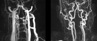
Duplex scanning of the vessels of the neck and brain is a diagnostic method that is based on the ability of ultrasonic waves to be reflected from moving blood cells (red blood cells) and vessel walls. This allows you to get a “picture” of the vessel and evaluate the characteristics of blood flow. This procedure allows you to provide a medical assessment of the condition of the subclavian, carotid, and vertebral arteries, and identify diseases or high risks of their formation.
The importance of the diagnostic procedure in the early diagnosis of vascular diseases is difficult to overestimate: it helps prevent blockage of blood vessels, therefore it is resorted to in case of high risks and probabilities of developing pathology.
What is duplex (triplex) scanning of blood vessels?
Duplex or triplex scanning - synonyms, duplex of vessels and triplex of vessels, ultrasound duplex angioscanning or simply UZDAS .
This is a non-invasive ultrasound research method that allows you to obtain a two-dimensional image of blood vessels with the ability to assess the condition of the vascular wall, the nature and speed of blood flow through them. Simply put, duplex scanning allows the diagnostician to see the vessels being examined, evaluate the places of their narrowings, blockages or, on the contrary, dilated areas, and also determine the presence of blood clots, atherosclerotic plaques, and blood flow disorders. The traditional duplex (triplex) scanning technique uses high-frequency sound waves that are not perceptible to the human ear. The computer receives information from the ultrasonic sensor and converts it into a two-dimensional dynamic image on the monitor screen.
Duplex (triplex) has great advantages over its outdated predecessor - Doppler or Dopplerography of blood vessels, a popular technique in the past that allows one to evaluate only the direction and speed of blood flow, but does not evaluate structural changes in the wall and lumen of blood vessels.
Decoding the results
If a comprehensive ultrasound is performed correctly, the doctor will receive complete information about the condition of the patient’s veins and arteries, in particular about:
- cross-country ability;
- anatomical structure;
- blood flow speed;
- the presence of stenosis or any formations that narrow the lumen of the vessel.
Duplex scanning is a collection of data that the doctor compares with normal values.
Duplex scanning rate
Artery diameter:
- general carotid – 4.2-6.9 mm;
- internal carotid – 3-6.3 mm;
- external carotid – 3-6.0 mm;
- vertebral – 2-4 mm.
When performing duplex scanning with color mapping, healthy vessels are colored in one tone, there are no gray areas. Any deviations from normal values indicate a potential violation of vascular functions.
Where can duplex be used?
A doctor may recommend vascular duplex for the following diseases and conditions:
- Atherosclerosis, endarteritis and diabetic angiopathy of the vessels of the lower extremities
- Atherosclerosis of the visceral branches of the abdominal aorta (vessels supplying the gastrointestinal tract, liver, spleen and kidneys)
- Aneurysm of the abdominal aorta and other vessels
- Varicose veins of the lower extremities
- Vasculitis (inflammatory vascular disease)
- Vascular diseases of the brain and neck
- Control of performed surgical intervention on blood vessels
- Postthrombophlebitic disease
- External vessel compression syndrome
- Screening examination (a study to identify asymptomatic forms of the disease)
- Thrombophlebitis and phlebothrombosis of the veins of the extremities
- Thrombosis of intestinal vessels
- Vascular trauma and its consequences
Indications for use
The main factor that has a negative impact on the treatment of vascular diseases is untimely contact with a neurologist and late diagnosis of the disease. The circulatory system is closed in a cycle, so disruption of the blood supply to tissues in one area inevitably causes problems in another. Main indications for duplex scanning of the great arteries:
- problems with vision, coordination, understanding, or speech production;
- headache and frequent dizziness;
- fainting or attacks of impaired consciousness;
- unsteadiness of gait;
- numbness of the limbs.
Duplex scanning of the main arteries of the head is also carried out if any pathologies of the vascular system are detected during other studies. Diagnostics are necessary so that the neurologist can assess the effect of the disease on the brain and prescribe adequate treatment.
There are two types of duplex scanning: two-dimensional mode and Doppler effect. The two-dimensional mode is used for visualization of blood vessels. The doctor receives information about the direction and speed of blood flow using the Doppler mode. These studies are indicated in the presence of the following pathologies or symptoms:
- fainting;
- sharp and frequent headaches;
- a feeling of coldness in the extremities or their swelling, cyanosis;
- development of trophic ulcers;
- sudden changes in blood pressure;
- tinnitus;
- weakness in the legs when walking.
The Neurology Clinic of the Yusupov Hospital employs neurologists with extensive experience who treat patients even with serious illnesses that other specialists have refused. An individual approach, modern treatment methods, high-precision and effective equipment allow us to promptly identify problems and develop ways to eliminate them.
Make an appointment
Which technique to choose - duplex or triplex scanning?
There is no big difference in information content between duplex and triplex scanning. The only advantage of triplex scanning (triplex) is the ability to assess the condition of the vessel in three different ultrasound modes simultaneously, while duplex scanning allows this to be done using only two modes. Thus, the quality of the study performed is primarily determined by the quality of the ultrasound equipment and the experience of the diagnostician, and not by the chosen technique.
What is it and how to prepare for the procedure
Ultrasound examination of blood vessels in combination with the Doppler effect allows us to identify various problems. The principle of the technique is the obvious differences in the reflection of waves by moving and stationary objects. Ultrasound is sent to the area of interest in the main vessels of the head through sensors, which makes it possible to determine such important indicators as the speed of blood flow, the vector of blood movement (depending on the changed frequency). The technique is indispensable in determining damage to the walls of blood vessels.
The main feature of the diagnosis is the absence of any preparatory measures and age restrictions. The procedure is prescribed for pregnant and nursing mothers, children (including babies) and adults. The patient can take the most comfortable position - lying or sitting. No sedation required.
For maximum diagnostic accuracy, you should give up alcohol, caffeine and salty foods the day before the procedure. It is forbidden to take a hot bath 2 hours before the diagnosis; it is also necessary to avoid cigarette smoke - passive smoking affects the results of MAG ultrasound. It is better to stop taking any medications and folk remedies that affect vascular tone.
Indications and contraindications
There is a large list of diseases, the development of which requires periodic (annually or every two years) MAG duplex scanning. It is important to follow your doctor's recommendations. Patients 40 years of age and older clearly need preventive studies. Office workers with more than 15 years of experience are at risk for developing disorders of the blood supply to the brain and other problems with the vessels of the cervical spine.
The doctor can prescribe ultrasound examinations of the MAG and when identifying symptoms that may indicate the development of dangerous diseases. The most common are dizziness and pain, lightheadedness and fainting. Visual disturbances (veil, floaters, etc.) that are observed constantly or periodically can also be a reason for ordering a study. Patients who have problems with memory, coordination, concentration, or weakness in the limbs are referred for a MAG duplex scan.
There are no contraindications to the procedure, except that if the cervical spine is injured, diagnosis may be delayed.
How should I prepare before duplex scanning?
Most often, specialists prescribe one of the following studies - duplex scanning of the arteries or veins of the lower extremities, as well as duplex scanning of the brachiocephalic arteries (arteries of the head and neck). Before these types of examinations, no special preparation is required from the patient.
However, in the case of a duplex examination of the vessels of the abdominal cavity and small pelvis (for example, duplex of the visceral branches of the abdominal aorta), especially in obese people, a 3-day preparation in the form of a diet is prescribed (meat, milk, black bread, cabbage and other vegetable foods rich in fiber) and taking medications that prevent gas formation in the intestines (Espumizan capsules, etc.).
Advantages of the method
Duplex scanning of vessels allows you to obtain with high accuracy important information about the vessels and those processes that occur both in the lumen of the vessel and outside it. The method is a safe and painless way to identify disorders in the arteries and veins of the lower extremities even at the earliest stages, when there are no clinical signs of the disease.
The main advantage for the patient is the simplicity and ease with which this examination is carried out. Duplex scanning does not require special preparation, has no contraindications and does not lead to complications.
For a doctor, in turn, this method is an indispensable diagnostic method, as it allows one to detect possible problems in the early stages of their development, even in the smallest, so-called peripheral vessels of the legs (microcirculation difficulties).
The ability to assess blood flow, its dynamics and characteristics in real time makes it possible to analyze not only structural malformations, but also possible functional disorders.
Also, if the study is carried out several times over a certain period of time, a tendency towards the development of certain conditions or diseases can easily be identified, which allows timely treatment to be prescribed if necessary.
What happens during the study?
The procedure for duplex (triplex) vessels is absolutely painless and does not require the use of local or general anesthesia. Depending on the area of the body being examined, diagnostics can be carried out while lying down, sitting or standing. After applying a special gel to the skin, the doctor places the sensor in the projection of the vessels being examined. The gel improves the conduction of ultrasound signals between the transducer and the patient's body. During the procedure, the researcher may ask the patient to help him by changing his body position, briefly holding his breath, and other simple actions. Duplex scanning lasts about 30-40 minutes on average and, as a rule, does not cause any discomfort to the patient.
Who is the test prescribed for?
There are many indications for duplex scanning of leg vessels and they are very diverse. The patient may be concerned about the condition of his lower extremities, and then a duplex scan is prescribed to specifically search for the problem, or the general state of health suggests the likely development of vascular complications - in this case, the attending physician will also definitely recommend examining the vessels of the legs.
So, this type of diagnosis is prescribed for the following suspicious conditions of the lower extremities:
- a feeling of pain in them at rest or during physical activity
- swelling of the feet and/or legs
- thickening of the veins, sensation of “harnesses” under the skin of the legs
- pain in the muscles of the lower extremities
- occurrence of seizures
– change in skin color on the legs (also sudden hair loss on the legs)
- tingling sensation in the toes, feet or coldness
- impaired sensitivity, numbness in the legs constantly or during physical activity.
The doctor will also recommend a duplex scan of the lower extremities if the patient:
- diabetes
- trophic (vascular) ulcers on the body
- constantly increased cholesterol levels in the blood
- arterial and/or venous insufficiency
- Previous injuries or operations performed on the legs.
What is Doppler?
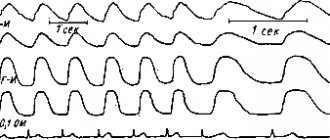
This diagnostic method got its name based on the discovery of a physical phenomenon made by the Austrian scientist K. Doppler. Its essence is to reflect changes in the frequency of the ultrasonic beam signal from blood cells moving in the vessels. This gives you the opportunity to evaluate:
- speed and direction of circulating blood;
- minute blood flow volume;
- the presence of atherosclerotic stenosis (narrowing) and blockage of the vessel;
- collateral (side) circulation;
- pulsation of blood vessels.