An intracerebral hematoma (ICH) is an accumulation of blood in a limited area of the brain substance that occurs due to a violation of the integrity of a cerebral vessel. The pathological condition is provoked by weak or injured intracerebral arteries or veins, which, having ruptured, began to release blood into the intracranial space. The VMH cavity can be filled with liquid or coagulated blood, or a bloody substance mixed with brain detritus (crushed fragments of brain tissue).
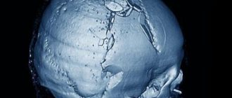
Schematic representation of a large hematoma.
Cerebral hematomas can be single or multiple, unilateral and bilateral, acute, subacute and chronic. They become chronic just 21 days after the hemorrhage occurs. Triggers of pathogenesis are often the consequences of arterial hypertension and atherosclerosis (70%-80% of cases), aneurysms and vascular malformations, mechanical intracranial damage due to TBI.
Intracerebral hematomas pose a serious threat to the health and life of the patient, since the structure-forming tissues of the brain at the site of the lesions are subject to compression, dislocation, edema and necrosis. Without adequate therapy, pathology can lead to severe morphological and functional brain damage, often irreversible. With ICH of any origin, the risks of disability and mortality are too high: disability affects up to 70% of surviving patients, the probability of death is 25%-50%.
Classification of intracerebral hematomas
Intracerebral hematomas in neurosurgery and neurology are classified by location and volume. The following types of VMH are diagnosed by location:
- medial (10%) – located medially from the internal capsule, determined in the zone of the thalamus and hypothalamus with possible spread to the midbrain and ventricles of the GM;
- lateral (the most common, accounting for about 50%-55% of all hematomas) – located outward from the internal capsule, localized mainly in the area of the shell, often extending to the level of the semioval centers and insular lobe;
- lobar (15%) – concentrated under the cerebral cortex in the subcortical white matter, usually within one lobe (occipital, temporal, frontal or parietal);
- cerebellar (10%) – located in the area of the right/left hemisphere of the cerebellum or worm-like structure (if the worm is damaged, a hematoma often breaks out with the contents entering the 4th ventricle, which causes the development of occlusive hydrocephalus);
- intra-stem (6%-9%) – formed in the brain stem, as a rule, more often such hematomas affect the structures of the pons by the type of hemorrhagic impregnation;
- mixed - simultaneously cover several intracerebral anatomical structures (for example, subcortical white matter, basal ganglia of the telencephalon and thalamus), or VMH is combined with subdural types of hematomas.
An important diagnostic indicator that will play a predetermining role in the choice of treatment algorithm is the size or volume of the formed hematoma. The following types of intracerebral hematomas are distinguished by volume:
- small – less than 40 cm³;
- medium – 40-60 cm³;
- large – 60-80 cm³;
- gigantic - more than 80 cm³.
Secondary complications in the form of increasing compression, deformation, swelling and displacement of the brain can occur with blood clots of not only large but also medium sizes. In some cases, especially if accompanied by a brain contusion, a hemorrhagic component of 30 cm³ may be sufficient to create a mass effect.
Contusion of a child's face
Bruises and other injuries in the chin area can lead to damage to the ligamentous apparatus. With this phenomenon, any movements of the lower jaw provoke pain in the child - one of the reasons to suspect a fracture of the condylar process. To clarify the diagnosis, an X-ray examination is mandatory.
A facial bruise in a child is characterized by the same causes and symptoms as in an adult. But let's not forget that children with great fear endure pain caused by injury, especially if it is accompanied by blood.
In addition, a bruise on a child’s face is serious because the child cannot always explain what exactly hurts and how. From a biological point of view, cell division in children occurs somewhat differently than in adults, just as a child’s body is a growing organism. Accordingly, the processes responsible for the natural development of the skin and muscle parts of the face may be disrupted. But there is a small, but positive side: scars on children heal faster and better than on adults.
But, nevertheless, during the period of screaming or crying, children may develop laryngospasm or may experience breathing problems.
For children, first aid is emergency. Whatever the conditions or setting, the child should be sitting or lying face down. Then you should turn the child on his side for convenience when emptying the oral cavity from the contents. The contents are removed using any safe means: a cotton swab or your hand. It happens that such actions are ineffective, and intubation is performed; tracheotomy is not recommended.
But, no matter what happens, the most important thing is not to panic, but to provide first aid in a timely manner (and not lose consciousness or become hysterical, like some mothers) and call an ambulance.
[17], [18]
Clinical signs of ICH
The clinical picture of intracerebral hematomas begins with the sudden onset of a crisis, characteristic of a cerebral hemorrhage. Hemorrhage is often preceded by high blood pressure or head trauma. During the acute period, typical symptoms are observed:
- sharp, intense headache;
- dizziness, loss of consciousness;
- nausea, vomiting;
- heavy wheezing breathing;
- abnormal heart rate;
- development of hemiparesis on the side opposite to the VMH with possible development into hemiplegia (usually in the leg, arm, facial muscles);
- coma.
The severity of symptoms depends on the size and location of the hematoma. As the pathology progresses, neurological deficits progress with a predominance of motor, sensory, speech, and thinking disorders. Patients with intracerebral hematomas are often bothered by epileptic-type convulsions, stiffness of the muscles of the back of the head and neck, impaired coordination and stunned consciousness syndrome of varying severity. Visual disturbances (mydriasis, loss of fields, unilateral hemianopsia, ptosis, etc.), bradycardia, memory problems, and mental disorders often occur.
To give a clear rationale for the symptoms occurring, it is necessary to conduct high-quality instrumental diagnostics of the brain using cerebral imaging methods. Without a normal examination of the brain, it is impossible to make a final conclusion on the diagnosis, its severity and location, it is impossible to choose a treatment method and form an idea about the prognosis of the outcome.
General information
The main cause of hematomas are bruises - closed injuries to soft tissues resulting from a blow or fall. A strong blow leads to rupture of the walls of small blood vessels, due to which blood begins to flow through the breakout sites into the subcutaneous tissue, soft tissues or body cavities. Hematomas form in different parts of the body - on the limbs, torso and even on the head. In addition to bruises, hematomas are caused by intense compression and stretching of tissues due to dislocations or fractures.
Small lesions, as a rule, do not require any treatment and resolve on their own within a few days. When large hematomas form, there is a risk of infection and the development of suppuration. Most often, hematomas form in representatives of younger age groups - children, adolescents and young people, who are characterized by high physical activity. Another “risk group” are people with increased fragility of the vascular wall, as well as with blood clotting disorders.
Diagnosis of hematoma in brain structures
Neuroimaging is of clinical value in making a diagnosis. A computed tomography scan is prescribed as an initial examination of the brain. This inexpensive method allows you to quickly and easily determine the presence of blood in the GM substance, the location and volume of the clot. The information value of CT is highest when 2-3 weeks have passed since the hematoma appeared (maximum 5 weeks). Until this time, the VMH area has the highest density, which facilitates diagnosis and can be limited to one CT scan.
MRI.
As the lesion ages (on average, after 14-21 days), the density of the hemorrhagic mass decreases, it becomes isodense, that is, close to normal brain tissue. During this period and later, purely magnetic resonance imaging can provide qualitative information about the intracerebral hematoma and the state of the brain.
Additionally, the patient is often recommended to undergo cerebral angiography. This technique does not have the necessary potential for verification of VMG. But angiography of cerebral vessels determines the intensity and distribution of vasospasm and allows one to exclude or confirm the involvement of a vascular malformation or arterial aneurysm in the development of ICH.
Causes of facial bruise
What are the causes of facial bruising? It is clear that a facial bruise will not occur out of nowhere in the same way as this phenomenon acquired as a result of mechanical action, that is, injury: a fall or blow. And here, many people immediately think: either a drunk fell down the stairs, or the husband raised his wife. Of course, such options are not excluded, but there are a number of household and work-related injuries, for example, we are all living people, and therefore each of us can stumble or stumble.
A facial bruise is not necessarily a “black eye”; it can relate to the jaw, cheekbones, nose, forehead, eyes, chin.
[3], [4], [5], [6], [7], [8], [9]
Treatment methods for intracerebral hematoma
Conservative therapy or neurosurgery are used to treat cerebral hematomas. Recommendations for conservative elimination of hematoma are:
- small size of the lesion (≤ 40 cubic cm) without pronounced symptoms, without clinical signs of herniation and dislocation;
- old age of the patient (75 years and older);
- inappropriateness of surgery due to the high risk of unfavorable outcome (for example, large-scale hemorrhage in the dominant hemisphere or with extensive neurological damage);
- severe condition of the blood coagulation system, sepsis;
- diabetes mellitus in the stage of decompensation, uncontrolled hypertension.
The problem is solved conservatively by competent neurologists and neuroreanimatologists. The treatment plan is developed strictly individually. It may involve basic anti-ischemic, antihypertensive treatment, reduction of cerebral edema with osmodiuretics, therapy with hemostatics and neuroprotectors.
Preventing facial bruises
Frankly, there is no way to protect yourself from injury at all, including facial bruising. We are all living people, and we can be inattentive or careless. Even if you put a helmet on your head, there is no guarantee that some emergency will not occur that has to do with the safety of your face.
The only thing that can be said is that possible injuries should be avoided, both at work and at home. Regarding children: do not leave your child alone even for a minute where there are “corners”, sideboards with glass, household items (which can cause injury), with a spoon in his hand, and so on. And, as for children, there should always be medicines at hand: ointments, tablets; bandages. These are children, they will always find adventure.
If a facial injury has already occurred, then in order to prevent the formation of bruises and swelling, it is necessary to apply a cold compress; to prevent other problems, it is necessary to do an ultrasound of the hematoma and an X-ray of the head.
Surgical methods for removing VMG
Surgery is indicated in most cases. Its main goal is to save a person’s life and normalize the neurological status. The use of surgery is justified in the following conditions:
- hematomas of any size, accompanied by a pronounced mass effect, cerebral edema, dislocation of the central structures by more than 5 mm;
- lobar and lateral VMGs from 50 cm³ or more in volume;
- medial formations from 20 cm³ and above;
- cerebellar hematomas ≥ 15 cm³;
- progressive deterioration of the patient’s well-being (exception – stage 2-3 coma);
- young age of the patient;
- severe intracranial hypertension when it is impossible to correct ICP conservatively.
The options for surgical treatment are transcranial surgery with direct inspection under microscope control (the standard, most common technique), stereotactic aspiration, and endoscopic removal of the hematoma.
- Transcranial removal. The operation involves direct craniotomy (classical trepanation, often extended) in the projection of the lesion. Next, an encephalotomy is performed in the area of the closest apposition of the VMG to the cerebral cortex. Then they begin to remove the pathological formation, which is carried out by aspiration with washing out the cavity with a stream of saline solution. Heavily condensed elements are removed using special windowed tweezers. Hemostasis is carried out by bipolar coagulation of blood vessels, the use of antihemorrhagic agents (sponges, fibrin-thrombin glue, pads with hydrogen peroxide). A drainage is installed on the wound. The surgical session lasts approximately 3 hours, manipulations are performed under general anesthesia. Direct craniotomy is more common for lobar and cerebellar VMH.
- Stereotactic procedure. Stereotactic aspiration is considered a minimally invasive tactic. However, despite the gentle effect, recurrences of hematomas occur more often after such treatment than after craniotomy. This is explained by the inability to perform thorough hemostasis during stereotactic surgery. The basic type of anesthesia is neuroleptanalgesia. Intervention is performed more often for hematomas of medial and mixed types. The patient's head is pre-fixed in a special stereotactic frame. Next, after placing a small burr hole in the skull, a thin cannula (diameter about 5 mm) is inserted into the hematoma cavity. Through an installed cannula, to which an electric aspirator is connected, pathological contents are evacuated from the brain. Intraoperative control of cannula insertion and therapeutic manipulations is carried out using navigation systems and X-ray equipment. Session duration is 1-3 hours.
- Endoscopic surgery. This is a minimally aggressive neurosurgery tactic that involves removing blood accumulated in the brain tissue under endoscopic control. Anesthesia is usually used by intubation. Access is performed using a surgical device such as trephine, which serves to create a small burr hole in the skull of a round shape. A rigid endoscopic tube is placed into the formed trefination hole. The endoscope tube has a video system that transmits images of brain structures to a color intraoperative screen. The endoscope is carefully brought to the area of interest, then its tidal working system suctions the hematoma and rinses the cavity. Hemostasis is also carried out endoscopically, using monopolar coagulation and defocused laser irradiation. The duration of the procedure is 45-90 minutes.
Russian clinics offer direct craniotomy at a price of 60 thousand rubles or more, stereotactic surgery - 40-60 thousand rubles, endoscopic removal of a hematoma - from 50 thousand to 62 thousand rubles.
After any surgical intervention, a full course of rehabilitation is prescribed, including deep prevention of all possible postoperative complications (infections, thrombosis, pneumonia, etc.). In the majority of cases, after neurosurgery, preventive therapy to prevent epileptic seizures is advisable.
On average, it takes about 6 months for an adult patient to recover after removal of an intracranial hematoma. Depressed functions can be restored completely, but there is no absolute guarantee of this, since everything depends on the initial condition of the patient, age, and the nature and consequences of the intracerebral hematoma at the preoperative stage. In children, rehabilitation usually progresses at a faster pace, and much more often it ends in full recovery.
Symptoms of a facial bruise
Facial bruise is characterized by clinical symptoms, which include: swelling, pain, dysfunction, hemorrhage.
- the first characteristic sign is pain. It appears immediately after injury. The pain may intensify after an hour or three. The increase in pain depends on the appearance of edema or hematoma;
- “puffy face” or swelling in the area of the grass is an almost instantaneous phenomenon. During palpation, a painful compaction is detected that does not have clear boundaries and affects healthy tissue. Typically, swelling occurs within an hour to a day after injury. After which inflammatory changes and traumatic swelling are already noticeable;
- the bruise is explained by the fact that the skin and subcutaneous tissue are saturated with spilled blood. It is impossible to say exactly how quickly a bruise may form, since the depth of the hemorrhage can affect the speed of its development. If the skin or subcutaneous tissue is injured, then a bruise is possible in the first minutes, sometimes hours. If we are talking about muscles, then a bruise may appear even on the third day, and far from the bruise area itself. Late-onset bruising, especially those located away from the area of the bruise, is a serious sign that requires a thorough examination, for example, an x-ray, to exclude the possibility of cracks or bone fractures. As for the color characteristics, the bruise is initially red, after 5-6 days it becomes green and then turns yellow. This process is caused by the breakdown of hemoglobin. Thanks to this, you can determine when a facial injury occurred.
Where is the best treatment?
Intracerebral hematomas are a very serious problem, associated with high risks of mortality and complete disability. Patients with this diagnosis need to be observed and treated in medical institutions of the highest global level. Some of the best neurosurgical specialists in Europe practice in the Czech Republic.
Central Military Hospital of Prague.
Clinics in the Czech Republic follow only advanced diagnostic principles and cutting-edge technologies for the safe treatment of central nervous system lesions. In addition, the famous republic offers impeccable rehabilitation care for patients after brain surgery.
Czech neurosurgery means the quality of the services provided, as well as their affordable prices. In the Czech Republic, high-tech treatment programs for intracerebral hematomas cost 2-3 times lower than in Israel and Germany.
Recovery prognosis
A mild brain injury has a favorable outcome, in which mental and neurological functions are completely restored. A moderate bruise with timely and correct treatment also ends in complete recovery. In some cases, after it there is a slight lack of coordination of movements, vegetative-vascular dystonia and hydrocephalus.
Severe traumatic brain injury causes death in approximately 30% of cases. In addition, there is a high percentage of disability among surviving patients.