| This article includes a list of references, related materials or external links. but its sources remain unclear as it lacks embedded quotations . |
| Alpha motor neuron | |
| Alpha motor neurons originate from the basal lamina (basal plate) of the developing embryo. | |
| Identifiers | |
| NeuroLex ID | sao1154704263 |
| H2.00.01.0.00008 | |
| F.M.A. | 83664 |
| Anatomical terms of neuroanatomy [edit in Wikidata] | |
Alpha
(
α
)
motor neurons
(also called
alpha motor neurons
), large, multipolar lower motor neurons of the brainstem and spinal cord. They innervate the extrafusal muscle fibers of skeletal muscles and are directly responsible for initiating their contraction. Alpha motor neurons are different from gamma motor neurons, which innervate the intrafusal muscle fibers of the muscle spindles.
Although their cell bodies are found in the central nervous system (CNS), α-motoneurons are also considered part of the somatic nervous system, a branch of the peripheral nervous system (PNS), because their axons extend peripherally to innervate skeletal muscles. .
The alpha motor neuron and the muscle fibers it innervates are a motor unit. Motor neuron pool
contains the cell bodies of all motor neurons involved in the alpha contracting muscle.
Location[edit]
Alpha motor neurons (α-MNs), innervating the head and neck, are found in the brainstem; the remaining α-MNs innervate the rest of the body and are found in the spinal cord. There are more α-MNs in the spinal cord than in the brainstem because the amount of α-MNs is directly proportional to the degree of fine motor control in that muscle. For example, muscles of one finger have more α-MN per fiber and more α-MN overall than muscles of the quadriceps, allowing more precise control of the force exerted by the finger.
Typically, alpha MNs on one side of the brainstem or spinal cord innervate muscles on the same side of the body. An exception is the trochlear nucleus in the brainstem, which innervates the superior oblique muscle of the eye on the opposite side of the face.
Brain stem[edit]
In the brainstem, α-MNs and other neurons are located in clusters of cells called nuclei
, some of which contain cell bodies of neurons belonging to cranial nerves.
Not all cranial nerve nuclei contain α-MNs; those that are present are motor nuclei
, and others are
sensory nuclei
. The motor nuclei are found throughout the brainstem—the medulla, pons, and midbrain—and for developmental reasons are located near the midline of the brain.
Typically, motor nuclei located higher in the brainstem (i.e., more rostral) innervate muscles located higher on the face. For example, the oculomotor nucleus contains α-MNs that innervate the eye muscles and is located in the midbrain, the most rostral component of the brainstem. In contrast, the hypoglossal nucleus, which contains α-MNs that innervate the tongue, is located in the medulla most caudal (i.e., inferior) to the brainstem structures.
Spinal cord[edit]
The corticospinal tract is one of the major descending pathways from the brain to the alpha MNBU of the spinal cord.
In the spinal cord, α-MNs are located in the gray matter, forming the ventral horn. These α-MNs provide the motor component of the spinal nerves that innervate the muscles of the body.
Alpha motor neurons are located in lamina IX according to the Rexed plate system.
As in the brainstem, higher segments of the spinal cord contain α-MNs that innervate muscles higher up on the body. For example, the biceps brachii, a muscle of the arm, is innervated by α-MNs in spinal cord segments C5, C6, and C7, which lie rostral to the spinal cord. On the other hand, the gastrocnemius muscle, one of the leg muscles, is innervated by α-MNs in segments S1 and S2, which are located caudally in the spinal cord.
Alpha motor neurons are located in a specific region of the gray matter of the spinal cord. This region is designated lamina IX in the Rexed lamina system, which classifies gray matter regions based on their cytoarchitecture. Lamina IX is located predominantly in the medial portion of the ventral horn, although there is some contribution to lamina IX from a recruitment of motor neurons located more laterally. As in other areas of the spinal cord, the cells of this plate are somatotropic organized, meaning that the position of neurons in the spinal cord is related to which muscles they innervate. Specifically, α-MNs in the medial zone of lamina IX tend to innervate proximal muscles of the body, whereas those in the lateral zone tend to innervate more distal muscles. A similar somatotopy is associated with α-MNs that innervate the flexor and extensor muscles: α-MNs that innervate the flexors are usually located in the dorsal part of lamina IX; those innervating the extensors are usually located more ventrally.
Spinal cord
Introduction
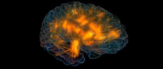
Guess which structure is the oldest in the human central nervous system?
Of course, the spinal cord! It was with him that everything began in the evolutionary development of the brain. It is with this that the development of the nervous system begins in every person, when he is still in the embryonic state. And it is from the spinal cord that we begin to consider the structure and functioning of the central nervous system. If we understand well the principles of the structure and operation of the spinal cord, then it will be easier for us to understand everything else.
Definition
The spinal cord (in Latin Medulla spinalis) is the most ancient tail part of the brain, stretching in the spinal canal along the body and providing control of muscles and internal organs, as well as communication with the brain.
The spinal cord is the most phylogenetically (evolutionarily) ancient and ontogenetically (embryonic) early structure of the central nervous system, formed from the neural tube and having a metameric structure. Functionally, the spinal cord provides control of the executive organs (muscles and glands), as well as communication of the entire central nervous system, including the brain, with the perceptive and executive structures of the body. © Sazonov V.F., 2012. © kineziolog.bodhy.ru, 2012.
The spinal cord is the “executive department” of the central nervous system; almost all actions go through it.
"Memory" (metaphor for memory)
The spinal cord is the workhorse of the central nervous system, and all other parts of the brain want to ride on it. Do you think the comparison is too free? But then what about the fact that the spinal cord officially has a cauda equina? © Sazonov V.F., 2012. © kineziolog.bodhy.ru, 2012.
The spinal cord has the shape of a long cord and is located along the spine.
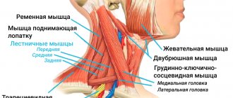
In cross section it looks like a butterfly. Only the “wings” of this butterfly are called “horns” of the spinal cord, and there are 3 pairs of them: anterior (“motor”), posterior (“sensory”) and small lateral (“vegetative”). This is how a six-horned miracle of nature turns out.
Its length is 42-45 cm, and then it is no longer the brain itself, but the “horse tail” - a bundle of nerves. You definitely need to pay attention to this: the fact that the spinal cord ends somewhere at the level of the lumbar region. It turns out a strange thing: the lumbar, sacral and coccygeal sections of the spinal cord are not located in the lower back, sacrum and coccyx, but much higher!
In the photo of the girl trying to show off her spinal cord, the bottom edge of the purse hanging over her shoulder marks the bottom edge of the spinal cord. And then down comes the spine, but without the spinal cord.
Essence
The spinal cord has a segmental structure - and this is its main feature. Those. its individual sections seem to be repeated and connected with certain areas of the body. There are 31-33 segments in total.
Another feature of the spinal cord is that the gray matter (neuron bodies) is located in its inner part, and the white matter (neuron processes) is located on the surface. In the brain, on the contrary, the gray matter lies on the surface in the form of a cortex, and the white matter is hidden inside the brain.
Sections of the spinal cord:
- Cervical.
- Chest.
- Lumbar.
- Sacral.
- Coccygeal
Thickenings:
- Cervical (controls the upper limbs).
- Lumbosacral (controls the lower limbs).
Neurons of the spinal cord
The number of neurons in the spinal cord is about 13 million, and of these, only 3% are motor neurons that control muscles. 97% of neurons are intercalary. As you can see, there are much fewer “working” neurons in the nervous system than “managers”!
Interneurons also belong to the somatic
, and to
the autonomic
nervous system.
Development[edit]
Under the influence of the sonic hedgehog protein, shown here, cells in the lower lamina of the developing spinal cord differentiate into alpha motor neurons.
Alpha motor neurons originate in the basal lamina, the ventral part of the neural tube of the developing embryo. Sonic hedgehog (Shh) is secreted by the adjacent notochord and other ventral structures (such as the floor plate), creating a gradient of highly concentrated Shh in the basal plate and less concentrated Shh in the alar plate. Under the influence of Shh and other factors, some basal lamina neurons differentiate into α-MNs.
Like other neurons, α-MNs send axon projections to reach their target extrafusal muscle fibers through axon guidance, a process regulated in part by neurotrophic factors released by the target muscle fibers. Neurotrophic factors also ensure that each muscle fiber is innervated by the appropriate number of α-MNs. Like most types of neurons in the nervous system, α-MNs are more abundant early in development than in adulthood. Muscle fibers secrete a limited number of neurotrophic factors that can support only a portion of the α-MNs that are initially projected into the muscle fibers. Those α-MNs that do not receive enough neurotrophic factors will undergo apoptosis, a form of programmed cell death.
Because they innervate many muscles, some α-MN clusters receive high concentrations of neurotrophic factors and survive this stage of neuronal pruning. This is true for α-MNs innervating the upper and lower limbs: these α-MNs form large columns of cells that contribute to cervical and lumbar spinal cord enlargement. In addition to receiving neurotrophic factors from muscle, α-MNs also secrete a number of trophic factors to support the muscle fibers they innervate. Reduced levels of trophic factors contribute to the muscle atrophy that follows α-MN damage.
Communication [edit]
Like other neurons, lower motor neurons have both afferent (incoming) and efferent (outgoing) connections. Alpha motor neurons receive input from a number of sources, including upper motor neurons, sensory neurons, and interneurons. The main output of α-MN is extrafusion muscle fibers. This afferent and efferent connection is necessary to achieve coordinated muscle activity.
Afferent input[edit]
Selected pathways between upper motor neurons and alpha motor neurons
| UMN origin | α-MN target | Path name |
| Cortex | Brain stem | Corticonuclear tract |
| Cortex | Spinal cord | Corticospinal tract |
| Red core | Spinal cord | Rubrospinal tract |
| Vestibular nuclei | Spinal cord | Vestibulospinal tract |
| Midbrain tectum | Spinal cord | Tectospinal tract |
| Reticular formation | Spinal cord | Reticulospinal tract |
Upper motor neurons (UMN) send inputs to the α-MN through several pathways, including (but not limited to) the corticonuclear, corticospinal, and rubrospinal tracts. The corticonuclear and corticospinal tracts are commonly encountered in studies of the interaction of upper and lower motor neurons in the control of voluntary movements.
The corticonuclear tract is so named because it connects the cerebral cortex to the cranial nerve nuclei. (The corticonuclear tract is also called the corticobulbar tract
, since the target in the brainstem—the medulla oblongata—is archaically called the “bulb.”) It is along this pathway that upper motor neurons descend from the cortex and synapse on α-MNs. brain stem. Similarly, the cortical UMNs directly control spinal cord α-MNs via the lateral and ventral corticospinal tracts.
Sensory input to α-MNs is extensive and originates from Golgi tendon organs, muscle spindles, mechanoreceptors, thermoreceptors, and other sensory neurons in the periphery. These connections provide the structure of the neural circuits underlying reflexes. There are several types of reflex circuits, the simplest of which consists of a single synapse between a sensory neuron and an α-MN. The knee jerk reflex is an example of such a monosynaptic reflex.
The most extensive input to α-MNs comes from local interneurons, which are the most abundant type of neuron in the spinal cord. Among their many functions is the synapse of interneurons onto α-MNs to create more complex reflex circuits. One type of interneuron is the Renshaw cell.
Efferent output[edit]
Alpha motor neurons send fibers, which mostly synapse, onto extrafusion muscle fibers. Other fibers from the α-MN synapse on Renshaw cells, that is, inhibitory interneurons, which synapse with α-MN and limit its activity to prevent muscle damage.
Alarm[edit]
Main article: Neuromuscular junction
Like other neurons, α-MNs transmit signals in the form of action potentials, rapid changes in electrical activity that propagate from the cell body to the end of the axon. To increase the speed at which action potentials travel, α-MN axons are large in diameter and are heavily myelinated by both oligodendrocytes and Schwann cells. Oligodendrocytes myelinate the part of the α-MN axon that is in the central nervous system (CNS), while Schwann cells myelinate the part that is in the peripheral nervous system (PNS). The transition between the CNS and PNS occurs at the level of the pia mater, the innermost and thinnest layer of meningeal tissue surrounding the components of the CNS.
The α-MN axon connects to its extrafusal muscle fiber through the neuromuscular junction, a special type of chemical synapse that differs in both structure and function from the chemical synapses that connect neurons to each other. Both types of synapses rely on neurotransmitters to convert an electrical signal to a chemical signal and back again. One of their differences is that synapses between neurons typically use glutamate or GABA as neurotransmitters, while neuromuscular junctions exclusively use acetylcholine. Acetylcholine is accepted by nicotinic acetylcholine receptors. on extrafusal muscle fibers, causing them to contract.
Like other motor neurons, α-MNs are named after the properties of their axons. Alpha motor neurons have Aα axons, which are large-caliber, heavily myelinated fibers that conduct action potentials rapidly. In contrast, gamma motor neurons have Aγ axons, which are thin, lightly myelinated fibers that conduct less rapidly.
Links
Afferent nerve/Sensory neuron GSA GVA SSA SVA Nerve fibers (Muscle spindles (Ia), Neurotendon spindle (Ib), II or Aβ fibers, III or Aδ fibers, IV or C fibers) Efferent nerve/Motor neuron GSE · GVE · SVE · Upper motor neuron · Lower motor neuron ( α motor neurons , γ motor neurons) Synapse Chemical synapse Neuromuscular synapse Ephaps (Electrical synapse) Neuropil Synaptic vesicle Touch receptor Meissner's corpuscle Merkel's corpuscle Pacini's corpuscle Ruffini's corpuscle Neuromuscular spindle Free nerve ending Olfactory neuron Photoreceptor cells Hair cells Taste bud Neuroglia Astrocytes (Radial glia) Oligodendrocytes Ependymal cells (Tanycytes) Microglia Myelin (White matter) CNS: Oligodendrocytes PNS: Schwann cells (Neurolemma · Node of Ranvier/Internodal segment · Myelin notch)
Connective tissue Epineurium Perineurium Endoneurium Nerve fiber bundles Meninges: dura mater, arachnoid membrane, pia mater
Clinical significance[edit]
Main article: Motor neuron disease
Poliomyelitis, caused by the poliovirus observed here, is associated with selective cell loss in the ventral horn of the spinal cord, where α-MNs are located.
a-MU lesion is the most common type of lower neuron motor lesion. Damage can be caused by trauma, ischemia, and infection, among other things. In addition, some diseases are associated with selective loss of α-MN. For example, polio is caused by a virus that specifically attacks motor neurons in the ventral horn of the spinal cord. Amyotropic lateral sclerosis is also associated with selective motor neuron loss.
Paralysis is one of the most pronounced consequences of α-MN damage. Because α-MNs provide the only innervation to extrafusal muscle fibers, loss of α-MN effectively severing the connection between the brainstem and spinal cord and the muscles they innervate. Without this connection, voluntary and involuntary (reflex) muscle control is impossible. Voluntary muscle control is lost because α-MNs transmit voluntary signals from upper motor neurons to muscle fibers. Loss of involuntary control results from interruption of reflex circuits, such as the tonic stretch reflex. The consequence of interruption of the reflex is a decrease in muscle tone, which leads to flaccid paresis. Another consequence is inhibition of deep tendon reflexes, causing hyporeflexia.
Muscle weakness and atrophy are also inevitable consequences of α-MN lesions. Because muscle size and strength depend on the degree of use, denervated muscles are prone to atrophy. A secondary cause of muscle atrophy is that denervated muscles are no longer supplied with trophic factors from the α-MNs that innervate them. Alpha motor neuron lesions also result in abnormal EMG potentials (such as fibrillation potentials) and fasciculations, the latter being spontaneous involuntary muscle contractions.
Diseases that impair signaling between α-MN and extrafusal muscle fibers, namely neuromuscular junction diseases, have features similar to those of α-MN disease. For example, myasthenia gravis is an autoimmune disease that interferes with signaling across the neuromuscular junction, resulting in functional denervation of the muscles.
Alpha motor neuron
Alpha motor neurons are large, multipolar lower motor neurons in the brainstem and spinal cord. They innervate the extrafusal muscle fibers of skeletal muscles and are directly responsible for initiating their contraction. Alpha motor neurons are distinct from gamma motor neurons, which innervate the intrafusal muscle fibers of the muscle spindles. While the cell bodies of alpha motor neurons are located in the central nervous system of the CNS, α motor neurons are also considered part of the somatic nervous system—a division of the peripheral nervous system of the PNS—because their axons travel as part of the peripheral nerves to innervate skeletal muscle. The alpha motor neuron and the muscle fibers it innervates form a motor unit. The motor neuron pool combines all the motor neurons that innervate one muscle.
Topographic human anatomy Head Brain Neurons of the brain
1. Location
Alpha motor neurons innervating the head and neck are located in the brain stem; The α-MNs that innervate the rest of the body are located in the spinal cord. There are more α-MNs in the spinal cord than in the brain stem, because the amount of α-MN is directly proportional to the accuracy of control over the work of an individual muscle. For example, the finger muscles have a higher number of α-MNs per fiber and a higher total number of α-MNs than the quadriceps femoris, allowing for finer control of the fingers. Typically, α-MNs located on one side of the brainstem or spinal cord innervate muscles located on the same side of the body. An exception is the trochlear nerve nuclei, located in the brainstem, which innervate the superior oblique muscle of the eye on the opposite side of the face.
1.1. Location
Brainstem
In the brainstem, α-MNs and other neurons are found within clusters of cells called nuclei, some of which contain cell bodies of neurons belonging to the cranial nerves.
Not all cranial nerve nuclei contain α-MNs; on this basis, the nuclei are divided into motor and sensory. Typically, motor nuclei located higher in the brainstem, i.e., more rostrally innervate muscles located higher on the face. For example, oculomotor nerve nuclei containing α-MNs that innervate the ocular muscles are located in the midbrain, the most rostral component of the brainstem. On the other hand, the nucleus of the hypoglossal nerve, which contains α-MNs innervating the tongue, is located in the medulla oblongata - the most caudal one, i.e. located to the bottom of the stem structures. 1.2. Location
Spinal Cord
In the spinal cord, α-MNs are located in the gray matter of the anterior horns.
These α-MNs provide the motor component of the spinal nerves that innervate the muscles of the body. As in the brainstem, the overlying segments of the spinal cord contain α-MNs that innervate muscles higher up on the body. For example, the biceps brachii muscle, a muscle of the arm, is innervated by α-MNs located in spinal cord segments C5, C6, and C7, which are located in the rostral upper portion of the spinal cord. On the other hand, the gastrocnemius muscle, one of the leg muscles, is innervated by α-MNs located within the S1 and S2 segments, which are located in the caudal lower part of the spinal cord. Alpha motor neurons are located in a specific region of the gray matter of the spinal cord. This region belongs to lamina IX of the Rexed lamina system, which classifies gray matter regions based on their cytoarchitecture. Plate IX is located predominantly in the medial part of the ventral anterior horn, although some of its parts lie within plates VII and VIII. Like other regions of the spinal cord, the cells of this plate are organized somatotopically, meaning that the position of neurons in the spinal cord is related to which muscles they innervate. In particular, α-MNs in the medial zone of lamina IX tend to innervate proximal muscles of the body, whereas neurons in the lateral zone typically innervate more distal muscles. In addition, there is somatotropia associated with α-MNs innervating the flexors and extensors: α-MNs innervating the flexor muscles are usually located in the dorsal part of lamina IX; those that innervate the extensors tend to be located more ventrally. 2. Connections
Like other neurons, lower motor neurons possess both afferent input and efferent output connections. Alpha motor neurons receive afferent innervation from a number of sources, including upper motor neurons, sensory neurons, and interneurons. In turn, α-MN innervate extrafusal muscle fibers. These afferent and efferent connections are involved in the coordination of muscle activity.
2.1. Connections
Afferent input
Upper motor neurons of the UMN send axons to the α-MN through several pathways, including but not limited to the corticonuclear, pyramidal, and red nuclear spinal tracts.
The corticonuclear tract connects the cerebral cortex with the nuclei of the cranial nerves. The corticonuclear tract is also called the corticobulbar tract. It is a bundle of upper motor neuron axons that descends from the cortex and terminates at synapses on brainstem α-MNs. Similarly, cortical UMNs directly control α-MNs in the spinal cord through the lateral and ventral corticospinal tracts. Afferent sensory input to α-MNs is extensive and originates from Golgi tendon organs, muscle spindles, mechanoreceptors, thermoreceptors, and other sensory neurons in the periphery. These connections form the structure of neural circuits that underlie reflexes. There are several types of reflex circuits, the simplest of which consists of a single synapse between a sensory neuron and an α-MN. The knee jerk reflex is an example of such a monosynaptic reflex. The most significant afferentation comes to α-MNs from local interneurons, which are the most abundant type of neurons in the spinal cord. In addition to the many other functions they perform, interneurons send axons to α-MNs, which create more complex reflex circuits. One type of interneuron is the Renshaw cell, which will be discussed below. 2.2. Connections
Efferent output
Alpha motor neurons send out axons that mainly terminate at synapses on extrafusal muscle fibers.
Some α-MN axons terminate on Renshaw cells, inhibitory interneurons that send axons to α-MNs and limit their activity to prevent muscle damage. 3. Signals
Like other neurons, α-MNs transmit signals in the form of action potentials, rapid changes in electrical activity that propagate from the cell body to the axon terminal. To increase the speed at which action potentials propagate, α-MN axons are large in diameter and heavily myelinated by both oligodendrocytes and Schwann cells. Oligodendrocytes myelinate the portion of the alpha motor neuron axon that lies in the central nervous system of the CNS, whereas Schwann cells myelinate the portion that lies in the peripheral nervous system of the PNS. The transition between the CNS and PNS occurs at the level of the pia mater. The α-MN axon connects to the extrafusal muscle fiber via the neuromuscular junction, a specialized type of chemical synapse that differs in both structure and function from the chemical synapses that connect neurons to each other. Both types of synapses use neurotransmitters to translate an electrical signal into a chemical signal and back again. However, they differ in that the synapses between neurons typically use glutamate or GABA as neurotransmitters, while neuromuscular junctions exclusively use acetylcholine. Acetylcholine binds to nicotinic acetylcholine receptors on extrafusal muscle fibers, causing them to contract. Like other motor neurons, α-MNs are named according to the properties of their axons. Alpha motor neurons have Aα-type axons that are large in diameter, heavily myelinated, and have a high rate of conduction of action potentials. In contrast, gamma motor neurons have axons of the Aγ type, thin, poorly myelinated, and with low conduction velocity.
4. Role in diseases
α-MN lesions are the most common type of lower peripheral motor neuron dysfunction. Damage can be caused by trauma, ischemia, infection, etc. In addition, some diseases are associated with selective loss of α-MN. For example, polio is caused by a virus that kills motor neurons in the anterior horn of the spinal cord. Amyotrophic lateral sclerosis is also associated with the death of motor neurons. Paralysis is one of the most pronounced effects of α-MN damage. Because only α-MNs provide voluntary innervation to extrafusal muscle fibers, loss of α-MNs effectively disrupts the connection between the brainstem, spinal cord, and the muscles they innervate. Without this connection, voluntary and involuntary reflex control of muscles is impossible. Voluntary muscle control is lost as α-MNs switch voluntary innervation from upper motor neurons to muscle fibers. Loss of involuntary control leads to interruption of reflex arcs such as the tonic stretch reflex. The consequence of interruption of reflex arcs is a decrease in muscle tone, resulting in flaccid paresis. Another consequence is inhibition of deep tendon reflexes, causing areflexia. Muscle weakness and atrophy are also inevitable consequences of α-MN damage. Because muscle size and strength are related to the intensity of use, denervated muscles are prone to atrophy. A secondary cause of muscle atrophy is that denervated muscles no longer receive trophic factors from the α-MNs that innervate them. Damage to alpha motor neurons also appears on the electromyogram, for example, in the form of fibrillation potentials, as well as in the form of fasciculations - spontaneous, involuntary muscle contractions. Diseases that impair signal transmission between α-MNs and extrafusal muscle fibers, called neuromuscular junction diseases, have similar features to those that occur in α-MN diseases. For example, myasthenia gravis is an autoimmune disease that interferes with the transmission of excitation at the neuromuscular junction, leading to functional denervation of the muscles.
5. Development
Alpha motor neurons originate from the basal lamina, located in the ventral part of the neural tube of the developing embryo. The Sonic the Hedgehog protein SHH is secreted by the nearby notochord and other ventral structures such as the floor plate, creating a gradient with a high concentration of SHH in the basal plate and a lower concentration of SHH in the pterygoid plate. Under the influence of SHH and other factors, some neurons from the basal lamina differentiate into α-MNs. Like other neurons, α-MNs send axons to their target extrafusal muscle fibers using axonal guidance, a process regulated by neurotrophic factors secreted by the target muscle fiber. Neurotrophic factors also ensure that each muscle fiber is innervated by the appropriate number of α-MNs. Like most types of neurons in the nervous system, α-MNs are more numerous early in development compared to adulthood. Muscle fibers secrete a limited number of neurotrophic factors that can support only a small part of α-MNs that are initially projected onto the muscle fiber. Thus, α-MNs that do not receive enough neurotrophic factors undergo apoptosis, a form of programmed cell death. Some α-MN clusters receive a high concentration of neurotrophic factors because they innervate many muscles, hence they survive this stage of embryonic development. This is true for α-MNs innervating the upper and lower extremities: these α-MNs are located in the cervical and lumbosacral enlargements of the spinal cord. In turn, α-MNs also secrete a number of trophic factors to maintain the muscle fibers they innervate. Decreased levels of trophic factors contribute to muscle atrophy that follows α-MN damage.