1. Application of minimally invasive operations in neurosurgery
In modern neurosurgery, the principle of using gentle methods of surgical interventions is practiced - the so-called minimally invasive operations
. Such operations significantly reduce trauma to the patient’s body, while maintaining the full effect of their implementation.
Traditional brain surgery is usually performed in 3 stages:
- organizing access to the surgical site (trepanation or craniotomy);
- removal of a hematoma, source of damage or accumulation of cerebrospinal fluid (cerebrospinal fluid);
- stopping bleeding (hemostasis), restoration (sealing) of the meninges and cranial membranes.
The use of minimally invasive operations makes it possible to reduce the volume of the 1st stage, shorten the time of surgical intervention and reduce the risk of developing postoperative complications. Among such techniques, mention should be made of shunting, stereotactic and drainage methods of surgery.
A must read! Help with treatment and hospitalization!
Symptoms of increased intracranial pressure in infants (from open sources)
Anxiety, vague moodiness, sleep disturbance, tearfulness and loudness. All children cry, but if a child constantly screams at the slightest irritation, sleeps poorly, is restless, then there is reason to think.
In addition, increased intracranial pressure can be assumed if the child eats poorly and little or eats small portions very often, spits up a lot and often, and perhaps periodically vomits.
Symptoms may also include increased muscle tone and tremors of the arms and legs when they tremble, for example, when excited or screaming.
There may be ophthalmological symptoms: constant or periodic strabismus, protrusion and drooping of the eyes and other changes.
2.Operations of external ventricular/lumbar drainage
This type of surgical intervention is used to accelerate the restoration of cerebrospinal fluid circulation in case of hydrocephalus in case of impossibility of bypassing the ventricles of the brain for one reason or another, with subarachnoid hemorrhages (hematomas), as well as after certain types of operations.
In addition, such external drainage is used as an independent manipulation during conservative treatment to normalize cerebrospinal fluid circulation
, and as an auxiliary, complementary surgical intervention. In the second case, external lumbar drainage is usually performed before major surgery, and is maintained for some time after the operation - usually several days.
Visit our Neurosurgery page
Hypertensive-hydrocephalic syndrome in children of the first year of life. What do you need to know about him?
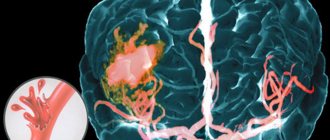
Firstly, it should be noted that a true increase in intracranial pressure occurs as a result of a fairly serious number of reasons: a volumetric formation of the brain (vessel aneurysm, tumor, etc.), inflammation of brain structures, increased production of cerebrospinal fluid, blockage of outflow cerebrospinal fluid (disturbance in the structure of cerebrospinal fluid containing spaces, hemorrhage into the ventricles of the brain, etc.).
Secondly, any of these conditions is characterized by a number of symptoms, accompanied by an increase in intracranial pressure, which is manifested by persistent expressed anxiety of the child, sleep disturbances, appetite, vomiting, bulging fontanelles and gradual separation of the sutures between the bones of the skull, a progressive increase in the size of the ventricles of the brain according to ultrasound data brain (NSG - neurosonography).
If you notice similar symptoms in a child, you must strictly contact a neurologist to rule out serious diseases manifested by these symptoms. At the same time, there are a number of symptoms that often serve as the basis for a diagnosis: intracranial hypertension. However, the presence of only one of them in a child casts doubt on the diagnosis of “increased intracranial pressure.”
Greffe's sign (the appearance of a strip of white (sclera) between the upper eyelid and the iris of the eye) is a fairly common phenomenon in children in the first months of life. This is due to some discrepancy between the sizes of the orbit and the eyeball. During growth, the volume of the orbit increases, the eyeball takes a deeper position, and the strip of sclera above the iris disappears.
An increase in head circumference serves as an indicative sign for a neurologist by which intracranial hypertension can be suspected, provided that other symptoms are present.
Normally, head circumference (CH) increases most actively in the first months of life: 1 month. – up to 3.5 cm; 2 months - 2 cm and 3 months. – 2 cm. Then the rate decreases and the increase averages 1-1.5 cm per month for up to 6 months. In premature babies, these rates are even higher. The idea that the increase is 1-1.5 cm per month is wrong!
There are no absolute standards for head circumference. It is correct to start from the indicators at birth, because if at birth the head circumference was 33 or 35 cm, and the increase in 1 month was 3 cm, then in the end the indicator is 1 month. will be 36 and 38 cm, respectively, and both values are normal.
Increase in the size of liquor-containing spaces according to NSG - neurosonography
Exceeding the size by 1-2 mm, without any other neurological symptoms and dynamics, should be considered as a possible individual norm for a particular child. After all, we do not treat data from brain ultrasound (NSG - neurosonography), it is necessary to look at the whole situation in its entirety.
Also, a slight increase in size does not at all indicate an increase in intracranial pressure. Intracranial pressure is reliably measured only during puncture, and the expansion of cerebrospinal fluid-containing spaces on neurosonography (NSG) only indicates their expansion, and not an increase in intracranial pressure. Liquor fills the expanded spaces compensatory, which ensures constant intracranial pressure and a shock-absorbing effect.
Nosko Anastasia Sergeevna, PhD, neurologist.
3.Method of lumbar drainage
Drainage installation
occurs with the patient lying on his side. The legs are bent at the hip joints, the knees are tucked to the stomach, the chin is pressed to the chest. First, a puncture is performed between the vertebrae (in the L3-L5 area), at the site of which anesthesia with novocaine or similar drugs is introduced layer by layer, taking into account the individual reaction of the patient. Using a special needle with a diameter of 2.5 mm, a puncture of the subarachnoid space is performed, passing through the skin, subcutaneous fat, epidural fat, dura mater and arachnoid mater. Upon reaching the desired area, the mandrin is removed from the needle, and the cerebrospinal fluid begins to flow out. Then a synthetic catheter with a diameter narrower than the needle (2 mm) is inserted into the needle and advanced into the subarachnoid space. The needle is then removed, the catheter is secured to the skin with an adhesive tape and a special sterile fluid collection system is attached to it. By changing the height of the liquor collector, they try to maintain the pressure at a relatively stable level.
To prevent such complications
, such as radicular pain, inflammation and hypotension syndrome,
strict control of the pressure and volume of drained fluid, the prescription of painkillers and antibacterial measures are recommended.
About our clinic Chistye Prudy metro station Medintercom page!
ICP norms of intracranial pressure in children and adults (from open sources)
Intracranial pressure in newborns is normally lower than in older children and adults. It should be taken into account that the normal numbers can change both up and down, but this will not mean that the child is sick. Increased or slightly decreased intracranial pressure in newborns and children may be due to exposure to some external irritants. For example, a cold or, conversely, hot room, emotional state (when, say, the baby was upset about something, the mother scolded or did not buy the desired toy, when the child did not get enough sleep) and even the posture when measuring ICP. Normally, intracranial pressure in infants, children and adults is measured in the supine position.
- Intracranial pressure in children is normally 3–7 mmHg. Art.
- Intracranial pressure in newborns is 1.5–6 mmHg. Art.
- Intracranial pressure in adults is 3–15 mmHg. Art.
Symptoms of external hydrocephalus
The clinical picture in each specific case will be different, and the nature of the manifestation of the disease depends on the severity of the pathological process and the state of the central nervous system. Common symptoms are frequent headaches, blurred vision, nausea, vomiting, and weakness. By the way, pain is more localized in the frontoparietal region and in the area of the eyeballs. A person with dropsy experiences pain in the first half of the day, with sudden movements, coughing, sneezing, or severe physical exertion.
Symptoms may vary depending on the severity of the disease. Scientists distinguish 3 stages, and each has its own characteristics:
Mild external hydrocephalus. With a minimal amount of dropsy, the human body will try on its own to cope with such a problem as impaired circulation of the cerebrospinal fluid. In this case, you will feel a slight malaise, periodic dizziness, short-term darkening in the eyes, and a tolerable headache.
The middle stage of development of the disease. At this stage of the spread of the disease, symptoms appear intensely and are more pronounced. Due to an increase in intracranial pressure, severe headaches occur during physical activity, swelling of the optic nerve and facial tissues, increased fatigue, nervousness, depression, and surges in blood pressure.
Severe form of the disease. Signs of pathology in severe forms of external hydrocephalus are reduced to convulsive seizures, frequent fainting, a state of apathy, loss of intellectual abilities, memory loss and inability to care for oneself. Progressive dropsy can even lead to death, so there is no need to delay going to the doctor. It is better to do this at the first suspicion and a slight deterioration in health.
With chronic accumulation of cerebrospinal fluid, symptoms such as unsteady gait, paralysis of the upper and lower extremities, urinary incontinence, nighttime insomnia and daytime sleepiness, depressed mood, and a complex of psychoneurological disorders can be observed.
What types of external hydrocephalus of the brain are there?
External hydrocephalus of the brain refers to the accumulation of cerebrospinal fluid (cerebrospinal or cerebrospinal fluid) outside the cerebral hemispheres - in the subarachnoid space. Due to the large accumulation of fluid, the subarachnoid fissures widen, which causes increased pressure on the cerebral cortex and the resulting negative consequences.
The nature and level of complexity of the disease directly depend on the specific type of dropsy. Several criteria are used for classification. The most common ones are:
- intensity of manifestation (pronounced - accumulation of a large amount of cerebrospinal fluid, causing neurological symptoms; moderate - minimal amount of fluid, no signs);
- degree of impact on brain structures (compensated - cerebrospinal fluid does not affect the brain; decompensated - there is a deterioration in the functioning of the nervous system and brain);
- causes of occurrence (replacement - more often diagnosed in older people and is accompanied by the death of brain cells; acquired - occurs due to the spread of infections and mechanical traumatic brain injuries);
- nature of the course (chronic form - a gradual increase in neurological disorders; acute form - a sharp deterioration in the patient’s well-being).
Employees of the Department of Neurosurgery of the City Clinical Hospital named after. Eramishantsev will first determine the type of external hydrocephalus and only then begin treatment. Particular attention is paid to diagnostic data and a thorough study of the symptoms of the identified disease.
Diagnosis of external hydrocephalus in adult patients
Studying the symptoms and visual examination of the patient is not a sufficient condition for determining external hydrocephalus of the brain. Indirect signs, of course, are important, but you can’t do this without professional diagnostics. Today, 6 methods for detecting dropsy are used:
- Ultrasound examination (US) of the neck and head to assess the condition of blood vessels;
- Magnetic resonance imaging (MRI) helps to identify changes in soft tissues and most accurately determine the type of hydrocephalus and the stage of development of the pathology;
- computed tomography (CT) is intended to determine the degree of damage to brain tissue, the size of subarachnoid fissures, and the presence of neoplasms;
- X-rays with the introduction of a contrast agent are aimed at identifying disturbances in the outflow of venous blood and damage to the vascular bed;
- a spinal puncture is prescribed if there is a suspicion of the development of dropsy after encephalitis or meningitis and you need to find out what level of cerebrospinal fluid pressure;
- ophthalmological examination - an opportunity to determine whether the patient has swelling of the optic nerve and atrophy of the tissues of the ocular apparatus.
IMPORTANT! If the diagnosis of “chronic external hydrocephalus of the brain” is confirmed, it is advisable to carry out an additional diagnostic examination after 6 months. The intensity of further visits to the doctor depends on the data obtained and is determined individually.