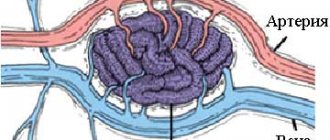
Cavernous hemangioma (cavernoma) is a benign vascular tumor. It consists of deformed venous vessels separated by cavities (cavities). As a rule, these are congenital neoplasms and much less often post-traumatic. They can be located in any area of the brain. They are more common in the hemispheres, but can also form in the trunk (a particularly problematic localization), and in one case out of ten they are multiple. The sizes of caverns range from a few millimeters to several centimeters. Despite their benign nature, hemangiomas, when located in the brain, often lead to serious consequences:
- they can compress and irritate brain structures, manifesting like any volumetric intracranial process with general cerebral and focal symptoms;
- Cavernomas are a source of intracerebral hemorrhages not associated with hypertension in two to four cases out of ten. Moreover, if the patient does not die after the first episode, the risk of a recurrent vascular accident reaches 30%, and a fatal outcome – 70%.
Small cavernous hemangiomas can exist for a long time without symptoms. Most often, the first symptoms appear in middle age. Clinical manifestations depend both on the size of the cavernoma and on its location. It can be:
- headache;
- dizziness;
- double vision;
- nausea;
- vomit;
- noise in the head;
- lack of coordination;
- seizures of a convulsive nature;
- paresis;
- sensitivity disorders;
- speech disorders;
- mental disorders.
Terminology
There are many synonyms for this pathology, including the terms cavernous hemangioma , cerebral cavernous malformation , or simply cavernoma . This pathology does not relate to neoplastic processes, therefore the terms hemangioma and cavernoma should be avoided. Additionally, it is important to note that according to the latest ISSVA nomenclature for classifying vascular anomalies, these changes correspond to low-velocity venous malformations. cavernous in the description or conclusion is useful, since this term is best known to most clinicians.
Clinical picture
Cavernomas are often asymptomatic.
Symptoms begin to appear with hemorrhages, as well as with growth into the surrounding tissues or their compression.
Focal neurological symptoms are associated with the amount of hemorrhage, epileptic seizures or increased intracranial pressure.
The most common headaches are of various types.
When the cavernoma is located supratentorially, epileptic seizures occur in more than 55%.
After the hematomas resolve, the symptoms decrease significantly.
Morophology
Macroscopically, cavernous venous malformation looks like a mulberry. On histological examination, cavernous venous malformations are thin-walled cavities of irregular shape, the walls of which are formed by endothelium. The cavities may be closely adjacent to each other, or be separated by collagen fibers or fibrous tissue, unlike AVMs, which may have medulla between the loops. In some cases, they are closely associated with developmental venous anomalies (DVA) and in this case it is a mixed vascular malformation.
Prevalence
Cavernomas can remain asymptomatic throughout a person’s life, so it is quite difficult to get an idea of the prevalence of the pathology. According to a few studies, cavernomas occur in 0.3%–0.5% of the population. It is not possible to estimate what proportion of these cavernomas manifest clinically, since such studies are lacking. However, it is safe to say that the vast majority of cavernomas remain asymptomatic. Cavernomas occur in two main forms: sporadic and hereditary. Until recently, it was believed that the sporadic form of the disease was the most common. Studies in recent years have shown that the ratio of sporadic and familial cavernomas depends on the quality of examination of relatives of patients with clinically manifested pathology - the wider the scope of those examined, the higher the percentage of hereditary forms. According to some data, the frequency of hereditary forms reaches 50%. Cavernomas of the central nervous system can manifest clinically at any age, from infancy to the elderly. Among those examined at the institute, in two cases the first symptoms of the disease appeared in the first weeks of life, and in several patients - at the age of over 60 years. The most typical development of the disease is between the ages of 20 and 40 years. According to our data, with a hereditary form of the pathology, the first signs of the disease appear in childhood more often than with sporadic cavernomas. The ratio of men and women among patients with cavernomas is approximately the same.
Diagnostics
Cavernous venous malformations of the brain are localized supratentorially in ~80% of cases, but in general can be localized in any part of the brain, including the brainstem. Most often they are single, but at the same time, in one third of patients with sporadic formation, more than one malformation may occur.
Computed tomography Large malformations are visualized by computed tomography as areas of increased density, in the presence of subacute hemorrhage, surrounded by perifocal edema, which do not enhance with contrast. Small malformations are difficult to distinguish.
Magnetic resonance imaging Magnetic resonance imaging is the method of choice. The classic sign is of popcorn or berries, surrounded on the periphery by a ring-shaped zone of signal loss due to the presence of hemosiderin and characterized by increased magnetic susceptibility.
- T1 and T2 MR signal is variable depending on the stage of breakdown of hemoglobin products in the malformation and fluid-liquid levels may sometimes be visualized.
- Gradient echo or T2* sequences are better at detecting malformations than T1 and T2 weighted images. In patients with a hereditary form of multiple cavernous malformations, they are important for counting the number of lesions that may be missed on spin echo sequences.
- Magnetic susceptibility-weighted images (SWI) have a sensitivity comparable to gradient sequences in detecting capillary telangiectasia, and they are also highly sensitive to detecting calcifications compared with T1- and T2-weighted images.
In the presence of recent hemorrhage, moderate perfiocal edema may be detected. Basically, the T1 MR signal intensity does not change after the administration of a contrast agent, but at the same time this is possible.
Selective angiography Kvernous malformations are not visualized during angiography, and arteriovenous shunts are not detected.
Dimensions and location
| Cavernoma of the spinal cord at the Th2 level |
The sizes of caverns can be very different - from microscopic to gigantic. The most typical cavernomas are 2–3 cm in size. Cavernomas can be located in any part of the central nervous system. Up to 80% of cavernomas are found supratentorially. Typical localization of supratentorial cavernomas is the frontal, temporal and parietal lobes of the brain (65%). Rare cases include cavernomas of the basal ganglia and thalamus opticum - 15% of cases. Even less common are cavernomas of the lateral and third ventricles, the hypothalamic region, the corpus callosum, and the intracranial sections of the cranial nerves. In the posterior cranial fossa, cavernomas are most often located in the brain stem, mainly in the tegmentum of the pons. Isolated midbrain cavernomas are quite rare, and medulla oblongata cavernomas are the least common. Cerebellar cavernomas (8% of all cavernomas) are most often located in the cerebellar hemispheres, less often in the vermis. Cavernomas of the medial parts of the cerebellar hemispheres, as well as the vermis, can spread to the fourth ventricle and to the brain stem. Spinal cord cavernomas in our series accounted for 2.5% of all cavernomas. Taking into account the location of cavernomas in terms of difficulty of access and the risk of surgical intervention, it is customary to divide supratentorial cavernomas into superficial and deep. Among superficial cavernomas, there are those located in functionally important zones (speech, sensorimotor, visual cortex, insula) and outside these zones. All deep cavernomas should be considered as located in functionally significant zones. According to our data, cavernomas of functionally significant areas of the cerebral hemispheres make up 20% of supratentorial cavernomas. For cavernomas of the posterior cranial fossa, all localizations, with the exception of cavernomas of the lateral parts of the cerebellar hemispheres, should be considered functionally significant. CNS cavernomas can be single or multiple. The latter are detected in 10–20% of patients. According to our data, patients with multiple cavernomas made up 12.5% of those examined. Single cavernomas are typical for the sporadic form of the disease, and multiple ones are typical for the hereditary form. The number of cases of multiple cavernomas in the hereditary form reaches 85%. The number of cavernomas in one person varies from two to 10 or more. In some cases, the number of cavernomas is so large that it is difficult to count.
Differential diagnosis
Kvernous venous malformations should be differentiated from numerous other pathologies leading to multiple microhemorrhages, including:
- cerebral amyloid angiopathy
- chronic hypertensive encephalopathy
- diffuse axonal injury
- cerebral vasculitis
- radiation damage to the brain
- hemorrhagic metastases
- Parry-Romberg syndrome
Large malformations can simulate:
- hemorrhage into metastases
- hemorrhage into a primary tumor (eg glioblastoma)
What needs to be done to prepare for this procedure?
A few days before the procedure, the patient must contact the Humanitas Clinic for a preliminary appointment and undergo a blood test, ECG of the heart, eye and hearing examinations, as well as a medical examination to ensure a complete compilation of all medical records. It is important that the patient is informed of any allergic reactions to medications or contrast agents, as well as claustrophobia. On the day of the procedure, the patient must arrive accompanied by a family member or friend. You cannot eat food from midnight the night before; You should shower with a disinfectant shampoo containing products such as chlorhexidine. It is better not to wear jewelry: brooches, hair clips, and also not to use hair cosmetics and artificial nails; wash off makeup; Wear comfortable clothes and shoes, preferably with buttons or zippers.
Cavernous angiomas on CT
When using visualization methods, it is useful to separate the cavity into 3 components. These include (1) a peripheral pseudocapsule, consisting of glial tissue impregnated with hemosiderin, (2) an irregularly structured intermediate connective tissue separating the cavities, and (3) a central vascular part, consisting of vascular cavities with slow blood flow.
On CT images without contrast enhancement, a cavernoma appears as a focal formation of an oval or nodular shape, characterized by slightly or moderately increased X-ray density and does not have a volumetric effect on the surrounding parenchyma. Areas of calcification and hemosiderin deposition in the walls of fibrous septa, along with stagnation of blood in the cavities, contribute to increased x-ray density on non-contrast-enhanced images. On CT images, calcifications are detected in approximately 33% of all cavernomas. If the formation is old, then it may contain central non-contrasting areas of reduced density, which corresponds to cysts from resorbed hematomas.
Contrast enhancement can be minimal or maximal, although 70-94% of cavernous malformations are weakly or moderately enhanced after intravenous contrast. In most cases, good contrast is the result of increased blood flow in the vascular component of the mass. The heterogeneous “speckled” enhancement is caused by intravascular fibrous septa, and the low-density rim along the periphery is caused by a pseudocapsule of glial tissue surrounding the formation.
Mass effect is not typical for cavernomas, unless they are associated with recent hemorrhage. On CT images without contrast enhancement, cavernomas may not be detected at all. In hemorrhages and the formation of intracerebral hematoma, cavernomas are visualized as areas of focal signal enhancement in the area adjacent to the hematoma.
Any hemorrhage detected on CT in a relatively young patient should be carefully investigated, and cavernous angioma should always be considered as a possible cause. When evaluating a patient with a seizure disorder, cavernous angioma should also be considered as a likely etiological factor, especially if the patient is between 20 and 40 years of age.
Cavernous malformations identified by CT may also include other rare vascular malformations (thrombosis of arteriovenous malformation, capillary telangiectasia), glioma (poorly differentiated astrocytoma or oligodendroglioma), and metastatic melanoma.
Cavernous angioma or tumor?
Brain scanning using CT and MRI in most cases allows one to clearly distinguish a cavernoma from other brain formations, including tumors of varying degrees of malignancy. However, in some cases, differentiation of these formations represents a diagnostic problem, the solution of which requires extensive experience. In this regard, the ability to attract a highly qualified diagnostician is critical. In addition, high-quality interpretation of CT and MRI images provides a solution to other diagnostic problems: exclusion of surrounding cerebral edema, identification of the severity of hemorrhage, description of details affecting the operability of the cavernoma. If you are in doubt about the diagnosis, you should consult a radiologist from a leading center specializing in brain pathology. A second opinion from such a diagnostician can be very valuable in the differential diagnosis of angiomas and other pathological conditions.