Synonyms: nerve compression syndrome, compression neuropathy, peripheral nerve compression.
A pinched nerve occurs when a peripheral nerve loses mobility, flexibility, or is compressed by surrounding tissue. Nerve compression can cause neuropathic/neurogenic pain, which can be either acute or chronic.
Pinched nerve syndromes (referring to a general group of signs and symptoms) occur in individuals as a result of swelling of surrounding tissues or anatomical abnormalities.
Compression neuropathies occur at the level of the peripheral nerves and are typically characterized by pain and/or loss of function (motor and/or sensory) of the nerves as a result of chronic compression. To adequately diagnose a pinched nerve, it is important to know the neural pathways and areas of peripheral nerve innervation/responsibility.
Friends, Georgy Temichev’s seminar “Diagnostics and treatment of pain in the lumbar region” will take place very soon. Find out more...
The most common types of nerve entrapments include (but are not limited to):
- Carpal tunnel syndrome (pinched median nerve).
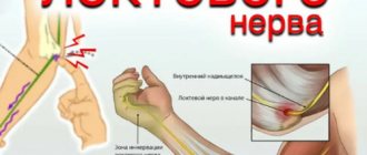
- Cubital tunnel syndrome (compression of the ulnar nerve).
- Superior outlet syndrome (brachial plexus lesion).
- Suprascapular nerve entrapment.
- Compression of the obturator nerve.
- Tarsal tunnel syndrome (compression of the posterior tibial nerve).
- Damage to the interdigital nerves (especially between the 3rd and 4th fingers), known as Morton's neuroma.
- Muscular arcade of the supinator (also known as the arcade of Froese).
- Pinching of the posterior interosseous nerve of the forearm.
It should be noted that chronic mild nerve irritation can lead to increased nerve tension. This may be the underlying cause of a person's symptoms.
Finally, it is important to keep in mind that damage or compression of nerve structures often occurs due to joint or muscle injuries. A thorough assessment of the affected area is necessary to make an accurate diagnosis.
Etiology
Pinched nerve syndrome can result from chronic damage to the nerve as it passes through the osseous-ligamentous tunnel. Compression usually occurs between the ligamentous canal and adjacent bony surfaces.
In cases of nerve entrapment, at least one part of the compressive surface is movable. This results in either repeated compression or friction/sliding against sharp or hard edges, resulting in chronic trauma. Immobilizing the nerve with a splint or lifestyle modifications may relieve symptoms. Compression neuropathies can also be caused by systemic disorders such as rheumatoid arthritis, acromegaly, or hypothyroidism.
Diagnosis of neuropathies of the ulnar, radial and median nerves
The most successful treatment for neuropathy is treatment directly at the point of nerve damage. For successful treatment, your treating doctor will find out:
- At what exact point is the nerve damaged (compressed); this helps us provide targeted treatment;
- What exactly led to the nerve suffering (trauma, scar, compression);
- The degree of nerve suffering (complete or partial damage, the presence of a recovery process, the presence of complete death of the nerve, etc.).
Often, to establish the cause of nerve pain, a detailed neurological examination is sufficient , during which the strength of the muscles controlled by the nerve, the possibility of certain movements, sensitivity, the presence of pain points and seals along the nerve are assessed. Auxiliary diagnostic methods are electroneuromyography, radiography and computed tomography.
Electroneuromyography allows you to assess the speed and volume of impulses along the nerve, detect the location of damage/compression, and determine the prognosis for recovery. Electromyography helps us evaluate the effect of certain types of treatment and choose the most suitable ones. The Echinacea clinic uses a modern computer electroneuromyograph.
X-ray and computed tomography of the joints will provide complete information about the deformation of the joints and bone canals of the nerves, the causes and points of nerve compression.
Pathophysiology
By its nature, nerve damage can be either ischemic or mechanical.
Repeated injury and trauma to the nerve can lead to microvascular (ischemic) changes, swelling, damage to the outer layers of the nerve (the myelin sheath) that facilitate the transmission of nerve impulses, and structural changes in membranes at the organelle level (both the myelin sheath and the nerve axon) . Focal segmental demyelination in the area of compression is a common feature of compression syndromes. Full restoration of function after surgical decompression is associated with remyelination of the injured nerve. Incomplete recovery occurs due to Wallerian axonal degeneration and persistent fibrotic changes at the neuromuscular junction, which may prevent complete reinnervation and restoration of function.
Prevention
Prevention of scalene muscle syndrome is aimed at preventing the circumstances that lead to the occurrence of the syndrome. What are these circumstances?
Firstly, scalene muscle syndrome very often occurs as one of the links in the pathology of the intervertebral discs of the cervical spine. Therefore, timely consultation with a doctor and elimination of any pathology of the spine and discs in the early stages is the most important preventive action.
Secondly, since the main provoking factor in the development of the syndrome is prolonged forced positioning of the head and arms, which usually happens during sedentary work, it means that it is necessary to take regular breaks. Keep in mind that the optimal time for continuous work is 45 minutes, like a school lesson (plus or minus 15 minutes). In other words, try to change your body position - it doesn’t matter how exactly - get up, stretch, walk - the main thing is to make a short-term change in position. In general, approach this issue creatively and act according to the circumstances.
Thirdly, very often scalene muscle syndrome occurs against the background of sports overtraining, so people who are actively involved in sports need to be aware of this and be attentive to the signals of their body.
In general, this wish is not only for lovers of an active life, but for everyone - do not ignore “body language”, because many problems arise from neglecting these signals. As practice shows, almost all patients who consult a doctor with scalene muscle syndrome describe the onset of the disease in the same way: “It all started a long time ago and at first it didn’t bother me much.” This is a classic onset - gradual and not very painful. This is how the body sends us signals, giving us the opportunity to quickly correct everything. And we jam the signals with analgins, thinking that “everything will resolve itself.” Alas, this is not true. From a certain point, the disease will rush like a rapid avalanche, and it will be very difficult to stop it and eliminate the consequences. Therefore, we draw your attention once again - do not ignore “body signals”, do not let yourself go and do not bring the situation to a crisis.
Risk factors
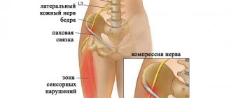
Certain factors put people at greater risk of experiencing nerve compression or pinching. These factors include:
- Previous fracture or dislocation in the area.
- Scar tissue and/or myofascial tension.
- Bone spurs/arthritis.
- Hematoma or swelling.
- Cysts.
- Repetitive or prolonged activity requiring repeated movements.
- Systemic diseases such as diabetes.
Because compression neuropathies are so varied in signs, symptoms, causes, and locations, it is imperative for the physician to perform a thorough history and physical examination to rule out more serious conditions.
Symptoms
Symptoms of a pinched nerve may vary from person to person. Often they will be localized or referred sensations along the affected nerve. The most common symptoms include:
- Localized or referred pain.
- Numbness, tingling, or electric sensation.
- Paresthesia.
- Burning.
- Impaired movement of the affected part of the body.
- Muscle weakness.
- Dry thin skin (in case of chronic pinching of motor and sensory nerves).
Causes of piriformis syndrome
Pain in the buttock occurs with sciatica - irritation of the sciatic nerve, which is most often observed with lumbosacral osteochondrosis and its consequences - disc protrusion, disc herniation or proliferation of osteophytes in the vertebral bodies, as well as with spondylosis and spondylolisthesis. Sudden pain causes a reflex spasm of the piriformis muscle, namely, in its thickness or under it passes the sciatic nerve, which is formed by the fibers of the spinal nerves emanating from the spinal canal.
Thus, pain in the buttock can have two sources - compression of the roots of the spinal nerves at the spine and compression of the trunk of the sciatic nerve by the piriformis muscle. Other causes of piriformis syndrome include:
- Injury to the piriformis muscle - when falling on the buttocks, it is bruised, a hematoma is formed, a painful spasm - all this contributes to irritation of the sciatic nerve in its thickness;
- Consequences of injury - the damage heals, but the muscle fibers in the area of the bruise are replaced by a connective tissue scar, which compresses the sciatic nerve - and a pain syndrome is formed again;
- Inflammation - myositis of the piriformis muscle - can also cause pain in the buttock;
- Neoplasms, bone tuberculosis, osteomyelitis, arthritis or arthrosis of the hip joint can cause reactive inflammation of the piriformis muscle and pain.
The pain is often unilateral, but sometimes both sides are affected. At the same time, it is quite pronounced, radiating to the back of the thigh, lower leg and foot. Skin sensitivity disorders on the leg are usually not detected; sometimes there may be a slight tingling sensation. But the patient tries to take care of his leg; it is uncomfortable for him to sit on the sore side - as a rule, the person holds his leg in an elevated position and is not able to sit straight.
Inspection
Visual assessment
Visual inspection and palpation of the entire upper/lower limb, cervical or lumbar spine is mandatory. The examination should focus on limb asymmetry, muscle atrophy, or abnormal posture. The limbs should also be compared for changes in skin temperature and pulse quality in provocative positions.
Palpation
By applying pressure to the area where the nerve is believed to be compressed, the doctor can reproduce the patient's symptoms.
Upper limb
Sensorimotor testing and provocative maneuvers such as the Spurling sign, Tinel sign, median nerve compression test (upper limb nerve tension tests), Phalen test, and elbow flexion test may be helpful in diagnosing cervical radiculopathy or specific nerve entrapment syndromes.
Lower limb
Evaluation of the lower extremity should also include detailed sensorimotor testing of the peripheral nervous system of the lower back and lower extremity. Below are common nerve tension tests that may be performed in a clinic. These tests may provoke symptoms in the patient or, alternatively, symptoms such as tingling or numbness.
- Straight leg raise test.
- Slouch test.
- Thomas test.
Anesthetics and electroneuromyographic study
- If you inject a few milliliters of local anesthetic into the area of suspected nerve damage, you can reduce or completely eliminate the existing symptoms.
- Electroneuromyography can also confirm nerve damage. However, there is no consensus on the “gold standard” for diagnosis. Some experts believe that there are no ideal diagnostic methods (X-ray, ultrasound, MRI, scintigraphy) that could detect a pinched nerve.
- It is also important to note that a normal ENMG does not exclude symptomatic compressive neuropathy, especially in the early stages.
Diagnostics
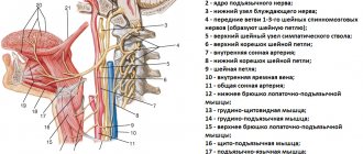
Diagnosis of anterior scalene syndrome is based on a typical clinical picture and always begins with an examination. During such an examination, most patients will experience swelling in the supraclavicular region. This is the so-called pseudotumor (pseudotumor) of Kovtunovich. It occurs due to compression of the lymphatic vessels by the anterior scalene muscle. Also, during the examination, pay attention to the hand - it may be swollen and slightly bluish. In advanced cases, more pronounced changes will be noted, even trophic ones.
When palpating the anterior scalene muscle, it will be sharply tense and painful, and this will also cause increased pain in the neck with possible spread to the arm.
Neurological examination - checking sensitivity, reflexes, etc. - identifies sensory, motor and autonomic disorders.
Spasm test. It is carried out to identify myofascial syndrome of the scalene muscles. You need to turn your head as far as possible in the painful direction and firmly press your chin to your collarbone. The scalene muscles will contract and trigger points in them will be activated. As a result, the patient will feel increased pain.
Adson's test: if you raise your arm on the side of the anterior scalene muscle syndrome and tilt your head here, then where you usually check the pulse at the wrist, there will be no pulse.
Doppler ultrasound (USD) of the neck vessels also has a certain diagnostic value, especially taking into account that the vertebral artery, which supplies the back of the brain, departs from the subclavian artery. And this entire vascular network may suffer from compression of the subclavian artery, which in turn threatens cerebrovascular accident or thrombosis.
Electroneuromyography (ENMG) is considered a generally accepted diagnostic method. Although, at the initial stages of pathology, its information content is not high enough, and sometimes even fraught with erroneous conclusions.
Therefore, the best test for all stages of anterior scalene syndrome is good old manual muscle testing.
Sign up for scalene muscle diagnostics
- Undergo a comprehensive diagnostics of the scalene muscles. We will conduct palpation, neurological examination, spasm test, Adson test and manual muscle testing: we will test the muscles for the presence of active and latent trigger points.
- Diagnostic duration is 30 minutes. This is a full-fledged examination, and not a 2-minute “feeling” for show.
- The diagnosis is carried out personally by Dr. Vlasenko A.A., a doctor with 30 years of experience, an expert in the treatment of myofascial and radicular syndromes.
Dr. Vlasenko Alexander Adolfovich, neurologist, manual therapy doctor
Sign up for diagnostics
Treatment
Treatment for a pinched nerve requires the doctor to consider many variables specific to the individual. The degree of compression, the location of the compression, and the current stage of the recovery process (acute, subacute or chronic stage) all must be taken into account.
Since this condition is usually not life-threatening, it is important to keep moving. A physical therapist or health care professional can tell you what exercises and movements to avoid to prevent further nerve irritation. Clinicians should begin with nonsurgical treatment options and also consider surgery if conservative approaches have failed.
Non-surgical treatment
- To eliminate the underlying mechanisms causing nerve irritation, postural and biomechanical correction are important. The practitioner should also be aware of existing movement patterns that may cause the nerve to rub against or become pinched by surrounding tissue.
- The doctor may consider using non-steroidal anti-inflammatory drugs such as ibuprofen or naproxen to help reduce swelling around the nerve. Although steroids such as Cortisone are very effective anti-inflammatory drugs, their injections are not usually used because there is a risk of nerve damage associated with their use.
- Orthotics or splinting should also be considered as this allows the affected nerve to rest.
- Exercises to increase nerve glide may also be considered if the nerve is in an appropriate healing phase (this type of exercise is not used during the acute phase).
Surgery
Surgery should be considered as a last option for pinched nerve syndrome because... it may entail long-term rehabilitation treatment. Again, due to the variability that exists with this type of pathology, it is best to consult with a physician or neurosurgeon for the best options for the patient. Treatment may involve a simpler procedure such as local arthroscopic debridement or a more complex procedure such as neural decompression.
Advantages of treatment at the Spina Zdorova clinic
- Guarantee of complete and qualified treatment. The word “full” is key in our work.
- High qualifications and extensive practical experience - 30 years.
- We consider each case individually and comprehensively - no formalism.
- Synergy effect.
- Guaranteed fair treatment and fair price.
- The location is a stone's throw from the metro in the very center of Moscow.
Other methods of treatment. Physiotherapy
Physical therapy for scalene syndrome is a supportive treatment. The most commonly chosen procedures are:
Sinusoidal modulated currents (SMC) or – second name – amplipulse therapy. This procedure perfectly copes with pain and muscle spasms, while simultaneously restoring their blood supply and nutrition.
Electrophoresis is a classic of physiotherapy. A less powerful, but gentler method of treatment. Suitable where there are contraindications for SMT therapy.
Ultrasound (UT) is, in fact, a micromassage. Propagating deep into the tissue, ultrasound transmits its vibrations to these tissues, thereby improving their drainage and trophic functions. Deep swelling, stagnation and inflammation disappear, blood circulation and tissue nutrition are normalized. Due to this, the pain syndrome is reduced.
Magnetotherapy – increases microcirculation, thereby eliminating congestion, swelling, inflammation and reducing pain.
Surgical treatment
Surgical treatment of scalene muscle syndrome is used only in advanced cases of the neurovascular stage of the syndrome, when compression of the nerves and subclavian artery could not be eliminated by other means. As we said above, conservative treatment is not always able to solve the problem, and then surgery becomes the only option.
During surgical treatment of scalene muscle syndrome, the muscle itself and adjacent tissues compressing the nerve and artery are dissected. To prevent relapses, muscle resection is carried out over as large an area as possible.
Once again, we draw your attention to the fact that it is advisable to consider the issue of surgical treatment of scalene muscle syndrome only when all conservative treatment methods have been completely exhausted.
Don't let your illness get worse! Timely consultation with a doctor allows you to avoid surgery!