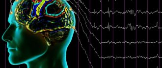
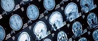
Anatomically realistic 3D brain model depicting localized activity in real time. Each color represents the source of bioelectrical activity and connectivity in a different frequency band (theta, alpha, beta, gamma); gold lines represent anatomical white matter fiber tracts. The supposed transfer of information between brain regions is visualized as pulses of light passing along fiber tracts connecting these regions
Bioelectric activity of the brain
(BEA) is the electrical activity of neurons associated with their excitation, i.e. with a shift in membrane potential.
The bioelectric activity of the brain is based on transmembrane ionic currents, which give rise to the phenomena of action potential and impulse activity of neurons. In this way, neural transmission of signals is carried out in the form of short electrical impulses, each of which represents the main information unit of the brain (quantum of information).12
Mechanism
The bioelectrical activity of the brain is explained by the physiology of the nervous tissue. Each neuron is covered by a membrane. Protein pumps built into the membrane create a difference in electrical potentials inside and outside the cell by pumping charged ions of sodium, potassium, chlorine, etc.
The excitation of a neuron is associated with the opening of ion channels and the passage of ions through the membrane, which leads to a change in voltage, which is recorded by various methods in the form of bioelectrical activity.
An individual neuron is in a continuous process of bioelectrogenesis. Excitation of a nerve cell can occur when it is irritated, i.e. when an impulse arrives from other neurons, and in some cases spontaneously. There are no other sources of the bioelectric field, except neurons, in the brain.
➥ Main article: Bioelectric potentials
Vertical integration of spatial scales from the molecular level (nanometer scale) to the entire brain (centimeter scale)
Levels of brain organization
Conventionally, there are three levels of brain organization at which different forms of bioelectrical activity can be studied: microlevel (activity of one neuron), mesolevel (activity of a local group of neurons) and macrolevel (activity of different areas of the brain).
➥ In more detail: Functional systems, structure and organization of the brain
Microscale is the lowest level of brain organization, reflecting the activity of single neurons due to synaptic and molecular mechanisms.
Mesoscale (English mesoscale) is a level located between the micro and macro levels, at which interactions between large neural ensembles occur.
Macroscale - used to define brain regions and large-scale connections between them.
Methods for recording bioelectrical activity of the brain
Registration of BEA at different levels of organization
(A) Scalp electrode (green), multicontact macroelectrode (red) and microelectrode (blue) (resolution: volume
The study of bioelectrical activity is carried out using electrophysiological methods based on recording fluctuations in the potential difference between two points in the medium, for example, between two electrodes located on the scalp.
Electroencephalography (EEG, English electroencephalography, EEG) is a non-invasive method that allows you to record the bioelectrical activity of the brain from the surface of the scalp.
➥ Main article: Electroencephalography
Magnetoencephalography (MEG, English magnetoencephalography, MEG) is a method based on recording a magnetic field that is formed due to the electrical activity of neurons.
Electrocorticography (ECoG, English electrocorticography
, ECoG) is an invasive method of recording bioelectrical activity using electrodes located on the surface of the cerebral cortex.
➥ Main article: Electrocorticography
Microelectrode methods are a set of methods that use microelectrodes for intra- or extracellular recording of biopotentials. These include recording the activity of a single neuron, multi-unit activity, local field potential (LFP), as well as patch clamp and dynamic clamp methods.
➥
Types of bioelectric activity
Bioelectrical activity, depending on the scale (from a single neuron to the coordinated activity of large neural ensembles), manifests itself in different forms.
➥ Main article: Functional systems, structure and organization of the brain
| Micro level of the brain | Meso level of the brain | Macro level of the brain | |
| Regular bioelectrical activity | spike trains; Regular pattern of subthreshold membrane potential oscillations (SMPO) | Oscillations of neural populations | Brain rhythms or waves generated by the largest neural ensembles |
| Irregular bioelectrical activity | thermal noise; Channel noise; burst noise; Synaptic noise; Spike | 1/f noise | Disorganized EEG 1/f noise |
| Transient bioelectrical activity | Irregular subthreshold membrane potential oscillations; Spindle-like subthreshold membrane potential oscillations | Non-epileptiform transient activity (SWR); Epileptiform transient activity (interictal epileptiform activity, IED) | Non-epileptiform (eng. nonepileptiform) transient activity; Epileptiform transient activity (eng. epileptiform discharges); Event-related potential (ERP) |
Rhythmic activity
Regular subthreshold oscillations of membrane potential
➥ Main article: Rhythmic activity of the brain
Rhythmic or regular bioelectrical activity is a repeating frequency pattern in the central nervous system. In other words, these are the same type or self-similar oscillatory events that repeat at equal intervals.
Neural oscillations generated by the bioelectrical activity of the brain are observed throughout the central nervous system at all levels of its organization and include spike trains/bursting, a regular pattern of subthreshold oscillations of the membrane potential, high-amplitude oscillations of the local field potential and large-scale oscillations (brain waves).
EEG is one of the most accessible electrophysiological methods for recording rhythmic activity. Since the electric field created by a single neuron or a collection of closely located neurons is so small that it cannot be recorded from the surface of the head, EEG is a method that reflects the total activity of a large pool of neurons, the magnitude of which is sufficient to generate a potential difference on the surface of the scalp. It should be emphasized that the electrical activity must be as powerful as is required for the signal to pass through the bones of the skull and skin, which have noticeable resistance to electric current. Again, based on the above necessary and sufficient conditions: the activity recorded from the scalp is due to the relatively powerful total activity of the neuronal pool, it logically follows that the total EEG is primarily due to the activity of cortical neurons as those closest to the skin surface heads - place of registration.
Rhythmic processes play a key role in the functional activity of the brain.
Irrhythmic activity
Irrhythmic or irregular activity refers to chaotic or stochastic changes in the bioelectrical activity of the brain.
At the micro level
Irregular brain activity can be divided into molecular, synaptic and 1/f noise,3 and spiking activity by mechanisms.
Spikes
(English spikes) can also be extremely irregular, both during constant, spontaneous activity, and during evoked activity at a high frequency of impulse emission. There is uncertainty about the source of this irregularity, ranging from intrinsic sources of noise in neurons to collective effects in large-scale cortical networks. Cortical interneurons show highly irregular spike timing in response to direct current injection in vitro. This is in sharp contrast to cortical pyramidal cells, which exhibit irregular bioelectrical activity in vivo but regular activity in vitro. In vitro recordings and computational models have shown that this is associated with the rapid activation kinetics of interneuronal K ion currents. In this case, an arrhythmic spike can contribute to the irregular activity of the entire cortical network.45
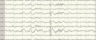
Above is a normal EEG, below is an EEG of a patient with hypsarrhythmia.
At higher levels of organization, nonrhythmic activity can manifest itself in the form of disorganization of the bioelectrical activity of the brain. Types of disorganization:
- Disorganization of bioelectrical activity with a predominance of alpha activity. On the EEG, alpha activity is the main one, but it is not regular enough or is completely irregular in frequency. This more or less disorganized alpha rhythm has an insufficiently high amplitude and may even dominate in all areas of the brain. Beta activity is also often enhanced, often represented by low-frequency oscillations of increased amplitude. Along with this, theta and delta waves with a fairly high amplitude may be present in the EEG.
- Disorganization of bioelectrical activity with a predominance of theta and delta activity. The structure of this type is characterized by a weak representation of alpha activity. Fluctuations in biopotentials in the alpha, beta, theta and delta ranges are recorded without any clear sequence. This non-dominant type of curve can have both medium and high amplitude levels.
- Hypsarrhythmia - manifests itself as disorganization of the background bioelectrical activity of the brain with irregular slow waves of high voltage; multifocal peaks and polyspikes can be superimposed during sleep.6
Paroxysmal activity
➥ Main article: Paroxysmal activity
Paroxysms or transients are short intervals (10 to 1000 ms)7 during which the signal changes abruptly and becomes atypical or relatively unpredictable.
(A) Spindle-shaped subthreshold oscillations of membrane potential; (B) Irregular subthreshold fluctuations in membrane potential
Transient activity reflects the functional integration of brain structures and is a fundamental element of neural interaction. Transients generated jointly by different neural populations indicate the connection of these populations. This communication may be synchronous or asynchronous.8
Examples of such activity include spontaneous synchronous synaptic transients generated by Ca2+ ion currents between adjacent cortical neurons.9 Spikes and sharp waves that constitute seizure or interictal activity in individuals with epilepsy or a predisposition to it, or transients in the form of vertices and sleep spindles , which are the norm. Irregular subthreshold membrane potential oscillations and spindle-shaped subthreshold membrane potential oscillations can also be recorded using in vivo intracellular recording.
Event related potential
➥Main article:ERP
Event related potentials
(EPS, English
event-related potential
- ERP) are electrical voltage fluctuations recorded from the scalp, generated in brain structures in response to certain events or stimuli.
Also refers to transient brain activity. These are EEG changes time-locked to sensory, motor or cognitive events that provide a safe and non-invasive approach to studying the psychophysiological correlates of mental processes. They are believed to reflect the total bioelectrical activity of postsynaptic potentials that occur when a large number of similarly oriented cortical neurons (on the order of thousands or millions) fire synchronously during information processing. ERP is measured using electroencephalography (EEG) and magnetoencephalography (MEG). The magnetoencephalographic (MEG) equivalent of ERP is the ERF or event -related
. 10 ERP subtypes include evoked and evoked potentials.
Evoked potentials
➥ Main article: Evoked potentials
Cognitive evoked potentials (EP, EP)
represent event-related activity that occurs as an electrical response from the brain to various types of sensory stimulation of neural tissue; auditory and visual stimulation are most often used. Recording of such electrical potentials is a non-invasive objective test that provides information, for example, about sensory pathway disorders, the location of lesions affecting sensory pathways, and disorders related to language and speech. Evoked potentials are recorded from the scalp using EEG. Potentials typically appear as a transient waveform, the morphology of which depends on the type and strength of the stimulus and the location of the electrodes on the scalp. The mental state of the subject, such as attention, wakefulness, and anticipation, also influences the morphology of the waveform.
Induced activity
Along with evoked activity, stimulus-related neural activity can lead to induced activity
. The induced response is associated with processes in the brain that are not directly related to the stimulus, but are caused indirectly by the nonlinear interaction of neurons after the stimulus. The functional component of the induced response is likely to represent corticothalamic feedback (top-down) processes linking external stimuli to the internal cortical model of the environment, i.e., combining externally controlled sensory information with internal brain activity.1112 The induced response may be related with cognitive brain functions such as perception, attention and learning, which can be considered higher order processes. A well-studied type of induced activity is a change in the amplitude of oscillatory activity. For example, gamma activity often increases during times of increased mental activity, such as during the presentation of an object. Since induced responses are not phase related and will therefore be excluded during averaging, they can only be obtained using time-frequency analysis. Induced activity typically reflects the activity of multiple neurons: amplitude changes in oscillatory activity are thought to result from synchronization of neuronal activity, such as spike timing or fluctuations in the membrane potential of individual neurons.
Changes in bioelectrical activity of the brain
➥ Similar article: EEG changes
The frequency of bioelectrical activity can indicate various functional states of the brain: sleep, consciousness, cognition and some mental disorders. Decreased bioelectrical activity may appear on the EEG as slow waves, which are most often observed in conditions such as sleep, coma,13 brain death, depression,14 autism,15 brain tumors, obsessive-compulsive disorder (OCD),16 attention deficit disorder and hyperactivity (ADHD),17 encephalitis.18
In contrast, when there is increased bioelectrical activity in the brain, rapid waves are observed on the EEG, in conditions such as epilepsy,19 anxiety, post-traumatic stress disorder (PTSD) and substance use.20
It should be noted that the use of the terms “decrease” and “increase” of bioelectrical activity is incorrect, since the bioelectrical activity of the brain at different levels of organization manifests itself differently, and in the context of the whole brain one can only evaluate parameters (amplitude, power, index ) signals obtained during EEG recording. Then, based on numerous experimental data, one can already interpret changes in one or another parameter and talk about any changes in the functional activity of the brain.
Classification of BEA changes
Based on localization, changes in bioelectrical activity can be:21
- Focal – limited area (up to 3 leads);
- Regional – zone of the brain lobe (3 or more leads);
- Lateralized - pathological activity detected above the hemisphere;
- Generalized - general pathological activity, affecting the entire brain, recorded in all leads according to the mechanisms of primary and secondary generalization;
- Diffuse (unspecified) – cannot be classified into the above groups. According to the degree of severity, diffuse changes in bioelectrical activity can be divided into mild, moderate and pronounced.
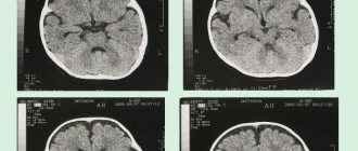
Diffuse EEG changes
A. Flat EEG with burst suppression at intervals of 5–10 s.
B. Continuous generalized acute and slow complexes. C. Diffuse delta activity with peaks in the frontotemporal areas, spreading and predominant in the left hemisphere. A characteristic feature of pathological activity with diffuse changes in bioelectrical activity is the lack of locality, instability of spatial distribution (mosaicity), and violation of bilateral synchrony.
Diffuse brain damage is often associated with the development of encephalopathy, that is, widespread small-focal lesions. Due to the polymorphism of small focal changes and their prevalence, pathological changes are very diverse and do not form any organized activity, which is manifested by mosaic changes in the bioelectrical activity of the brain.
EEG disorganization
When the brain is damaged, pathological diffuse changes in bioelectrical activity are characterized by disorganization and the absence of regular dominant activity (alpha rhythm). Diffuse pathological activity can manifest itself in different ways (for example, in the form of slow-wave rhythms or epileptiform activity is detected), and also have different amplitudes (low-amplitude, medium-amplitude or high-amplitude).
Recording video EEG with single-channel ECG
The EEG demonstrates "slow" and "flat" phases during cardiac asystole, followed by a second "slow" phase after cardiac sinus rhythm resumes. Myoclonic seizures (MS) occur during asystole and after resumption of sinus rhythm. Tonic posture (tone) occurs during asystole. Pay attention to the artifact of tonic muscle contraction recorded on the ECG channel
Disorganization of bioelectrical activity is usually associated with a decrease in EEG amplitude and absence of alpha rhythm, which is described in more detail in the article alpha rhythm depression. Dysrhythmic brain activity can manifest itself in the form of a flat EEG (desynchronous type III, considered as a borderline norm), as well as with a predominance of alpha activity (disorganized type IV) and with a predominance of θ- and δ-activity (disorganized EEG type V). More details: classification of EEG disorders by type.
Slowing brain activity
A slowdown in the bioelectrical activity of the brain (when low frequencies predominate) does not always indicate pathological changes and may be associated with drowsiness. Slowing of the background rhythm in children is observed depending on the age group:
- 1 year – fundamental frequency less than 5 Hz
- 4 years – fundamental frequency less than 6 Hz
- 5 years – fundamental frequency less than 7 Hz
- over 8 years old – fundamental frequency less than 8 Hz
Slowing of cortical rhythms often occurs against the background of diffuse dysfunction of cortical and subcortical structures, due to vascular, metabolic or toxic lesions of the brain, and in children it can be a consequence of perinatal pathology.
Generalized slowdown
FIRDA pattern with slightly slowed background theta EEG in an elderly man with metabolic encephalopathy. Periods of well-formed occipital alpha activity are also noticeable
Irritative changes on the EEG
In the presence of irrigative changes against the background of disorganization of the alpha rhythm with a pointed shape and uneven amplitude of alpha oscillations, the voltage of beta oscillations increases 2-3 times. Pathological changes may appear in combination with diffuse epileptoid activity. In some patients, sharp waves are consistently recorded on the EEG, which coincide with the rhythm of the electrocardiogram. The totality of these EEG changes, expressed equally in all areas of the hemispheres, reflects irritative phenomena in the cerebral cortex. They are caused by an excessive influx of afferent impulses from the angioreceptive zones and from the richly innervated meninges, which are constantly exposed to the effects of a slowly growing tumor.
When EEG is recorded in such patients over time, as the tumor grows, the amplitude of frequent rhythms decreases and low-amplitude delta waves appear, equally pronounced in all areas of both hemispheres of the brain. The stage of irritative cerebral disturbances of biopotentials is more often observed when vascular neoplasms are located in the sagittal, perisagital and anteriobasal regions of the brain. In these areas, tumor nodes are directly connected to the venous sinus.
If patients suffering from brain tumors have symptomatic epilepsy, irritative cerebral changes are also recorded in the EEG in the early stages of the disease. They manifest themselves as a combination of sharpened waves of the alpha rhythm, increased beta oscillations and epileptoid diffuse potentials. Against the background of a general disturbance of cortical rhythm, the EEG may record an epileptogenic focus in the area of the cortex that is directly affected by the tumor. A mildly irritative type of EEG indicates minor damage to brain structures.