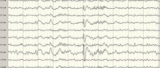
EEG (electroencephalography) of the brain is a highly informative method for diagnosing the state of the central nervous system, based on recording the bioelectric potentials of the cerebral cortex during its life. The results of the study are recorded on paper tape or displayed on a computer monitor. Neurophysiologists at the Yusupov Hospital interpret the EEG results of the brain in adults using a computer program.
The patient receives a conclusion on the second day. If the results of EEG decoding are interpreted ambiguously, they are discussed at a meeting of the expert council with the participation of professors and doctors of the highest category. Leading neurologists and neurophysiologists collectively make decisions regarding the diagnosis and further tactics of patient management. The results of an EEG of the brain performed at the Yusupov Hospital are always accurate, since the study is carried out using the latest European and American equipment, and the decoding is done by candidates of medical sciences who have been trained in the best domestic and foreign diagnostic centers.
Normal EEG in adults
Decoding the EEG results consists of three sections:
- description of leading types of activity and graphic elements;
- conclusion after description with interpreted pathophysiological materials;
- correlation of the indicators of the first two parts with the clinical picture of the disease.
The main descriptive term in EEG is "activity". He evaluates any order of waves. The main types of activity that are recorded during the study and subsequently subjected to decoding, as well as further study, are the frequency, amplitude and phase of the waves. Frequency is measured by the number of wave oscillations per second. It is expressed in units of measurement - hertz (Hz). In the description, the neurophysiologist indicates the average frequency of the activity being studied.
Amplitude is the range of wave oscillations of the electrical potential. It is measured by the distance between the peaks of waves in opposite phases, expressed in microvolts (µV). A calibration signal is used to measure the amplitude. When interpreting the results, neurophysiologists interpret the most common values, completely excluding the rare ones.
The phase evaluates the current state of the process and determines its vector changes. Using an electroencephalogram, functional diagnostic doctors evaluate certain phenomena and evaluate the phases they contain. Oscillations are monophasic, biphasic and polyphasic.
For a restful sleep
If you still find it difficult to achieve the theta wave for sleep, but you have enough zeal, then why not use healthy stimulation methods? Avoid stressful situations or reconsider your reaction to them. Develop your speech, which, by the way, also increases your IQ. Listen to more music. It brings emotions out and thereby increases the activity of theta waves. Adjust your sleep schedule. Quality sleep is important for your mental health. It's good to make time for meditation sometimes. This is the healthiest way to increase theta activity.
What about hypnosis or self-hypnosis? In the process, you facilitate the introduction of the necessary attitudes into the subconscious. The regularity of such actions will reduce stress by activating theta waves. Make time for yoga. It quickly leads to relaxation. And the simplest thing is, don’t forget about creative visualization. Every time you close your eyes, try to visualize what you see
Pay attention to small details. Practice and you can get in the right mood
Electrical rhythms of the brain
The concept of “rhythm” in the EEG is considered to be a type of electrical activity that relates to a certain state of the brain and is coordinated by appropriate mechanisms. When deciphering indicators of the EEG rhythm of the brain, neurophysiologists take into account its frequency corresponding to the state of the brain region, amplitude and characteristic changes during functional changes in activity.
The characteristics of brain rhythms depend on whether the patient is asleep or awake. Brain activity recorded on the EEG in an adult has several types of rhythms, which are characterized by certain indicators and the state of the body.
On the EEG, the alpha rhythm is characterized by a frequency of 8 to 14 Hz. It is present in most healthy individuals. The highest amplitude values are observed in the resting state of the subject, who is in a dark room with his eyes closed. The alpha rhythm is best determined in the occipital region. It can be fragmentarily blocked or completely subside during visual attention or mental activity.
The wave frequency of the beta rhythm on the EEG fluctuates in the range of 13–30 Hz. Its main changes are observed when the subject is active. Pronounced fluctuations are detected in the frontal lobes under the obligatory condition of active activity (mental or emotional arousal).
The gamma rhythm has an oscillation range from 30 to 180 Hz. It is characterized by a rather reduced amplitude - less than 10 μV. Exceeding the amplitude limit of 15 μV is considered a pathology that causes a decrease in intellectual abilities. Rhythm is determined when solving situations and tasks that require increased concentration and attention.
The kappa rhythm is characterized by an interval of 8–12 Hz. It is observed in the temporal part of the brain during mental activity by suppressing alpha waves in other areas. The lambda rhythm has a range of 4–5 Hz. It is triggered in the occipital region when it is necessary to make visual decisions (searching for an object with open eyes). The vibrations disappear completely after concentrating your gaze on one point. The mu rhythm has an interval of 8–13 Hz. It starts in the back of the brain and is best observed in a calm state. Suppressed when any activity is started.
A separate category of types of rhythms that manifest themselves in sleep conditions or in pathological conditions includes 3 types of this indicator:
- The delta rhythm is determined in comatose patients and in the deep sleep phase, and is recorded when recording signals from areas of the cerebral cortex located on the border with areas affected by malignant neoplasms;
- the theta rhythm has a frequency range of 4–8 Hz, manifests itself during sleep, is responsible for the high-quality assimilation of information, and underlies self-learning;
- The sigma rhythm has a frequency of 10–16 Hz, is considered one of the noticeable and main oscillations of the spontaneous electroencephalogram, and occurs during natural sleep at its initial stage.
Based on the results obtained from recording the EEG, an indicator is determined that characterizes a complete all-encompassing assessment of the waves - the bioelectrical activity of the brain. A functional diagnostics doctor checks EEG parameters - frequency, rhythm and the presence of sharp flashes that provoke characteristic manifestations. On these grounds, the neurophysiologist makes a final conclusion.
A few thoughts.
But I’ll digress a little from the topic. The quote from the Czech writer and aphorist Gabriel Laub, which I included at the beginning of the article, is not accidental: have you ever thought about how poorly we use our brain resources?
Already in the last century, G. Laub noticed the global brain “laziness” of humanity. And why bother: calculators, computers, robots, finally, will do everything for us. This fact leads to the fact that over the last few tens of millennia the human brain has only been shrinking. What threatens us next?
In our age of total computerization, the need for a fairly significant part of brain activity disappears.
Did you know that as a result of, for example, counting on a calculator, not a single new neural connection is formed? Accordingly, not a single new convolution appears.
At this rate, the human brain will soon “dry up” completely, and terrible science fiction films with the triumph of robots over humanity have a great chance of coming true.
Let's each try to start with ourselves? To do this, let’s take a closer look at how neural connections are formed and what brain rhythms are.
Decoding EEG indicators in an adult
In order to decipher the EEG and provide accurate results, without missing any of the smallest manifestations in the recording, neurophysiologists take into account all the important points that may affect the indicators being studied, such as:
- patient's age;
- the presence of certain diseases;
- possible contraindications.
After collecting all the EEG data and processing it, the functional diagnostics doctor conducts an analysis and generates a final conclusion, which he provides for making a further decision on the choice of therapy method. Any disturbance in activity may be a sign of diseases caused by certain factors.
EEG disorders are considered:
- constant fixation of the alpha rhythm in the frontal lobe;
- constant violation of wave sinusoidality;
- presence of frequency dispersion;
- exceeding the difference between the hemispheres by up to 35%;
- amplitude below 25 μV and above 95 μV.
The presence of violations of this indicator indicates possible asymmetry of the hemispheres. This may be the result of malignant neoplasms or cerebral circulatory disorders (ischemic or hemorrhagic stroke). A high frequency indicates traumatic brain injury or brain damage.
If a high amplitude of the delta rhythm is detected, the neurophysiologist can assume the presence of a space-occupying tumor in the brain. Inflated values of the theta and delta rhythm, which are recorded in the occipital region, indicate impaired circulatory function, inhibition and a delay in the development of the child.
Start from the basic level
So, you wanted to activate. The reasons can be very different. Someone is suffering from an illness. Some people are tired of living alone, while others just need meaning in life. Theta range waves can help solve your problem. In the process of training in special courses and seminars, you will learn about the role of brain frequencies in the treatment of illnesses, you will be able to recognize the manifestations of waves and vibrations in everyday life, and also improve your health, relationships and financial situation. A good practitioner will be able to guide you, but here you need to be careful, because scammers have not been canceled. Don’t rush to part with your money and find out the biography of your mentor. This, by the way, distinguishes a self-confident person from a fanatic - you know when you can get quality, and not zilch in the eyes.
Mastering the basic level of immersion allows you to stay on the theta wave for as long as you like. You manage to change certain attitudes and replace your beliefs, which were formed on the basis of ancestral memory, karmic moments and collective pressure.
Decoding the EEG of the brain in children
EEG in children has its own peculiarities. An EEG recording of a premature baby born at 25–28 weeks of gestation looks like a curve in the form of slow flashes of delta and theta rhythms, which are periodically combined with sharp wave peaks of 3–15 seconds in length with a decrease in amplitude to 25 μV. In full-term newborns, these values are divided into 3 types of indicators:
- when awake (with a periodic frequency of 5 Hz and an amplitude of 55–60 Hz);
- in the active phase of sleep (with a stable frequency of 5–7 Hz and a fast, low amplitude);
- during restful sleep with flashes of delta oscillations at high amplitude.
Over the course of 3-6 months of a baby’s life, the number of theta oscillations is constantly growing. The delta rhythm is characterized by a decline. From 7 months to one year, alpha waves are formed in the child, and delta and theta gradually fade away. Over the next 8 years, the slow waves on the EEG are constantly replaced by fast alpha and beta oscillations. Until the age of 15, alpha waves predominate. By the age of 18, the formation of the biological activity of the brain is completed.
In order to undergo an examination and decipher the EEG results, call the Yusupov Hospital. The contact center is open every day around the clock. Neurophysiologists analyze the EEG over time and compare the results of the study with the normal EEG.
Theta level
Deep meditation, or REM sleep, is slow brain waves called theta.
At the theta level, you experience a deep understanding of yourself, your intuition, spiritual connection, creativity and oneness with yourself. Physical sensations slip away, although the automatic functions of the body continue to work, but you do not feel your body; the main emotional state is euphoria and bliss.
As the right hemisphere becomes more active in theta, this level is characterized by a significant decrease in speech function, which is the prerogative of the left hemisphere. Young children spend a lot of time in the theta state, when imagination is as real as reality.
Theta is a state of “believing is seeing.”
What does being at the theta level give you:
Uninhibited imagination, creativity, intuition. Imagination is not limited to the left hemisphere of the brain in problem solving.
