Spinal cord - a rather complex system that is responsible for many processes in the body and which is quite difficult to understand on your own. Basic knowledge can be obtained by studying anatomy at school, but when it comes to a more in-depth analysis, many incomprehensible issues arise.
Let's try to figure out what the spinal cord is, how it is structured, what functions it performs, and just understand why it is needed at all.
Spinal cord as part of the nervous system
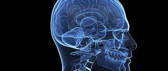
The spinal cord is one of the components of the human nervous system. In Latin its name looks like medulla spinalis.
It is a thick cylindrical tube with a narrow channel located inside it. It is located in the spinal canal, or, more simply, inside the spine.
This organ has a rather complex structure and segmental structure. The main function of this organ is the transmission of various impulses and signals from the human brain to specific organs. In addition, it performs reflex activity, that is, it is responsible for human reflexes, both simple and more complex reflexes.
Human nervous system
Risk of organ damage
Due to the characteristic structure of the brain, it is connected to most systems in the body. The integrity of its structure is extremely important for the correct functioning of the musculoskeletal system and the health of internal organs. Any injury, regardless of severity, can lead to disability. Sprains, dislocations, disc damage, vertebral fractures with or without displacement can cause spinal shock and paralysis of the legs, and disrupt the normal functioning of the cords.
Severe injuries result in shock lasting from several hours to several months. In this case, the pathological condition is accompanied by a number of neurological symptoms. These include numbness, sensory disturbances, pelvic organ dysfunction, and inability to control the process of urination and bowel movements.
Treatment of minor spinal injuries is carried out on an outpatient basis, using medications, therapeutic exercises and massage. Severe injuries require surgical intervention, especially if compression of the spinal cord is detected. Cells are quickly damaged and die, so any delay can cost a person’s health. The recovery period after such an intervention is up to two years. Various physiotherapeutic procedures help with this, for example, reflexology, ergotherapy, electrophoresis, magnetic therapy, etc.
The spinal cord is a key element of the human central nervous system, which is connected in one way or another with almost all internal organs and human muscle tissue. The specific structure allows you to transmit impulses and signals, ensure full motor activity, and perform a number of other functions.
The meaning of the spinal cord
There are only two main and most important functions:
- Reflex. To put it simply, a whole series of reflex arcs are closed on this organ. It is thanks to this that reflexes are carried out (the so-called spinal reflexes).
- Conductor. The organ in this case acts as a conductor. It conducts signals that come from various organs to the brain. It is through this organ that the brain receives all information and processes it. Everything works the same way in the opposite direction.
Spinal cord location
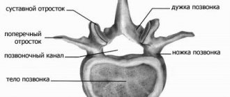
The organ is located in the spinal canal (located inside the human spine). This canal is quite long and practically reaches the lower vertebrae. In fact, this is a special canal, which is an oblong hole in which the spinal cord lies. From the sides it is protected by vertebrae, as well as intervertebral discs.
The organ is also located at the lower edge of the foramen magnum, where connections with the brain occur. It is in this place that there is a huge number of roots that directly connect to the human brain. This connection is called the left and right spinal nerves.
Spinal cord location
The bottom ends at the level of 1-11 vertebrae. Afterwards the organ turns into a thin terminal filament. In fact, it is still the spinal cord, because it contains nervous tissue.
Shells
The entire length of the organ is covered by 3 meninges:
- The inner (first) is soft. It contains veins and arteries that supply blood.
- Arachnoid (medium). It is also called arachnoid. Between the first and inner membranes there is also a subarachnoid space (subarachnoid space). It is filled with cerebrospinal fluid (CSF). When a puncture is performed, it is important to get the needle into this subarachnoid space. Only from it can liquor be taken for analysis.
- External (solid). It extends to the foramina between the vertebrae, protecting the delicate nerve roots.
In the spinal canal itself, the spinal cord is securely fixed by ligaments that attach it to the vertebrae. The ligaments can be quite tight, so it is important to take care of your back and not endanger your spine. It is especially vulnerable from the front and back. Although the walls of the spinal column are quite thick, it is not uncommon for it to become damaged. Most often this happens during accidents, accidents, or severe compression. Despite the well-thought-out structure of the spine, it is quite vulnerable. Its damage, tumors, cysts, intervertebral hernias can even provoke paralysis or failure of some internal organs.
There is also cerebrospinal fluid in the very center. It is located in the central canal - a narrow long tube. Along the entire surface of the spinal cord, grooves and fissures are directed deep into it. These recesses vary in size. The largest of all the slits are the back and the front.
In these halves there are also grooves of the spinal cord - additional depressions that divide the entire organ into separate cords. This is how pairs of anterior, lateral and posterior cords are formed. The cords contain nerve fibers that perform different but very important functions: they signal pain, movement, temperature changes, sensations, touches, etc. The fissures and grooves are penetrated by many blood vessels.
Topography and shape of the spinal cord
Let's look at the features of location (topography) and shape.
To do this, consider a number of features:
- The average length is 42-43 centimeters. In men, the length is often several centimeters longer, while in women, on the contrary, it is shorter.
- Weight 33-39 grams.
- There is a middle gap on the front part, it is clearly visible. You can notice that it seems to be growing into the organ. In essence, it creates a kind of partition that divides the brain into two sections.
- In the cervical and lumbosacral regions it is possible to
- mark two fairly serious thickenings. This is due to the fact that innervation of the upper and lower extremities occurs here. In simple terms, this is where the nerve endings from the limbs “attach” to the spinal cord, which
- Allows them to transmit the necessary signals.
- The spinal cord is topographically practically not connected with the vertebrae. The various sections are not located depending on a specific vertebra or several vertebrae.
The increase in volume in these areas is due to the fact that this is where the largest number of nerve cells are located, as well as fibers through which signals are transmitted from the limbs and back.
Despite the fact that the spine is a kind of “storage place” for the organ, the location of the nerve endings, especially in the lower part of the spine, does not correspond to specific vertebrae. This is due to the fact that the length of the spinal cord is less than the length of the human spine.
That is why it is necessary for doctors to know the exact location of each of the segments, because they will not be able to navigate along the spine.
Location in the spine
What are segments
In order for the spinal cord to reliably communicate with other parts of the body, nature created sections (segments). Each of them has a pair of roots that connect the nerve system with internal organs, as well as skin, muscles, and limbs.
The roots emerge directly from the spinal canal, then nerves are formed, which are attached to various organs and tissues. Movement is reported mainly by the anterior roots. Thanks to their work, muscle contractions occur. That is why the second name of the anterior roots is motor.
The dorsal roots capture all the messages that come from the receptors and send information about the sensations received to the brain. Therefore, the second name for the dorsal roots is sensitive.
All people have the same number of segments:
- cervical – 8;
- infants – 12;
- lumbar – 5;
- sacral – 5;
- coccygeal - from 1 to 3. In most cases, a person has only 1 coccygeal segment. For some people, their number can increase to three.
The intervertebral foramen contains the roots of each segment. Their direction changes because not the entire spine is filled with brain. In the cervical region the roots are located horizontally, in the thoracic region they lie obliquely, in the lumbar and sacral regions they lie almost vertically.
The shortest roots are in the cervical region, and the longest are in the lumbosacral region. Part of the lumbar, sacral and coccygeal segments form the so-called cauda equina. It is located under the spinal cord, below the 2nd lumbar vertebra.
Each segment is strictly responsible for its part of the periphery. This zone includes skin, bones, muscles, and individual internal organs. All people have the same division into these zones. Thanks to this feature, it is easy for a doctor to diagnose the place of development of pathology in various diseases. It is enough to know which area is affected, and he can conclude which part of the spine is affected.
The sensitivity of the navel, for example, is able to regulate the 10th thoracic segment. If the patient complains that he does not feel touch to the navel area, the doctor may assume that pathology is developing below the 10th thoracic segment. In this case, it is important that the doctor compares the reaction not only of the skin, but also of other structures - muscles, internal organs.
A cross section of the spinal cord will show an interesting feature - it has different colors in different areas. It combines gray and white shades. Gray is the color of the neuron bodies, and their processes, central and peripheral, have a white tint. These processes are called nerve fibers. They are located in special recesses.
The number of nerve cells in the spinal cord is amazing in its numbers - there can be more than 13 million of them. This is an average figure, there can be even more. Such a high figure once again confirms how complex and carefully organized the connection between the brain and the periphery is. Neurons must control movement, sensitivity, and the functioning of internal organs.
A cross section of the spinal column resembles the shape of a butterfly with wings. This bizarre median pattern is formed by the gray bodies of neurons. The butterfly has special protrusions - horns:
- thick front ones;
- thin rear.
Individual segments also have lateral horns in their structure.
The anterior horns contain securely located cell bodies of neurons that are responsible for motor function. The dorsal horns contain neurons that receive sensory impulses, and the lateral horns contain neurons that belong to the autonomic nervous system.
There are departments that are strictly responsible for the work of a separate body. Scientists have studied them well. There are neurons that are responsible for pupillary, respiratory, cardiac innervation, etc. This information must be taken into account when making a diagnosis. The doctor can determine cases where spinal pathologies are responsible for the disruption of internal organs.
Malfunctions in the functioning of the intestines, genitourinary, respiratory systems, and heart can be caused by the spine. This often becomes the main cause of the disease. A tumor, hemorrhage, injury, or a cyst of a certain section can provoke serious disorders not only of the musculoskeletal system, but also of the internal organs. The patient, for example, may develop fecal and urinary incontinence. Pathology can limit the flow of blood and nutrients to a certain area, causing nerve cells to die. This is an extremely dangerous condition that requires immediate medical attention.
Communication between neurons is carried out through processes - they communicate with each other and with different areas of the brain, spinal cord and brain. The shoots go down and up. The white processes create strong cords, the surface of which is covered with a special sheath - myelin. The cords combine fibers of different functions: some conduct signals from joints and muscles, others from the skin. The lateral cords are conductors of information about pain, temperature, and touch. They send a signal to the cerebellum about muscle tone and position in space.
The descending cords transmit information from the brain about the desired position of the body. This is how the movement is organized.
Short fibers connect individual segments to each other, while long fibers provide control from the brain. Sometimes the fibers intersect or move into the opposite zone. The boundaries between them are blurred. Crossings can reach the level of different segments.
The left side of the spinal cord collects conductors from the right side, and the right side collects conductors from the left. This pattern is especially pronounced in the sensory processes.
It is important to detect and stop the damage and death of nerve fibers in time, since the fibers themselves cannot be restored further. Their functions can only sometimes be taken over by other nerve fibers.
Characteristics of the spinal cord depending on age
Let's consider the features, depending on the age of the person:
- In a newly born child, the length of the organ is 13.5-14.5 centimeters.
- At 2 years, the length increases to 20 centimeters.
- At about 10 years old, the length can reach 29 centimeters.
- Growth ends in different ways, depending on the characteristics of a particular person’s body.
Let's look at the external features and changes depending on age:
- In infants, cervical and lumbar thickening is more noticeable than in adults. The same applies to the width of the central channel.
- The above features become almost invisible by the age of two.
- The volume of white matter grows many times faster than the volume of gray matter. This is due to the fact that the segmental apparatus is formed earlier than the pathways that connect the brain and spinal cord.
Otherwise, there are practically no age-related characteristics, because from birth the spinal cord performs almost all functions, as in an adult.
Blood supply
To ensure adequate nutrition of the brain, many large, medium and small blood vessels are connected to it. They originate from the aorta and vertebral arteries. The process involves the spinal arteries, anterior and posterior. The vertebral arteries supply the upper cervical segments.
Many additional vessels flow into the spinal arteries along the entire length of the spinal cord. These are radicular-spinal arteries, through which blood passes directly from the aorta. They are also divided into rear and front. The number of vessels may vary among different people, being an individual feature. Typically, a person has 6-8 radicular-spinal arteries. They have different diameters. The thickest ones feed the cervical and lumbar thickenings.
The inferior radicular-spinal artery (artery of Adamkiewicz) is the largest. Some people also have an additional artery (radicular-spinal) that arises from the sacral arteries. There are more radicular-spinal posterior arteries (15-20), but they are significantly narrower. They provide blood supply to the posterior third of the spinal cord along the entire cross section.
The vessels are connected to each other. These places are called anastomosis. They provide better nutrition to different parts of the spinal cord. The anastomosis protects it from possible blood clots. If a separate vessel is blocked by a blood clot, blood will still flow through the anastomosis to the desired area. This will save neurons from death.
In addition to the arteries, the spinal cord is generously supplied by veins, which are closely connected with the cranial plexuses. This is a whole system of vessels through which blood then flows from the spinal cord into the vena cava. To prevent blood from flowing back, there are many special valves in the vessels.
Features of the structure of the spinal cord
Now let's look at the structural features, one by one examining each segment separately that makes up the organ.
Spinal cord membranes
The spinal cord is located in a kind of canal, but at the same time it has protection, which also performs a huge number of functions.
The spinal membranes of the spinal cord, of which there are three in total:
- hard shell;
- arachnoid;
- soft shell.
All shells are interconnected, and below they fuse with the terminal filament.
Spinal cord membranes
White and gray matter
The spinal cord contains white and gray matter.
Let's try to figure out what it is:
- White matter is a complex system of pulpal and non-pulphate nerve fibers, as well as supporting nervous tissue.
- Gray matter is made up of nerve cells and their processes.
White and gray matter of the spinal cord
Spinal cord sections
There are five main sections of the spine, let’s look at them starting from the top:
- cervical;
- chest;
- lumbar;
- sacral;
- coccygeal
Features of anatomy
The spinal cord is a unique organ of the human body, so it is not surprising that it has its own characteristics. It is located along the spine, and this anatomy allows you to effectively protect this extremely important and sensitive organ from any mechanical influences from the outside. But it is shorter in length than the entire ridge, so the ratio of the spinal cord segments does not correspond to the numbering of the sections of the column itself. Complete agreement is observed only in preschool children.
The spinal cord begins at the back of the head and ends at the lumbar level, approximately at the level of L1-L2. In women the cord is slightly shorter than in men. In general, its length reaches 43–45 cm. The spinal cord ends in a cone, from which a terminal filament extends, surrounded by a nerve outlet.
The entire cord along its entire length is unequal in thickness, as it has two seals in the cervical and lumbosacral regions. This is due to the need to place a large number of neurons responsible for the functioning of the lower and upper extremities.
Functions of the spinal cord
Let's move on to looking at functions. For convenience, we will consider each separately.
Reflex and motor functions
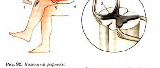
This function is responsible for human reflexes. For example, if a person touches something very hot, he will reflexively withdraw his hand. This is a reflex or motor function. But let’s take a step-by-step look at how it’s all tripled and how it’s connected to the spinal cord.
It’s best to look at everything using an example, so let’s imagine a situation where a person touched a very hot object with his hand:
- When touched, the signal is primarily received by receptors that are located throughout the human body.
- The receptor transmits a signal to the nerve fiber.
- The signal is sent along the nerve fiber to the spinal cord.
- On the approach to the organ there is a spinal ganglion, where the body of the neuron is located. It was along its peripheral fiber that the impulse transmitted from the receptors was received.
- Now the impulse is transmitted along the central fiber to the posterior horns of the spinal cord. At this moment, a kind of switching of the impulse to another neuron occurs.
- The processes of the new neuron transmit the impulse to the anterior horns.
- Now the return journey begins, because the anterior horns transmit the impulse to the motor neurons. They are responsible for the movement of the upper limbs.
- Through these neurons, the impulse is transmitted directly to the hand, after which the person removes it (motor function).
Reflex arc in the circuit
As a result of this whole process, a person withdraws his hand from the hot object and a reflex arc closes. The whole process takes a split second, so when touching any object, a person immediately feels its temperature, consistency and other features.
Conductor function
In this situation, the organ acts as a conductor. The conductor in this case is between the receptors and the brain. The receptors receive an impulse that is transmitted to the spinal cord and then to the brain. The information is analyzed there and transmitted back.
Thanks to this function, a person receives sensitivity, as well as a sense of himself in space. This has been proven many times, and this becomes especially evident in cases of serious spinal injuries.
Integrative function
This function is often forgotten, but it is no less important for a person than others. The integrative function manifests itself in reactions that cannot be classified as simple reflexes. In order for the body to respond, it is necessary to involve other parts of the human nervous system. This is how the spinal cord can form a connection between organs.
These include reflexes of chewing, swallowing, regulation of digestion, breathing and much more. In fact, this is an invisible function that ensures normal functioning.
Consequences of central nervous system injury
Even partial damage to the spinal cord can cause disruption of the functionality of a wide variety of systems and organs.
Bones and joints
A spinal cord injury can impair the absorption of calcium and some other important minerals into bone tissue. But at the same time, these substances can accumulate in the urinary system, causing the formation of stones. Experts usually explain such processes by a decrease in human motor activity. In addition, decreased mobility can lead to stiffness in joints, including the knees, elbows, and shoulders. To prevent this, there are special sets of exercises for people with disabilities.
Urinary system
The urinary system consists of the kidneys, which filter the blood and produce urine, and the bladder, which collects and then removes it from the body. After a spinal cord injury, the kidneys continue to produce urine, but the bladder may not work as well as before. After an injury, a person may not feel when his bladder is full of fluid, or his urinary system, due to a lack of necessary impulses, may lose the ability to eliminate urine naturally. As a result, a catheter may be needed to empty the bladder. Another option: urine may, on the contrary, come out involuntarily and the person loses the ability to control this process.
Respiratory system
Even after a spinal injury, human lungs continue to function. However, the ability to inhale and exhale air is controlled by the muscles. Depending on the level of injury, a person may lose the ability to cough or take deep breaths. And they are necessary for complete straightening of the lungs and deep air circulation.
The most important muscle for breathing is the diaphragm. It is a large arcuate muscle that is located directly under the lungs. And if the diaphragm is paralyzed due to damage to the spinal cord, special equipment may be needed to maintain respiratory function.
Leather
Damage to the spinal cord can also negatively affect the skin. Skin plays a very important role for humans. It protects the body from germs and other pathogens from the outside world. The spinal cord is an important organ that helps protect the skin from damage. For example, when sitting in one position for a long time, the brain sends signals in the form of a feeling of discomfort, which encourages you to change your position and prevent damage or pressure on the skin. In the same way, the central nervous system protects against burns, cuts and other possible damage to the skin (for example, a person reflexively withdraws his hand from a hot or cutting object). With spinal cord injuries, such signals may not appear, which greatly increases the risk of damage to the skin and, consequently, the penetration of microbes into the body.
Reproductive system
After a spinal cord injury in the lumbar or sacrococcygeal segment, the functionality of the reproductive system may be affected. Men usually experience erectile dysfunction, problems with ejaculation and fertility. In women, after injury, sensitivity in the genital area may be impaired, although fertility itself may not be directly affected.
Intestines
It is well known that the digestive system is responsible for the breakdown of food consumed, the absorption of nutrients from it and the removal of metabolic products. A fairly common consequence of central nervous system injury is impaired intestinal motility. That is, the digestive system continues to absorb and digest food, but the process of eliminating excrement is disrupted. In a healthy body, when the rectum is full, an exchange of impulses occurs between the intestines and the brain. If the spinal cord is damaged, such signals may not be received. As a result, a person loses the ability to control the process of defecation.
Autonomic nervous system
The autonomic nervous system regulates the activity of internal organs, blood and lymphatic vessels, as well as endocrine and exocrine glands. Body temperature, blood pressure, digestion and other important functions depend on it. After a spinal injury, some of its functions may be impaired. Quite often (especially if the damage occurs in the thoracic segment) after an injury the body loses the ability to thermoregulate. What does it mean? If the autonomic nervous system works correctly, then the body temperature remains stable regardless of the weather conditions outside or the indoor microclimate. Due to spinal cord damage, body temperature may rise or fall according to indoor or outdoor temperatures. Disturbances in thermoregulation are most noticeable in parts of the body located below the area of injury.
Spinal cord dysfunction
Impaired function can lead to serious consequences and often even death. Disorders often occur either due to injury or due to various diseases.
For example, due to dysfunction of the spinal cord, a person may lose sensitivity, in which case he may, for example, stop feeling temperature. In the worst case, the disorder can lead to uncontrollable actions of the limbs (or paralysis), disruption of the functioning of internal organs and the nervous system as a whole.
Spinal cord puncture
Puncture of cerebrospinal fluid (CSF) is a procedure that pursues diagnostic, anesthetic and therapeutic purposes. The procedure itself consists of an injection of an angle under the arachnoid membrane between the 3rd and 4th vertebrae, and then a certain amount of cerebrospinal fluid is extracted for research.
During the procedure, the brain itself is not affected, so there is no need to worry about violations. However, this procedure is quite serious and painful.
Spinal cord puncture

