Quick transition Treatment of vestibular neuronitis
Vestibular neuronitis (VN) is a disease of the peripheral vestibular system. It is characterized by the acute onset of severe systemic (rotational) dizziness and severe imbalance, which are accompanied by nausea and vomiting.
VN ranks third among all causes of peripheral vertigo after benign paroxysmal positional vertigo (BPPV) and Meniere's disease and first among causes of rotational vertigo lasting more than 24 hours.
The annual incidence of VN ranges from 3.5 to 15.5 people per 100,000 population. Both men and women are susceptible to the disease; the peak of VL occurs at 40-50 years of age.
Pathogenesis
Normally (from the vestibular receptors at rest), the same sensory signal travels along the vestibular nerves in both directions. It is transmitted to the nuclei of the brain stem through the vestibular nerves. In LN, the pathological process reduces the signal from the affected side, causing an asymmetry in the tone of the brainstem nuclei. This leads to oculomotor disorders (nystagmus - involuntary oscillatory movements of the eyes), disturbances of perception (rotational vertigo), postural disorders (imbalance at rest and during movement), autonomic disorders (nausea, vomiting).
Symptoms and course of the disease
VL is characterized by a monophasic course—an acute or subacute onset followed by subsidence of symptoms. The duration of clinical manifestations ranges on average from 1 week to 3 months (maximum manifestations range from several days to several weeks), after which the disease disappears without a trace. The probability of VN recurrence is 2-5%.
The disease begins with severe dizziness, accompanied by a certain type of nystagmus. Dizziness increases with head movements and decreases with fixed gaze. In 78% of cases, it appears suddenly; in 22% of patients, 1-2 days before the onset of the disease, phenomena in the form of instability and slight loss of coordination of movements are noted. Oscillopsia is often observed - unclear visual perception of surrounding objects when performing passive or active head movements due to instability of the image on the retina. Coordination of movements and balance are sharply impaired. Almost all patients experience nausea, and 65% have vomiting.
During the first day, the patient lies in bed - the slightest movements of the head provoke nausea and vomiting. On the 2-5th day, the patient is able to move within the room (ward), but the gait remains unsteady, and sudden head movements or walking long distances provoke nausea. On the 10-14th day, the patient complains of “heaviness” in the head and fatigue, but overall he feels healthy and can return to his usual lifestyle.
In the first days of the development of the disease, the brain “understands” that the vestibular apparatus is transmitting an incorrect sensory signal: the healthy labyrinth is always in an active state, the other (affected) labyrinth is depressed, which is interpreted by the brain as a constant rotation in the healthy direction. Since it is impossible to restore conduction along the affected vestibular nerve in a short time, the brain blocks the activity of the healthy labyrinth, aligning the flow of sensory signals from both vestibular apparatuses. As the vestibular nerve recovers, the control system returns to its original, healthy state.
Causes
The reasons for the development of vestibular neuronitis are unknown. According to the most common theory, there is an inflammation of the vestibular nerve of an infectious-allergic or viral nature. The presence of a virus (usually type I herpes virus, as well as a new coronavirus infection) is supported by such features as the frequent development of neuronitis after a respiratory viral infection, the presence of epidemiological outbreaks in late spring, as well as noted cases of familial development of neuronitis (when the carriers of the infectious pathogen were one family member.Clinical cases of the development of herpetic encephalitis in this disease speak specifically for the herpes virus.
With vestibular neuronitis, the upper branch of the vestibular nerve is most often affected, which innervates the anterior semicircular and horizontal canals of the vestibular apparatus, along with them the elliptical sac of the labyrinth, as a result of which the disease is often combined with BPPV, which develops as a result of otolithiasis of the posterior semicircular canal, innervated, respectively, by the inferior branch of the vestibular nerve, damage to which in vestibular neuronitis is extremely rare.
Diagnostics
In the vast majority of cases, diagnosis is based on a typical clinical picture. The diagnosis can be confirmed using videonystagmography (recording the movement of the eyeballs during a series of tests), the head impulse test (a test that evaluates the functionality of 6 semicircular canals at once - a component of the inner ear responsible for coordination) and the Fuduko test (a marching test that evaluates deviation of the patient from the original position). In addition, patients with VN exhibit spontaneous horizontal rotatory nystagmus, which intensifies when looking towards the healthy ear.
Other tests and studies, including audiometry (hearing tests) and MRI of the brain, are performed as indicated to rule out other causes of dizziness.
Differential diagnosis
VN is also differentiated (distinguished) from labyrinthitis, Meniere's disease, BPPV, neuroma (tumor) of the VIII pair of cranial nerves, multiple sclerosis, stroke, vestibular migraine.
Consequences
Restoration of impaired functions depends on many factors. Among them are the degree of nerve damage, the speed of compensation of the central nervous system mechanisms, the adequacy and timeliness of the prescribed treatment, and the usefulness of vestibular gymnastics. It should also be noted that often sick people for a long time experience attacks of mild dizziness when changing body position and sharp turns of the head after an illness. A more complete restoration of nerve function, according to various authors, is observed in approximately half of the patients after 1 year, in 30% there is a partial restoration of function, while in the rest, unilateral vestibular areflexia remains, which, however, does not cause significant discomfort, due to the mechanisms of vestibular compensation. Moreover, the presence of ongoing complaints may be a sign of a person’s psychogenic state.
According to the latest data, relapses are not very common, about 5-15% according to various authors, and a healthy nerve can be affected, as well as an already affected nerve (if it is completely restored). (According to early data, relapses are extremely rare (no more than 1-2%), and a healthy nerve is more often affected). It is more logical to look for other causes in case of repeated attacks of systemic dizziness (BPPV, vestibular migraine, Meniere’s disease, etc.).
Treatment of vestibular neuronitis
The main directions of treatment for patients with VN are reducing dizziness, nausea and vomiting (symptomatic treatment) and accelerating vestibular compensation.
Symptomatic treatment includes the use of drugs that suppress vestibular function: antihistamines (dimenhydrinate); benzodiazepine tranquilizers (diazepam); antiemetics (metoclopramide hydrochloride, thiethylperazine).
Symptomatic treatment is recommended for no more than 3 days, since it slows down vestibular compensation, which leads to long-term dizziness.
The effectiveness of glucocorticosteroids, which were previously actively used in patients with LN, is currently being questioned. We do not recommend their use, as there is no convincing evidence of accelerated vestibular nerve recovery in these patients.
Despite the presumed viral etiology of VN, associated in particular with herpes simplex virus type 1, the use of antiherpetic drugs also does not accelerate the recovery process, and they should not be used in the treatment of VN.
Piracetam, Trental, Mexidol, Actovegin, Cavinton, glycine and other drugs with unproven effectiveness are not recommended for the treatment of VN.
From about the third day of the disease, patients with VL are recommended to perform vestibular gymnastics - exercises that accelerate compensatory processes, from simpler to more complex. These are exercises for fixing the gaze, training oculomotor reactions, and maintaining balance in various postures while standing and in motion.
Peripheral vestibular syndrome as a cause of dizziness and balance disorders
Recently, interest in the problems of dizziness and balance disorders has increased significantly. This is due not only to the growing number of patients turning to doctors with similar complaints, but also to the emergence of new diagnostic methods, the creation of both specific pharmacological drugs and methods of rehabilitation therapy. It is still very important for the doctor to accurately establish the cause of the disease and its form in order to further prescribe adequate therapy. Based on the experience gained over five years in a specialized center for dizziness and balance disorders, we concluded that in more than 50% of patients at the time of treatment, the appearance of such symptoms is due to a whole range of reasons. This is explained by the complex structure of the balance system and the vestibular system.
For a practicing physician, it is important to familiarize yourself in detail with the complaints presented by the patient and the history of the development of the disease. The use of additional modern diagnostic methods and consultation with specialists significantly increases the efficiency of diagnosis. First of all, among additional diagnostic methods, vestibulometry and posturography (possibly stabilometry) should be noted. If the first method, which is a set of tests with computer data processing, allows you to answer questions about the functional state of the vestibular system, including changes in the vestibular receptor, central vestibular structures, brain stem, cerebellum; state of the oculomotor system; cervical receptors, etc., then posturography makes it possible to assess the state of balance (functional composition of components - vision, vestibular system, proprioceptors), including for individual selection and rehabilitation therapy. Duplex ultrasound examination of the main blood flow of the transcranial and cervical sections allows, in addition to conventional procedures, to conduct studies with functional tests and obtain information about venous blood flow. In combination with rotary tables, this method makes it possible to obtain information about increased intracranial pressure.
In practice, we are faced with vestibular pathology, which can be divided into peripheral, central and mixed vestibular syndromes. The choice of the attending physician in the existing system of specialized care most often occurs as follows. If the patient has cochleovestibular syndrome, he is referred to an audiologist; vestibular syndrome without hearing changes - see a neurologist. In our opinion, at the first stage of the examination, consultations with a neurologist and an otoneurologist (otorhinolaryngologist) are mandatory, since peripheral vestibular syndrome without audiological symptoms can be the cause of the development of diseases of the inner ear, which should be discussed in more detail.
The group of diseases of the inner ear without changes in hearing includes: benign paroxysmal positional vertigo (otolithiasis), vestibular neuronitis, labyrinth fistula, vestibular form of Meniere's disease and secondary labyrinthine hydrops.
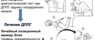
Benign paroxysmal positional vertigo (BPPV) is one of the most common causes of vertigo associated with pathology of the inner ear. Patients with this pathology develop short-term attacks of dizziness and nystagmus (usually lasting less than 30 seconds) when changing the position of the body and head: usually when turning in bed, getting in and out of bed, bending and straightening, or stretching the neck to look up.
The basic features of BPPV and associated positional nystagmus were described by Robert Barany (Barany, 1921), but the term did not exist until 1952, when Dix and Hallpike described the provocative positional test and clearly defined the syndrome (Dix and Hallpike, 1952).
The diagnosis of BPPV continues to rely on the finding of a characteristic positional nystagmus (also called paroxysmal positional nystagmus) in a patient with a typical history of positional vertigo. Nystagmus can be observed during visual inspection in the Dix-Hallpike test, however, since it occurs due to pathology of the vestibular receptor (i.e., it is peripheral), it is susceptible to depression when fixating gaze, so diagnosis involves the use of Frenzel glasses or videonystagmography. Currently, we have accumulated experience in treating more than 100 patients with a similar pathology. Treatment allows particles to be moved from the semicircular canals into the vestibule of the labyrinth and thus relieve dizziness. The effectiveness of the procedures, according to our and foreign sources, ranges from 90-95%.
Acute vestibular neuronitis
This is a disease with an as yet unknown etiology. The most popular hypothesis is about viral etiology. It is characterized by attacks of vestibular dizziness, often accompanied by autonomic reactions (nausea and vomiting). Any movement of the head increases the feeling of dizziness. The duration of the disease ranges from several hours to several days. An important sign is the absence, as with all diseases of this group, of hearing changes. During the examination, typical vestibular peripheral nystagmus can be observed against the background of an asymmetric lesion, which will be combined with a corresponding deviation of the hands and posture when performing statokinetic tests. Treatment is usually symptomatic with the use of vestibular suppressants in the first stage. Special mention should be made of toxic neuronitis caused by exposure to aminoglycoside antibiotics, especially when it comes to gentamicin. The introduction of gentamicin, one or two times, through the eardrum, in its effectiveness, successfully replaces surgery to destroy the labyrinth (if it needs to be performed when other methods of treatment are absolutely ineffective).
A labyrinthine fistula can be no less difficult to diagnose. This disease is characterized by the appearance of a communicating opening or canal between the middle and inner ear, resulting in the leakage of fluid from the inner ear into the cavity of the middle ear. The hydrodynamic system is disrupted, and then the state of the sensory cells. Most often, a fistula is formed due to barotrauma (for example, diving, coughing, sneezing). A characteristic symptom is the appearance of dizziness during the Valsalva maneuver (or similar loads - straining or lifting heavy objects). Fistulas tend to close on their own, but due to the frequently occurring increased intralabyrinthine pressure (hydropses), they can become chronic. Electrophysiological, radiological and other research methods cannot give a reliable answer to the question of the presence of a fistula. Therefore, after an examination and with certain test results, the issue of diagnostic myringotomy (surgical separation of the eardrum for revision of the tympanic cavity) is decided. When a fistula is detected, it is repaired.
The vestibular form of Meniere's disease has been the subject of separate studies, conducted mainly abroad. Typical Meniere's disease is characterized by the following symptoms. Usually the disease begins at the age of 25-45 years, men are more often affected. The disease is accompanied by attacks of vestibular dizziness, lasting up to 6-12 hours, tinnitus, fluctuating hearing loss of the sensorineural type with a tendency to progress in the degree of hearing loss, a feeling of discomfort, fullness in the affected ear.
The existence of a large number of patients with secondary hydrops of the labyrinth is noteworthy. The term “secondary” suggests that the increase in intralabyrinthine pressure occurs due to causes that are systemic, and what occurs in the inner ear reflects the manifestation of these changes. In one of the issues of “The Treating Doctor” (No. 9, 2000) we dwelt in detail on the features of diagnosis and treatment of this pathology.
Such changes require appropriate therapy, including at least two components: normalization of the state of venous outflow and treatment of labyrinthine hydrops. At this stage, work with the patient should also be carried out with the direct participation of a neurologist-vertebrologist (chiropractor) and an otoneurologist. An individual combination of physical and physiotherapeutic methods, as well as combination therapy of hydrops (dehydration, vestibular suppression, vasoactive effect on regional blood flow in the inner ear) make it possible to achieve a cure for the patient. Electrocochleography also remains a method of stage-by-stage assessment of the state of pressure in the inner ear.
Recent research in the field of inner ear immunology suggests that labyrinthine hydrops may be a consequence of an autoimmune lesion of the inner ear. Morphological changes are characterized by: degeneration of spiral ganglion cells, atrophy of the organ of Corti, arteritis around the cochlear nerve and stria vascularis, and the development of endolymphatic hydrops. For vestibular disorders, dizziness is not a constant symptom. If it is present, it is similar to Meniere-like dizziness, combined with ataxia and attacks of sudden falling. A vestibulometric study reveals bilateral symmetrical peripheral vestibular dysfunction. The clinical picture of the disease includes various combinations of auditory and vestibular symptoms, as well as their complete absence against the background of only sudden falls. For a practicing physician, the ability to determine the cause of the development of inner ear pathology serves as an indication for prescribing corticosteroid anti-inflammatory therapy.
Treatment of peripheral vestibular syndrome can be divided into two stages - the active manifestation of the disease and the subacute state. At the first stage, it is important to carry out pathogenetic therapy in combination with the prescription of vestibular suppressants and antiemetic drugs when dizziness is combined with vegetative manifestations.
Vestibular suppressants occupy the main place in the treatment of dizziness. The term “vestibular suppressant” is a collective term: this is usually the name given to drugs that reduce nystagmus and the feeling of dizziness, vestibular instability, or stop motion sickness (motion sickness).
At least four major neurotransmitters of the vestibular system are involved in the formation of the vestibulo-ocular reflex: glutamate, acetylcholine, gamma-aminobutyric acid and glycine, histamine.
Glutamate is the main excitatory neurotransmitter. Acetylcholine is both a peripheral and central agonist acting at muscarinic receptors. These receptors are found in the brain stem and bone marrow. There are reports of their possible participation in the formation of dizziness. Gamma-aminobutyric acid and glycine are inhibitory neurotransmitters found in second-order vestibular neurons and oculomotor neurons. Histamine is found in various central vestibular structures, where it is present diffusely.
Anticholinergics act on muscarinic receptors. An important feature for this group of drugs is that they do not cross the blood-brain barrier and are therefore ineffective in treating motion sickness. Also, unlike antihistamines, anticholinergic drugs are not effective if used after dizziness has already begun.
All anticholinergics used to treat dizziness have side effects such as dilated pupils and sedation. The most commonly used drugs in this group are scopolamine and atropine.
There is evidence that centrally acting antihistamines prevent motion sickness and reduce the severity of vestibular manifestations, even when symptoms have already appeared. Most antihistamines also have a calcium channel blocking effect. However, the sedative effect that antihistamines have has an adverse effect on the processes of vestibular adaptation, so this group of drugs should not be recommended to patients for long-term use.
Benzodiazepine drugs - GABA modulators - act centrally to suppress vestibular responses. These drugs are used in small doses. Their main disadvantages are addiction, memory impairment, increased risk of falls, and negative effects on vestibular compensation.
Betaserc (betahistine dihydrochloride) is characterized by a successful combination of the properties of a pathogenetic drug with the effect of suppressing the feeling of dizziness. When exposed to two groups of H-receptors, an increase in blood flow in the labyrinthine artery and suppression of information passing through the vestibular nuclei simultaneously occur. Betaserc can be considered as the drug of choice. Depending on the pathogenesis of the disease, betaserc can be combined with other therapeutic agents (with the exception of antihistamines, the combination with which leads to a weakening of the therapeutic effect of both drugs).
Peripheral vestibular pathology against the background of persistent ataxia in the future, as a rule, is compensated independently. However, in order to speed up the patient's recovery, vestibular rehabilitation can be used. This is a set of exercises performed independently or on special installations (posturographs or stabilographs) with biofeedback. The inclusion of vestibular rehabilitation in the complex therapy of peripheral vestibular syndrome, in our opinion, is justified and should be performed under the supervision of a physician. It is recommended that such exercises be prescribed after the patient has finished using vestibular suppressants. Perhaps the betaserc is an exception, since recently works have appeared in the domestic literature indicating the effectiveness of vestibular rehabilitation when using the betaserc. With age, the balance system deteriorates due to changes in the structure of sensory systems and nervous tissue. Therefore, in older people, even in the presence of peripheral vestibular syndrome, it is recommended to use vestibular rehabilitation.
In conclusion, I would like to emphasize that the success of working with patients suffering from dizziness and balance disorders depends on the diagnostic capabilities of the medical institution, the interaction of doctors of various specialties and the use of all means effective for the treatment and rehabilitation of patients.
O. A. Melnikov, Candidate of Medical Sciences, ANO "GUTA-Clinic", Moscow