Every year in Russia, about 600 thousand people suffer traumatic brain injury (TBI). 50 thousand of them die, and another 50 thousand become disabled. The incidence of TBI in men is twice as high as that in women, with this relationship maintaining in all age groups.
The most common causes of TBI
Typical clinical manifestations of traumatic brain injury.
TBI is characterized by a three-phase change in consciousness: a primary short-term loss of consciousness at the time of injury, subsequent recovery ( light interval ) and, after a certain period of time, repeated loss of consciousness . However, such a classical development of changes in the state of consciousness is not always observed. Often there are cases that occur without a light gap, or it is erased. Sometimes there may be no primary loss of consciousness. The duration of the clear interval (both with full and partial restoration of consciousness) in most patients with acute EDH is measured in several hours or even minutes.
Patients who have suffered a trauma and are accessible to contact usually complain of a headache with increasing intensity. In many cases, the headache has a bright shell-like hue, radiates to the eyeballs, jaws, and is accompanied by photophobia, hyperesthesia with characteristic facial reactions. The headache is usually constant with periodic crisis-like exacerbations, often accompanied by repeated vomiting . In severe pain, victims moan, demand help, grab their heads with their hands, toss in bed, and lose sleep.
After a traumatic brain injury, patients often experience bradycardia (in almost 1/2 of patients, the pulse rate does not exceed 60 beats per minute); in approximately 1/4 of the observations, an increase in maximum blood pressure above 150 mm Hg is noted. Art.
In victims with traumatic hematomas, it is almost always possible to identify one or another focal symptomatology . Among the signs of damage to the cerebral hemispheres, the first place belongs to motor disorders (weakness in the arm and leg, most often opposite the side of the hematoma). The severity of these disorders varies - from anisoreflexia to hemiplegia (paralysis).
Occasionally, the clinical picture of intracranial hematomas includes symptoms of irritation of the cerebral cortex in the form of general or focal epileptic seizures .
Among craniobasal symptoms, the most important role in the clinic of TBI is given to the dilation of one pupil with a decrease or loss of its reaction to light .
Clinical classification of TBI.
According to the clinical picture, traumatic brain injury is divided into:
1. Focal • brain contusions (mild, moderate, severe), • intracranial hematomas (epidural, subdural, intracerebral), • subdural hygromas, • depressed fractures, • head compression;
2. Diffuse • concussion, • diffuse axonal damage, • subarachnoid hemorrhage.
Diagnostics.
Currently, the gold standard when examining patients with traumatic brain injury is the following set of examinations: 1. Clinical examination by a neurosurgeon. 2. X-ray of the skull in two (at least) projections. 3. Echoencephalography. 4. CT (computed tomography) of the brain. The issue of additional examination methods is decided individually.
Very often, epidural hematomas are accompanied by a fracture of the skull bones.
Types of intracranial hematomas.
Brain contusions include focal macrostructural damage to its substance as a result of trauma. According to the unified clinical classification of traumatic brain injury adopted in Russia, focal brain contusions are divided into three degrees of severity: 1) mild, 2) moderate, and 3) severe.
| CT scan of the brain. Severe brain contusion, intracerebral hematomas of both frontal lobes. |
Epidural hematomas are a collection of blood caused by trauma, located between the inner surface of the skull bones and the dura mater, causing local and general compression of the brain.
The frequency of epidural hematomas among observations of initially hospitalized victims with TBI varies widely from 0.54% to 9%.
Causes of epidural hematomas. A temporary local deformation of the skull that occurs during an injury, often with a depressed fracture of the skull bones and rupture of the vessels of the dura mater, leads to the release of blood from the damaged vessel, which, exfoliating the dura mater, spreads within the cranial sutures, where the shell is tightly fused with the internal bone plate. For this reason, epidural hematomas have a smaller area of distribution and greater thickness than subdural hematomas.
| CT scan of the brain. Acute epidural hematoma in the left occipital region with brain dislocation and compression. |
Subdural hematomas are a volumetric accumulation of blood caused by trauma, located between the dura and arachnoid maters and causing local and general compression of the brain.
Subdural hematomas are more common than epidural hematomas. Isolated SDH accounts for approximately 2/5 of the total number of cases of brain-compressing intracranial hemorrhages; they occupy first place among various types of hematomas.
| CT scan of the brain. Acute subdural hematoma in the right fronto-parietal-temporal region with brain dislocation and compression. |
Post-traumatic chronic subdural hematomas are encapsulated volumetric hemorrhage located under the dura mater and cause local and general compression of the brain. The incidence of these hematomas ranges from 2 to 13 cases per 100,000 population per year, increasing significantly in elderly and senile people.
Chronic subdural hematomas differ from acute and subacute traumatic hematomas by a demarcation capsule that usually appears 2 weeks after the injury, which determines all the features of their pathogenesis, clinical course and treatment tactics. The volume of CSH ranges from 50 ml to 250 ml and is more often 100-150 ml.
If previously chronic subdural hematomas were detected exclusively in elderly and senile people, now they have become significantly “younger”, occurring quite often in young and middle-aged people, as well as in children.
The distinctive clinical sign of these hematomas is that the clear interval can last for weeks, months and even years. Clinical manifestation is extremely polymorphic. There is both a gradual development of compression syndrome and a sudden sharp deterioration in the patient’s condition to stupor and coma, spontaneously or under the influence of various additional factors (mild repeated head injury, overheating in the sun, alcohol consumption, colds, etc.).
The clinical picture may resemble various diseases of the central nervous system: benign and malignant brain tumors, etc. During the period of a detailed clinical picture of chronic subdural hematoma, changes in consciousness in the form of stupor or amentive confusion with impaired memory and orientation are frequent.
| CT scan of the brain. Bilateral chronic subdural hematomas. |
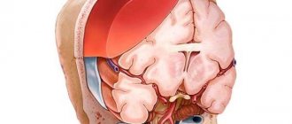
Damage to the structures of the posterior cranial fossa (PCF) is one of the severe types of traumatic brain injury (TBI). Their peculiarity lies in their extremely difficult clinical diagnosis and high mortality. Before the advent of computed tomography, the mortality rate for PCF injury was close to 100%.
The clinical picture of damage to the structures of the PCF is characterized by a severe condition that occurs immediately after the injury: depression of consciousness, a combination of cerebral, meningeal, cerebellar, and brainstem symptoms due to rapid compression of the brainstem and impaired cerebrospinal fluid circulation. If there is significant damage to the substance of the cerebrum, hemispheric symptoms are added.
The proximity of the location of damage to the PCF structures to the liquor-conducting pathways causes their compression and disruption of liquor circulation by a small-volume hematoma. Acute occlusive hydrocephalus, one of the most severe complications of damage to the structures of the posterior canal, is detected in 40%.
Treatment.
All patients with clinical signs of compression of the brain, as well as when this compression is detected on CT or MRI in severely ill patients, are advised to undergo urgent surgery - removal of the hematoma.
Symptoms and clinical manifestations of the disease meningeal hematomas
As a rule, meningeal hematomas are characterized by general cerebral signs, which are inherent in almost every brain pathology. A person with a meningeal hematoma will experience intense headaches, weakness, lethargy and apathy, nausea and vomiting that does not bring relief, drowsiness, dizziness, confusion and loss of consciousness, difficulty speaking and swallowing, different pupil sizes and weakness in the limbs, impaired coordination and fine motor skills. Often, before falling into a state of apathy, patients experienced psychomotor agitation. Further clinical manifestations depend on the location of the meningeal hematoma and the degree of brain damage. In advanced and severe stages of brain pathology, convulsive manifestations, coma and even a lethargic state may be observed.
Etiology of subdural hematomas
This type of hematoma is most often a consequence of traumatic brain injuries and ruptures of blood veins that develop as a result of them, passing in the subdural space. However, the reasons do not necessarily lie in injuries; sometimes hematomas can be provoked by pathological conditions characterized by the destruction of the walls of blood vessels and a decrease in their plasticity. Other reasons are as follows:
- Blood diseases that change its properties;
- Diseases of the blood vessels of the brain;
- Increased intravascular and blood pressure;
A hematoma formed on the side of the injury appears with a small contact area and even without direct contact. This can happen when stopping too suddenly or changing the direction of movement, as well as when falling on the lower limbs or legs. In the process, a sharp shaking of the head occurs, provoking a displacement of the cerebral hemispheres inside the skull, leading to rupture of the blood veins.
As for a hematoma formed on the opposite side of the injury, it can form when the head hits a large, inactive object, causing a large contact area. Much less often, hematomas are formed due to injuries directly to the veins and arteries when the integrity of the dura cerebral membrane is violated.
Symptoms
With chronic subdural hematoma, cerebral and focal symptoms occur. Most often, patients complain of impaired consciousness and headaches, which may be accompanied by vomiting. These manifestations are in one way or another connected with a previous head injury. Memory disorders may occur in which a person does not remember some events from a past life. In most cases, generalized epileptic seizures recur.
Patients complain not only of a headache, but also of a painful sensation in the area of the eyeballs. Discomfort increases when moving the eyes. The pain may radiate to the back of the head. There is also increased sensitivity to light. Decreased vision, the appearance of mydriasis of the opposite pupil, and a sharp reaction to light sources are possible. Oculomotor disturbances are also possible.
4. Treatment of the disease
Depending on the patient’s condition and the volume of the hematoma formed, treatment can be either conservative or surgical. Both treatment methods have their own advantages and disadvantages. Modern medicine has new methods for treating this pathology, which combine higher efficiency compared to conservative ones and are less invasive compared to surgical ones (craniotomy). This method is called closed external drainage of the hematoma.
To eliminate the hematoma, the skull is perforated through the skin with an intraosseous needle. Then drainage
with a catheter diameter of no more than 3 mm with a fluid receiver located below the level of the head.
Practice has confirmed the high efficiency of the new method and shown a number of advantages of hematoma drainage:
- surgical removal of compression (squeezing) of the brain;
- reducing the risk of postoperative infection;
- maintaining the integrity of the skull;
- minimizing the likelihood of relapse.
Treatment of meningeal hematoma
Treatment of this brain pathology in the acute phase is possible exclusively through surgery. It is possible to avoid surgery and use conservative therapy as treatment, but for this the hematoma must be small in size and there must be no compression with brain dislocation. An established diagnosis of meningeal hematoma is an indication for emergency surgery. The patient is immediately sent to the operating room, where he is given general anesthesia. Then a craniotomy is performed and blood and blood clots are removed using an aspirator. After emptying the cavity of the meningeal hematoma, surgeons begin to search for the site of bleeding. Having discovered the cause of blood loss, doctors carry out thorough hemostatic measures (vascular ligation, etc.), which are aimed at stopping blood loss. Having eliminated the vascular defects, a bone flap is applied and the surgical access site is sutured. In the postoperative period, a person is prescribed drug therapy, which is aimed at preserving the vital functions of the entire body.
After the operation, the patient expects a short rehabilitation period and complete recovery.
Classification of types of hematomas
According to the timing of development, chronic, subacute, and acute hematomas are distinguished. The latter option is the main cause of death. Late detection of the formation in the absence of surgical decompression leads to death due to compression of the brain.
Practice shows a high risk of subdural hemorrhage due to bruises. The epidural form forms faster in terms of speed, but the mortality rate is less. Subdural accumulation of blood causes multiple dangerous secondary changes due to high intracranial hypertension.
The chronic form develops over several weeks. The absence of initial symptoms makes the nosology unrecognized for months. Hemorrhages occur cyclically and can stop on their own. Small foci measuring about one centimeter are not dangerous. Correct therapy leads to complete repair.
Separation of hematomas for reasons of occurrence:
- Vascular;
- Medicinal;
- Traumatic;
- Intoxicating;
- Infectious.
By location:
- Bilateral;
- Right-handed;
- Left-handed;
- Frontal (frontotemporal, frontoparietal);
- Parieto-occipital;
- Parietotemporal;
- Temporal.
By size:
- Large - more than one hundred centimeters (cubic);
- Average - from fifty to one hundred;
- Small - up to fifty centimeters
By structure:
- Two-chamber;
- Single chamber;
- Multi-chamber.
According to the clinical course:
- Decompensated;
- Compensated.
Features of the structure, course, and location are indicated in the diagnosis after a thorough diagnosis using CT and MRI. X-rays show only bone-destructive changes in the skull.
Subdural and epidural hematoma after skull contusion
The difference between an epidural and subdural hemorrhage is the location of the blood collection. The first variety is located between the hard shell and the bones of the skull. Subdural form - between the arachnoid and dura mater due to rupture of the bridging vein. The epidural appearance occurs due to rupture of the anterior ethmoidal, middle meningeal artery, sigmoid or superior sagittal sinus.
Types can be differentiated based on CT scans based on the shape of the crescent (subdural) or biconvex (epidural) lens.
Which method of diagnosing epidural hematoma to choose: MRI, CT, X-ray, angiography
Selection method:
- MSCT
What will a head x-ray show for an epidural hematoma?
- It allows you to detect only a fracture of the skull bones, but not intracranial hemorrhage.
What will CT scans of the brain show in the presence of an epidural hematoma?
- Lenticular shape
- Hyperdense
- Fresh, unclotted blood may also be hypodense
- The hematoma does not cross the suture lines, since the dura mater is tightly fused with the bones of the calvarium along the boundaries between them
- Pronounced volumetric impact: displacement of midline structures
- Blurred boundaries between gray and white matter
- Impaired cerebrospinal fluid circulation (occlusion of the interventricular foramen of Monroe)
- Narrowing of the surrounding cistern (possible herniation of the medial parts of the temporal lobe into the notch of the tentorium of the cerebellum)
- The volume of the hematoma can quickly increase
- There is usually a fracture of the calvarial bones with displacement
- After removal of ventricular tamponade, recurrent postoperative contralateral bleeding (epidural or intracerebral) may occur.
Is MRI of the brain informative for epidural hematoma?
- Not shown, since organizing and conducting the study takes a lot of time.