CEREBRAL CORTICAL HEMISPHERES
CORTICAL CORTEX OF THE BRAIN HEMISPHERES, a layer of gray matter (1–5 mm) covering the hemispheres. This is the part of the brain that has an ordered layered structure; develops in the later stages of evolution and plays a key role in the implementation of higher nervous activity; participates in the regulation and coordination of all body functions. In the course of evolution, a precursor to K. b. appears in cyclostomes and fish. p. g. m. - pallium (lat. pallium - cloak, cover), in which 3 structures are distinguished: paleopallium (ancient cloak), archipallium (old cloak) and neopallium (rudimentary new cloak). Starting with reptiles, the pallium differentiates and acquires layering (from now on it is called “cortex”, from the Latin cortex - bark). Thus, in higher vertebrates K. b. p.g.m. is represented by elements of the paleocortex, archicortex and neocortex; the latter reaches its greatest development in mammals.
Development of K. b. p.g.m. in the process of evolution reflects the basic. stages of improving the perceiving and integrating activity of the brain and controlling purposeful movement. behavior. In lower mammals the surface of the cortex is smooth (lissencephalic type); in the higher ones, it becomes folded, covered with grooves and convolutions (gyrencephalic type) due to the uneven growth of the sections. structures of the neocortex. In humans, the cortex makes up approximately 44% of the volume of the hemispheres and contains approx. 14 billion nerve cells (neurons); its surface area averages 1468–1670 cm2; The neocortex forms the outer surface of the hemispheres (in humans there are 6–7 layers of cells), and it accounts for 95.6% of the brain. p.g.m., on the archicortex (its main part is represented by the hippocampus) - 2.2%, on the paleocortex, located on the lower and inner. surfaces of the frontal and temporal regions, - 0.6%, on the so-called. transitional formations (between ancient, old and new crust) - 1.6%.
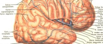
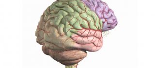
The lateral surface of the cortex of the right hemisphere of the human brain: 1 – frontal lobe; 2 – Sylvian fissure; 3 – temporal lobe; 4 – cerebellum; 5 – occipital lobe; 6 &ndas…
The composition of the cortex includes unmyelinated and thin myelinated fibers (see Myelin sheath), formed by axons, and neuroglia (performs the main supporting and trophic functions); The gray color of the tissue is due to the almost complete absence of the myelin sheath around the fibers. The structure of the cortex is characterized by an ordered distribution of neurons (cytoarchitecture) and fibers (myeloarchitecture) along horizontal and vertical layers. It is believed that the main The functional unit in the neocortex is a column - a vertically located group of cells with many connections among themselves.
Differences in the structure of the department. areas of the cortex (location density, size of neurons, their organization into layers and columns) determine its cytoarchitectonics. The cortex interacts closely with the underlying structures of the brain, which are connected to it by nerve fibers and are themselves under the control of certain cortical zones, receiving regulatory influences from them through descending nerve pathways.
Basic fissures - the Sylvian fissure (separates the temporal lobe of the hemisphere from the frontal and parietal) and the Rolandic fissure, which separates the frontal lobe from the parietal lobe. In addition to these basic grooves, there are a large number of others separating the convolutions of the cortex. The grooves and convolutions increase the surface of the cortex without increasing the volume of the skull. So, a person has approx. 2/3 of the surface of the entire cortex is located deep in the grooves.
Representation of sensory functions in the posterior central gyrus of the cerebral cortex (a) and motor functions in the anterior central gyrus (b).
In each of the lobes of the hemisphere, fields (zones) are distinguished that differ in structure and function. There are projection (sensory), associative and motor zones. Various information processing occurs in sensory areas. types of sensitivity, and each type of sensitivity has its own cortical zone. For example, the visual zone is located in the occipital lobe, the auditory zone is in the temporal lobe, and the gustatory and olfactory zones are in the internal lobe. surfaces of the temporal lobes. The largest area of the cortex is in the area of musculocutaneous sensitivity in the parietal lobe. The zone of skin sensitivity is mapped in detail: it contains areas responsible for certain areas of the skin. The more receptors there are in a particular area of the skin, the more neurons there are in that area of the neocortex that corresponds to this area. Therefore, in the cortex, for example, there are disproportionately large areas of the lips and fingers and very small areas of the back and abdomen. The motor zone is located ch. arr. in the frontal lobe, just in front of the center. furrow. This is where the basics begin. the path through which voluntary movements are realized. Motor zone map sensitivity, as well as the map of the skin sensitivity zone, has distorted proportions: some muscles (for example, the muscles of the hand) are capable of performing much more subtle movements, and a larger number of neurons are needed to control them. During evolution, various body functions become increasingly clearly represented in the cerebral cortex (corticolization of functions). Association zones are located in the posterior half of the parietal lobe, the frontal lobe, and areas related to the limbic system, and occupy most of the brain. p.g.m. They are especially important for thinking and speech, higher motors. functions, selection and activation of complex programs of behavior, memory and emotions. Association areas of the cortex work asymmetrically. In most healthy people, the left hemisphere contains areas responsible for the interpretation and formation of speech and writing. speech and control over the actions of the right hand. If the speech center in the temporal lobe of the cortex is damaged, the understanding of audible speech is impaired, and if the speech center in the frontal lobe of the cortex is damaged, a person hears and understands speech, but cannot speak. The right hemisphere cortex provides orientation in space and time, and is also involved in music. and artist creativity; it stores information about the individual characteristics and details of the department. objects, it instantly processes incoming signals and creates a holistic image. Some patients with damage to the right hemisphere cannot recognize familiar faces. There are also gender differences in the activity of associative zones of the cortex. For example, men, as a rule, solve spatial problems better in their minds and navigate space more easily, while women express their thoughts more accurately in words and perceive changes in the environment more quickly. See also Cerebrum, Telencephalon.