Useful articles
Vegetovascular dystonia is a disease based on changes of a psycho-neurological nature, manifested by complaints of functional disorders of organs. The consequences of the disease are best addressed in a specialized clinic for headaches and autonomic disorders.
Treatment of headaches with VSD
Treatment measures for this pathological process should begin after an accurate diagnosis has been established and other diseases have been excluded. After diagnosis and the absence of any pathologies that can provoke headaches, a patient with characteristic complaints should contact a neurologist or psychiatrist. Treatment of headaches associated with VSD involves eliminating the influence of pathological impulses that cause pain.
For these purposes the following should be prescribed:
- nootropic drugs (Nootropil, Piracetam), which will normalize metabolic processes in brain cells;
- tranquilizers (Sibazon, Phenazepam). This group will eliminate unnecessary fears and suspiciousness in patients;
- antidepressants – in the presence of developed depression.
An effective way to treat headaches with dystonia is psychotherapy, which allows patients to eliminate suspiciousness, fears and anxiety on a conscious level. Treatment of headaches with vascular dystonia must be supplemented with acupuncture, laser treatment of active points, and special types of massage.
The text was checked by expert doctors: Head of the socio-psychological service of the Alkoklinik MC, psychologist Yu.P. Baranova, L.A. Serova, a psychiatrist-narcologist.
CAN'T FIND THE ANSWER?
Consult a specialist
Or call: +7 (495) 798-30-80
Call! We work around the clock!
Causes and symptoms of VSD
Adolescents and women are at risk for developing vegetative-vascular dystonia. In men, VSD rarely occurs as an independent syndrome; dystonia is usually associated with other diseases. The causes of the pathology are most often unknown, but its appearance is associated with the following provoking factors:
- stress – constant nervous tension causes the release of stress hormones, which stimulate the autonomic nervous system;
- hormonal changes - physiologically in women during the menstrual cycle, with the onset of pregnancy, menopause, the concentration of sex steroids changes, which indirectly affect the functioning of the nervous system;
- maturation of the nervous system – in adolescents leads to pronounced signs of VSD;
- bad habits - smoking, drinking alcohol, caffeinated drinks cause disturbances in the functioning of blood vessels and the conduction of nerve impulses.
The first symptoms of vegetative dystonia may appear in childhood. They are associated with heredity and characteristics of pregnancy. If the expectant mother suffers from vegetative-vascular dystonia, smokes, and has been diagnosed with arterial hypertension, then the risk of pathology in the child increases significantly. The health of the baby is negatively affected by intrauterine hypoxia, disruption of feto-placental blood flow, as well as the stress experienced by the pregnant woman. Acute fetal hypoxia during childbirth can also lead to the formation of VSD at an early age.
The formation of vegetative-vascular dystonia in adults can be associated with osteochondrosis, head injuries, and poor nutrition. Prolonged life in poor environmental conditions and work in hazardous industries also leads to VSD. Weather-dependent people also often experience dystonia.
Symptoms of autonomic dysfunction are varied; doctors identify more than 40 signs of the disease, but not all of them occur in one patient. Usually this is a combination of 4-5 constant symptoms and several additional ones. Depending on the manifestations of the cardiovascular system, there are three types of vegetative-vascular dystonia:
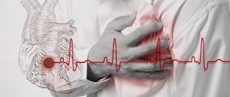
- cardiac - not accompanied by pressure surges, it is characterized by pain in the heart area or interruptions in its work;
- hypertensive type - a person suffers from high blood pressure, which is accompanied by panic attacks, chills, and increased physical activity;
- hypotensive type – characterized by low blood pressure, which is maintained at 100/50-90/45 mm Hg. Additional concerns include weakness, drowsiness, and dizziness.
Signs of dystonia include periodic shortness of breath, a feeling of shortness of breath, or a sensation of spasm in the throat. Many people complain to the doctor about pain in the heart, a feeling of increased heartbeat, pressure in the chest, interruptions in the heart, but it is rarely possible to record them using an ECG. Symptoms of autonomic dysfunction include decreased appetite, heartburn, flatulence and other digestive disorders. The disease manifests itself in the form of frequent urination or urinary retention, chills and cold extremities, and increased sweating. Many people complain of weather sensitivity, sleep disturbances, mood swings and irritability. In women with VSD, the menstrual cycle may be disrupted or symptoms of premenstrual syndrome may appear 1-2 weeks before menstruation.
The severity of signs of vegetative-vascular dystonia may vary. Depending on the frequency of symptoms, the following types of disease are distinguished:
- paroxysmal – attacks of vegetative-vascular dystonia periodically appear;
- permanent - the symptoms of dystonia are constant, usually mild, but can intensify under the influence of provoking factors;
- mixed - includes characteristics of the two previous types;
- latent type - signs of the disease appear only after severe stress, the rest of the time the symptoms do not bother.
Vegetative-vascular dystonia, if prolonged without treatment, can lead to disturbances in the functioning of the heart. People who are overweight, eat poorly, and suffer from physical inactivity have an increased risk of developing coronary heart disease and hypertension. In women aged 45-50 years, VSD aggravates the course of menopause.
Main Factors
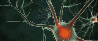
The main factors influencing the development of vegetative-vascular dystonia: natal damage to the central nervous system, somatic pathologies, diseases of the central nervous system, hormonal dysfunctions, damage to the central nervous system, personal characteristics of the patient. In most cases, psychogenic diseases are provoked by situations that contribute to maladaptation of the nervous system and the subsequent formation of autonomic disorders, such as conflicts at school and at home. Damage to the autonomic nervous system occurs when there is an inadequate response to stressful situations, neurotic and mental disorders in children, and emotional instability. Thus, autonomic dysfunction is formed.
Ear congestion
Fungus
Allergy
19979 04 February
IMPORTANT!
The information in this section cannot be used for self-diagnosis and self-treatment.
In case of pain or other exacerbation of the disease, diagnostic tests should be prescribed only by the attending physician. To make a diagnosis and properly prescribe treatment, you should contact your doctor. Ear congestion: causes, diagnosis and treatment methods.
Definition
Congestion in the ear or ears occurs as a result of a violation of sound perception and is characterized by various sensations, which may include deafness, a feeling of squeezing and heaviness, and the sound of one’s own voice being too strong. Ear congestion, regardless of the causes of its occurrence, is difficult for the patient to tolerate and, as a rule, requires the help of a specialist.
Types of ear congestion
Blockage in one or both ears may be accompanied by pain, tingling, noise or ringing in the ears, and dizziness. In some cases, congestion disappears after swallowing.
A dangerous symptom is ear congestion accompanied by fever, headache, discharge from the ear (purulent or bloody), and foreign body sensation.
Ear congestion does not always indicate a pathological process.
This condition can be caused by water getting into the ear, pressure changes
during air travel or diving to depth.
Sometimes too strong and incorrect blowing of the
nose from two nasal passages at the same time leads to a blocked ear (ears), which is associated with an increase in pressure in the middle chamber of the ear due to a sharp intake of air from the Eustachian tube.
Taking certain medications
(antibiotics, psychotropic substances) has a toxic effect on the ear, causing the development of congestion and hearing loss.
Diseases that can cause ear congestion.
Earwax
blocking the ear canal. Attempting to remove earwax yourself using improvised objects significantly increases the likelihood of pushing the plug deeper into the ear and sticking to the eardrum (this increases the risk of injury to the eardrum, which leads to complete or partial hearing loss). In these cases, the condition of congestion in the ears is accompanied by excruciating pain, noise, dizziness and nausea.
Mycotic, or fungal, infection of the external auditory canal
. Infection with fungi can be complicated by narrowing or blockage of the ear canal with a feeling of fullness in the ears. The spread of fungi in the ear is aggravated by hearing aids, in-ear headphones, and inflammatory diseases of the ear. The main signs of the disease are itching, ear congestion and resulting hearing loss, and increased sound of one’s own voice in the affected ear.
Damage to the external auditory canal and middle ear structures
may be accompanied by hearing loss and congestion. Bleeding and the formation of a blood clot that blocks the ear canal lead to deterioration in sound transmission. In addition, injury to the eardrum is possible during cleaning of the ear canal, a sudden change in pressure, or a strong blow to the outer ear. In this case, sharp pain occurs, which is replaced by congestion, ringing, noise and hearing loss.
Acute inflammatory diseases
accompanied by swelling and sometimes the formation of purulent contents.
They can lead to ear congestion and hearing loss. In particular, with otitis media of the middle ear (tympanitis),
the tympanic cavity and auditory tube are involved in the inflammatory process. Swelling, which narrows the lumen of the auditory tube, and suppuration of the soft tissues cause ear congestion and hearing impairment. As a rule, the infection enters this sterile cavity from the Eustachian tube, which is directly connected to the nasopharynx.
In children of the first and second year of life, acute otitis may occur when breast milk or formula enters the nasopharynx during regurgitation.
In older children, otitis media and congestion can be caused by
inflammation of the adenoids
, the lymphoid tissue responsible for the local immunity of the nasopharynx and closing the openings of the auditory tubes in the nasopharynx. The anatomical proximity of the adenoids and the auditory tube ensures the rapid transition of infection from the nasopharynx to the ears. In addition, enlarged adenoids can block the openings of the auditory tube, which causes a feeling of stuffiness.
Allergic reactions
can also lead to acute inflammation and swelling of the middle ear.
Otitis externa
characterized by inflammation of the external auditory canal. Congestion in the ear in this case occurs due to swelling of the tissues of the ear canal.
If the disease is caused by a foreign body entering the ear canal
, then swelling and congestion are complemented by a picture of severe irritation. The patient complains of severe itching, pain, a feeling of fullness, and heat in the ear area. The pain intensifies with chewing movements.
For furunculosis
In the external auditory canal, the picture of the disease is aggravated by a closed space where the inflammatory process develops. Increasing pain in the ear is complemented by its irradiation to the corresponding half of the head. The patient cannot lie on the painful side. Due to severe swelling of the tissues of the external auditory canal, sound transmission into the affected ear is disrupted, and a feeling of stuffiness occurs.
Among the anatomical and postoperative defects
that cause ear congestion include deviated nasal septum, narrowing of the nasal passage due to hypoplasia of the nasal wings, and stenosis of the external nasal valve.
Impaired nasal breathing leads to frequent runny nose, infection of the nasal sinuses and, as a consequence, to the transition of the inflammatory process to the auditory tube.
Ear congestion in these cases appears on the side of the narrow nasal passage. The same consequences occur after operations in the nose area.
Sensorineural hearing loss
occurs due to damage to any part of the auditory nerve. Most often, this is an irreversible phenomenon, the symptoms of which include imbalance, dizziness, nausea, fullness and noise in the ear, and poor perception of low sounds. The causes of sensorineural hearing loss can be previous infectious and vascular diseases, tumor processes, injuries, and toxic effects of various substances.
Meniere's disease
is a non-purulent disease of the inner ear, which is accompanied by congestion. An increase in the volume of lymph in the labyrinth of the ear leads to increased pressure and attacks of progressive deafness, tinnitus, and sudden dizziness. In most cases, one ear is affected first. The disease begins either with attacks of dizziness or with deterioration of hearing, which is completely restored between attacks. However, after a few years, hearing loss becomes irreversible.
Myofascial pain syndrome, temporomandibular joint diseases
. Patients with myofascial pain syndrome, which is associated with disruption of the masticatory muscles and limited mobility of the lower jaw, may also complain of ear congestion. In addition, the disease is accompanied by headaches and facial pain, difficulty opening the mouth, and clicking in the temporomandibular joint.
The root cause of the syndrome is spasm of the masticatory muscles.
A similar clinical picture is also given by diseases of the joint itself caused by malocclusion. Atherosclerosis of cerebral vessels, increased blood pressure
. Congestion in the ears due to damage or narrowing of blood vessels is explained by a deterioration in the blood supply to all tissues, as well as impaired circulation in the area of the inner and middle ear.
Vasomotor rhinitis, or runny nose during pregnancy
occurs under the influence of hormonal changes and is characterized by impaired vascular tone and the release of mucous secretion. With allergic rhinitis, the clinical picture of the disease is almost the same, but the provoking factor is not hormones, but a specific allergen. Swelling of the mucous membrane and narrowing of the nasal passages lead to obstruction of the auditory tube and cause ear congestion.
Tumors in the area of the auditory canal, auditory tube and inner ear
– the most serious cause of ear congestion. Among them should be called cholesteatoma - a tumor-like formation that consists of epidermal cells impregnated with cholesterol. Cholesteatoma is characterized by slow but steady growth. Forming in the middle ear, it can spread to the outer and inner ear, causing congestion and a feeling of heaviness in the ear, purulent discharge, swelling and redness of the auricle.
Which doctors should I contact if I have ear congestion?
If ear congestion occurs, you should contact an otolaryngologist. In the future, you may need to consult a neurologist, cardiologist, or allergist.
Diagnosis and examinations for ear congestion
To diagnose the disease that caused ear congestion, a careful questioning of the patient, examination of the outer ear and ear canal to the eardrum, and an audiometric examination are necessary. The infectious nature of the disease is determined on the basis of the clinical picture, otoscopy data and culture of the discharge.