Functions of a neuron
Despite its relatively simple structure, the neuron has many functions, the main ones of which are the following:
- perception of irritation;
- stimulus processing;
- impulse transmission;
- formation of a response.
Functionally, neurons are divided into three groups:
Afferent (sensitive or sensory). Neurons in this group perceive, process and send electrical impulses to the central nervous system. Such cells are anatomically located outside the central nervous system, but in spinal neuronal clusters (ganglia), or the same clusters of cranial nerves.
Intermediaries (also these neurons, which do not extend beyond the spinal cord and brain, are called interneurons). The purpose of these cells is to ensure contact between neurocytes. They are located in all layers of the nervous system.
Efferent (motor, motor). This category of nerve cells is responsible for transmitting chemical impulses to the innervated executive organs, ensuring their performance and setting their functional state.
In addition, another group is functionally distinguished in the nervous system - inhibitory nerves (responsible for inhibiting cell excitation). Such cells resist the propagation of electrical potential.
Neurons for dummies
Neurons are a special group of cells in the body that distribute information throughout the body. Using electrical and chemical signals, they help the brain coordinate all vital functions.
To put it simply, the tasks of the nervous system are to collect signals coming from the environment or from the body, assess the situation, decide how to respond to them (for example, change the heart rate), and think about what is happening and remember it. The main tool for performing these tasks is neurons, woven throughout the body in a complex network.
An average estimate of the number of neurons in the brain is 86 billion, each connected to another 1,000 neurons. This creates an incredible network of interactions. Neuron is the basic unit of the nervous system.
Neurons (nerve cells) make up about 10% of the brain, the rest are glial cells and astrocytes, whose function is to maintain and nourish neurons.
What does a neuron look like?
The structure of a neuron can be divided into three parts:
· Neuron body (soma) – receives information. Contains the cell nucleus.
· Dendrites are short processes that receive information from other neurons.
· An axon is a long process that carries information from the neuron body to other cells. Most often, the axon ends in a synapse (contact) with the dendrites of other neurons.
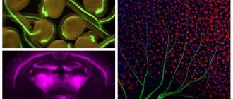
Diagram of the structure of a neuron (hereinafter, drawings from Wikipedia).
Dendrites and axons are called nerve fibers.
Axons vary greatly in length, from a few millimeters to a meter or more. The longest are the axons of the spinal ganglia.
Types of neurons
Neurons can be classified according to several parameters, for example, by structure or function.
Types of neurons depending on function:
· Efferent (motor) neurons – carry information from the central nervous system (brain and spinal cord) to cells in other parts of the body.
· Afferent (sensitive) neurons – collect information from the whole body and carry it to the central nervous system.
· Interneurons – transmit information between neurons, often within the central nervous system.
How do neurons transmit information?
A neuron, receiving information from other cells, accumulates it until it exceeds a certain threshold. After this, the neuron sends an electrical impulse along the axon - an action potential.
An action potential is generated by the movement of electrically charged particles across the axon membrane.
At rest, the electrical charge inside the neuron is negative relative to the surrounding intercellular fluid. This difference is called membrane potential. Typically it is 70 millivolts.
When the body of the neuron receives enough charge and it fires, depolarization occurs in the adjacent section of the axon - the membrane potential rises rapidly and then falls in about 1/1000 of a second. This process triggers depolarization of the adjacent section of the axon, and so on, until the impulse travels along the entire length of the axon. After the depolarization process, hyperpolarization occurs - a short-term state of rest; at this moment, impulse transmission is impossible.
The action potential is most often generated by potassium (K+) and sodium (Na+) ions, which move through ion channels from the intercellular fluid into the cell and back, changing the charge of the neuron and making it first positive, and then reducing it.
The action potential ensures that the cell operates according to the “all or nothing” principle, that is, the impulse is either transmitted or not. Weak signals will accumulate in the body of the neuron until their charge is sufficient for transmission along the processes.
Myelin
Myelin is a white, thick substance that covers most axons. This coating provides electrical insulation to the fiber and increases the speed of impulse transmission through it.
Myelinated fiber versus unmyelinated fiber.
Myelin is produced by Schwann cells in the periphery and oligodendrocytes in the central nervous system. Along the fiber, the myelin sheath is interrupted - these are nodes of Ranvier. The action potential moves from interception to interception, allowing rapid transmission of the impulse.
Multiple sclerosis, a common and serious disease, is caused by the destruction of the myelin sheath.
How do synapses work?
Neurons and the tissues to which they transmit impulses do not physically touch; there is always a space between the cells - a synapse.
Depending on the method of transmitting information, synapses can be chemical or electrical.
Chemical synapse
After the signal, moving along the neuron process, reaches the synapse, chemical substances - neurotransmitters (neurotransmitters) are released into the space between the two neurons. This space is called the synaptic cleft.
Scheme of the structure of a chemical synapse.
A neurotransmitter from a transmitting (presynaptic) neuron, entering the synaptic cleft, interacts with receptors on the membrane of the receiving (postsynaptic) neuron, triggering a whole chain of processes.
Types of chemical synapses:
· glutamatergic – the mediator is glutamic acid, which has an exciting effect on the synapse;
· GABAergic – the mediator is gamma-aminobutyric acid (GABA), has an inhibitory effect on the synapse;
· cholinergic – the mediator is acetylcholine, which carries out neuromuscular transmission of information;
Adrenergic – the mediator is adrenaline.
Electrical synapses
Electrical synapses are less common and common in the central nervous system. Cells communicate through special protein channels. The presynaptic and postsynaptic membranes in electrical synapses are located close to each other, so the impulse can pass directly from cell to cell.
The speed of impulse transmission through electrical synapses is much higher than through chemical ones, so they are located mainly in those sections where a quick reaction is necessary, for example, those responsible for protective reflexes.
Another difference between the two types of synapses in the direction of information transmission: if chemical synapses can transmit impulses only in one direction, then electrical synapses are universal in this sense.
Conclusion
Neurons are perhaps the most unusual cells in the body. Every action that the human body performs is ensured by the work of neurons. A complex neural network shapes personality and consciousness. They are responsible for both the most primitive reflexes and the most complex processes associated with thinking.
Aminat Adzhieva, portal “Eternal Youth” based on materials from Medical News Today: Neurons: The basics.
Functions of neurons
Without neurons, the human body cannot function. We saw that these nanocells are responsible for literally our every movement, every action. The functions they perform have not yet been fully studied and defined.
There are several classifications of neuron functions. We will focus on what is generally accepted in the scientific world.
Information dissemination function
This function
:
- is basic;
- studied better than others.
Its essence is that neurons process and transfer to the brain all impulses that come from the outside world or one’s own body. Next, they are processed, similar to how a search engine works in a browser.
Based on the results of scanning information from the outside, the brain, in the form of feedback, transmits the processed information to the senses or muscles.
We do not suspect that information is delivered and processed every second in our body, not only in the head and at the level of the peripheral nervous system.
To date, it has not been possible to create artificial intelligence that would come close to the operation of human neural networks. Each of the 85 billion neurons has at least 10 thousand experience-based connections, all of which work to transmit and process information.
Function of accumulating knowledge (saving experience)
A person has memory, the ability to understand the essence of things, phenomena and actions that he repeated once or many times. Neuronal cells, or more precisely neurotransmitters, connecting links between neighboring neurons, are responsible for the formation of memory.
Thus, it is not any separate part of the brain that is responsible for memory, but small protein bridges between cells. A person can lose memory when these neural connections collapse.
Integration function
This function allows individual lobes of the brain to interact with each other. As we have already said, signals from different sense organs arrive in different parts of the brain.
Neurons transmit and receive impulses in different parts of the brain through “bursts” of activity. This is how the process of thoughts, emotions and feelings appears. The more such diverse connections there are, the more effectively a person thinks. If a person is capable of reflection and analysis in a certain direction, then he will think well in another matter.
Protein production function
Neurons are such useful cells that they are not limited only to transmission functions. Nerve cells produce proteins necessary for human life. Again, neurotransmitters that are responsible for memory play a key role in the production of proteins.
In total, about 80 proteins are induced in neurons, here are the main ones that affect human well-being
:
- Serotonin
is a substance that causes joy and pleasure. - Dopamine
is the leading source of vigor and happiness for humans. Activates physical activity, helps to wake up, an excess can lead to a state of euphoria. - Norepinephrine
is a “bad” hormone that causes attacks of rage and anger. Along with cortisol, it is called the stress hormone. - Glutamate
is a substance responsible for memory storage.
From the editor: How to develop memory in preschool children
Stopping the production of proteins or releasing them in insufficient quantities can lead to serious illnesses.
Neuroglia (glia): general information
In addition to neurons, nervous tissue contains another type of cells - glial cells, glial cells, or glia (from the Greek "glia" - glue). They perform support and protective functions, and are also involved in neuronophagy. They are 10 times more numerous than neurons (10 to the 13th power and 10 to the 12th power, respectively) and they occupy half the volume of the central nervous system (CNS). Glial cells surround nerve cells and play a supporting role. Glial cells are more numerous than neurons: they make up at least half the volume of the CNS (Fig. 1-18).
There are several types of glia. Thus, some glial cells participate in maintaining the composition of the intercellular environment around neurons, others form the myelin sheath around axons, due to which the speed of action potentials increases. Consequently, without directly participating in short-term communication processes in the nervous system, neuroglial cells contribute to the implementation of this function by neurons.
Thus, glia not only performs support functions, but also ensures a variety of metabolic processes in nervous tissue, and also contributes to the restoration of nervous tissue after injury and infection.
Between neurons and glial cells there are interconnected gaps measuring 15-20 nm, the so-called interstitial space, occupying 12-14% of the total brain volume.
Glial cells are nonexcitable: during depolarization of glial cells, the conductivity of their membranes does not increase.
Neuroglial cells are divided into several types. Ependymal cells line the ventricles of the brain and the spinal canal and form the epithelial layer in the choroid plexus. They connect the ventricles with the underlying tissues.
Macroglia cells are divided into two categories - astrocytes and oligodendrocytes.
Protoplasmic astrocytes are localized in the gray matter; highly branched, short and thick processes extend from the cell body, containing an oval nucleus and a large amount of glycogen.
Fibrillary astrocytes are localized in the white matter. Their nucleus is also oval, and the cell body contains a lot of glycogen, but the processes are long and less branched, some branches literally rest against the walls of blood vessels. These cells carry nutrients from the blood to neurons.
The two types of astrocytes are interconnected and form a vast three-dimensional space in which neurons are embedded. They often divide, forming scar tissue in case of damage to the central nervous system.
Oligodendrocytes are localized in gray and white matter. They are smaller than astrocytes and contain one spherical nucleus. A small number of thin branches extend from the cell body, and it itself contains cytoplasm with a large number of ribosomes. Schwann cells are specialized oligodendrocytes that synthesize the myelin sheath of myelinated fibers.
Microglial cells are localized in both gray and white matter, but there are more of them in gray matter. From each end of the small oblong body of the cell, containing lysosomes and a well-developed Golgi apparatus, a thick process extends. Smaller side branches extend from all its branches. When the brain is damaged, these cells turn into phagocytes and, moving using amoeboid movement, resist the invasion of foreign particles.
Glia is a trophic supply system for the nervous system, and also takes an active part in the specific functioning of nervous tissue: normally it inhibits neuronal hyperactivity, promotes active absorption from the synaptic cleft and utilization of mediators and other agents involved in neuronal damage. Under ischemic conditions, microglial cells induce the synthesis of not only neurotoxic substances, but also signaling molecules, cellular regulators, trophic factors that promote the survival of neurons and reduce the processes of post-ischemic scarring
Editor's Note: Seven Telltale Symptoms of Dementia
Microglia are the only immunocompetent compartment in the central nervous system
In the central nervous system, neuroglia include astrocytes and oligodendrocytes, and in the peripheral nervous system, Schwann cells and satellite cells.
Microglial and ependymal cells are considered central glial cells (Fig. 32.7, Fig. 32.10).
How does our brain work or how to model the soul?
Hello Geektimes!
In a previously published article, a model of the nervous system was presented, I will describe the theory and principles that formed its basis. The theory is based on the analysis of available information about the biological neuron and nervous system from modern neurobiology and brain physiology. First, I will provide brief information about the modeling object; all information is presented below, taken into account and used in the model.
NEURON
A neuron is the main functional element of the nervous system; it consists of the body of a nerve cell and its processes. There are two types of processes: axons and dendrites. An axon is a long process covered with a myelin sheath, designed to transmit a nerve impulse over long distances. A dendrite is a short, branching process that communicates with many neighboring cells.
THREE TYPES OF NEURONS
Neurons can vary greatly in shape, size and configuration, despite this, there is a fundamental similarity of nervous tissue in different parts of the nervous system, and there are no serious evolutionary differences.
The nerve cell of the Aplysia mollusk can secrete the same neurotransmitters and proteins as a human cell. Depending on the configuration, there are three types of neurons:
a) receptor, centripetal, or afferent neurons, these neurons have a centripetal axon, at the end of which there are receptors, receptor or afferent endings. These neurons can be defined as elements that transmit external signals to the system.
b) interneurons (intercalary, contact, or intermediate) neurons that do not have long processes, but have only dendrites. There are more such neurons in the human brain than others. This type of neuron is the main element of the reflex arc.
c) motor, centrifugal, or efferent, they have a centripetal axon, which has efferent endings that transmit excitation to muscle or glandular cells. Efferent neurons serve to transmit signals from the nervous environment to the external environment.
Typically, articles on artificial neural networks stipulate the presence of only motor neurons (with a centrifugal axon), which are connected in layers of a hierarchical structure. A similar description is applicable to the biological nervous system, but is a kind of special case; we are talking about structures, basic conditioned reflexes. The higher the evolutionary significance of the nervous system, the less structures such as “layers” or a strict hierarchy prevail in it.
TRANSMISSION OF NERVOUS EXCITATION
The transfer of excitation occurs from neuron to neuron through special thickenings at the ends of dendrites, called synapses. Based on the type of transmission, synapses are divided into two types: chemical and electrical. Electrical synapses transmit nerve impulses directly through the site of contact. There are very few such synapses in nervous systems; they will not be taken into account in models. Chemical synapses transmit a nerve impulse through a special mediator substance (neurotransmitter, neurotransmitter), this type of synapse is widespread and implies variability in operation. It is important to note that changes constantly occur in a biological neuron, new dendrites and synapses are grown, and neuronal migrations are possible. At the points of contact with other neurons, neoplasms are formed, for the transmitting neuron it is a synapse, for the receiving neuron it is a postsynaptic membrane supplied with special receptors that respond to the mediator, that is, we can say that the neuron membrane is the receiver, and the synapses on the dendrites are transmitters signal.
SYNAPSE
When a synapse is activated, it releases portions of the transmitter; these portions can vary; the more transmitter is released, the more likely it is that the nerve cell receiving the signal will be activated. The transmitter, overcoming the synoptic cleft, enters the postsynaptic membrane, on which receptors that respond to the transmitter are located. Next, the mediator can be destroyed by a special destructive enzyme, or absorbed back into the synapse, this occurs to reduce the time of action of the mediator on the receptors. Also, in addition to the stimulating effect, there are synapses that have an inhibitory effect on the neuron. Typically, such synapses belong to certain neurons, which are designated as inhibitory neurons. There can be many synapses connecting a neuron with the same target cell. To simplify, we will assume that the entire set of influences exerted by one neuron on another target neuron is a synapse with a certain strength of influence. The main characteristic of a synapse will be its strength.
STATE OF NEURON EXCITATION
At rest, the neuron membrane is polarized. This means that on both sides of the membrane there are particles carrying opposite charges. At rest, the outer surface of the membrane is positively charged, the inner surface is negatively charged. The main charge carriers in the body are sodium (Na+), potassium (K+) and chlorine (Cl-) ions. The difference between the charges on the surface of the membrane and those inside the cell body is the membrane potential. The mediator causes polarization disturbances - depolarization. Positive ions from outside the membrane flow through open channels into the cell body, changing the charge ratio between the surface of the membrane and the cell body.
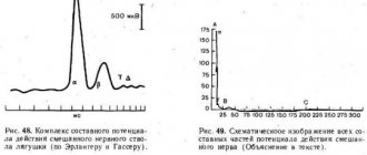
Changes in membrane potential during neuron excitation
The nature of changes in membrane potential during activation of nervous tissue is unchanged. Regardless of the force exerted on the neuron, if the force exceeds a certain threshold value, the response will be the same. Looking ahead, I would like to note that even trace potentials are important in the functioning of the nervous system (see graph above). They do not appear as a result of some harmonic oscillations balancing charges; they are a strict manifestation of a certain phase of the state of the nervous tissue during excitation.
THEORY OF ELECTROMAGNETIC INTERACTION
So, below I will give theoretical assumptions that will allow us to create mathematical models. The main idea is the interaction between charges formed inside the cell body during its activity, and charges from the surfaces of the membranes of other active cells. These charges are opposite, in connection with this we can assume how the charges will be located in the cell body under the influence of the charges of other active cells.
We can say that the neuron senses the activity of other neurons at a distance and strives to direct the spread of excitation in the direction of other active areas. At the moment of neuron activity, it is possible to calculate a certain point in space, which would be defined as the sum of the masses of charges located on the surfaces of other neurons. We will call this point a pattern point; its location depends on the combination of phases of activity of all neurons of the nervous system. A pattern in the physiology of the nervous system is a unique combination of active cells, that is, we can talk about the influence of excited areas of the brain on the functioning of an individual neuron. It is necessary to imagine the work of a neuron not just as a computer, but as a kind of excitation repeater that selects the directions of excitation propagation, thus forming complex electrical circuits. It was originally assumed that the neuron simply selectively turns off/on its synapses for transmission, depending on the preferred direction of excitation. But a more detailed study of the nature of the neuron led to the conclusion that the neuron can change the degree of influence on the target cell through the strength of its synapses, which makes the neuron a more flexible and variable computational element of the nervous system.
Which direction for excitation transmission is preferable? In various experiments related to the formation of unconditioned reflexes, it can be determined that paths or reflex arcs are formed in the nervous system that connect the activated areas of the brain during the formation of unconditioned reflexes, and associative connections are created. This means that the neuron must transmit excitations to other active areas of the brain, remember the direction and use it in the future. Let's imagine a vector whose beginning is located in the center of the active cell, and the end is directed to the pattern point defined for a given neuron. Let us denote as the vector of the preferred direction of excitation propagation (T, trend). In a biological neuron, the T vector can manifest itself in the structure of the neuroplasm itself, perhaps these are channels for the movement of ions in the cell body, or other changes in the structure of the neuron. A neuron has the property of memory; it can remember vector T, the direction of this vector, and can change and be rewritten depending on external factors. The degree to which the T vector can undergo changes is called neuroplasticity. This vector, in turn, influences the functioning of neuron synapses. For each synapse, we define a vector S, the beginning of which is in the center of the cell, and the end is directed to the center of the target neuron with which the synapse is connected. Now the degree of influence for each synapse can be determined as follows: the smaller the angle between the vector T and S, the larger the synapse will be, strengthened; the smaller the angle, the more the synapse will weaken and possibly stop the transmission of excitation. Each synapse has an independent memory property; it remembers the value of its strength. These values change with each activation of the neuron, under the influence of vector T, they either increase or decrease by a certain value.
MATHEMATICAL MODEL
The input signals (x1, x2,…xn) of the neuron are real numbers that characterize the strength of the neuron synapses that influence the neuron. A positive input value means an incentive effect on the neuron, and a negative value means an inhibitory effect. For a biological neuron, it does not matter where the signal that excites it came from, the result of its activity will be identical. A neuron will be activated when the sum of influences on it exceeds a certain threshold value. Therefore, all signals pass through the adder (a), and since neurons and the nervous system work in real time, therefore, the effect of the inputs must be assessed in a short period of time, that is, the effect of the synapse is temporary. The result of the adder passes the threshold function (b); if the sum exceeds the threshold value, then this leads to the activity of the neuron. When activated, a neuron signals its activity to the system, transmitting information about its position in the space of the nervous system and its charge, which changes over time (c). After a certain time, after activation, the neuron transmits excitation to all existing synapses, having previously recalculated their strength. During the entire period of activation, the neuron stops responding to external stimuli, that is, all effects of the synapses of other neurons are ignored. The activation period also includes the recovery period of the neuron. The vector T (r) is adjusted taking into account the value of the pattern point Pp and the level of neuroplasticity. Next, the values of all synapse strengths in the neuron(e) are reassessed. Note that blocks (d) and (e) are executed in parallel with block (c).
WAVE EFFECT
If you carefully analyze the proposed model, you can see that the source of excitation should have a greater influence on the neuron than another distant, active part of the brain. Therefore, the question arises: why does transmission still occur in the direction of another active site? I was able to determine this problem only by creating a computer model. The solution was suggested by a graph of changes in membrane potential during neuron activity.
Enhanced repolarization of the neuron, as mentioned earlier, is important for the nervous system; thanks to it, a wave effect is created, the tendency of nervous excitation to spread from the source of excitation. When working with the model, I observed two effects: if we neglect the trace potential or make it not large enough, then the excitation does not spread from the sources, but rather tends to localize. If you make the trace potential very large, then the excitation tends to “scatter” in different directions, not only from its source, but also from others.
COGNITIVE MAP
Using the theory of electromagnetic interaction, it is possible to explain many phenomena and complex processes occurring in the nervous system. For example, one of the latest discoveries that is widely discussed in brain science is the discovery of cognitive maps in the hippocampus. The hippocampus is the part of the brain responsible for short-term memory. Experiments on rats revealed that a certain place in the maze corresponds to its own localized group of cells in the hippocampus, and no matter how the animal gets to this place, the area of nervous tissue corresponding to this place will still be activated. Naturally, the animal must remember this labyrinth; one should not count on a topological correspondence between the space of the labyrinth and the cognitive map.
Each place in the maze is represented in the brain as a set of stimuli of a different nature: smells, the color of walls, possible remarkable objects, characteristic sounds, etc. These stimuli are reflected on the cortex, various representations of the sensory organs, in the form of bursts of activity in certain combinations. The brain simultaneously processes information in several sections; information channels are often separated, and the same information enters different parts of the brain.
Activation of place neurons depending on position in the maze (activity of different neurons is shown in different colors). source
The hippocampus is located in the center of the brain, the entire cara and its regions are located at equal distances from it. If we determine for each unique combination of stimuli the point of mass of charges on the surfaces of neurons, we can see that these points will be different and will be located approximately in the center of the brain. Excitation in the hippocampus will tend to these points and spread, forming stable areas of excitation. Moreover, alternating changes in combinations of stimuli will lead to a shift in the pattern point. Sections of the cognitive map will be associated with each other sequentially, which will lead to the fact that an animal placed at the beginning of a familiar maze can remember the entire subsequent path.
Conclusion
Many will have a question: where in this work are the prerequisites for the element of rationality or the manifestation of higher intellectual activity?
It is important to note that the phenomenon of human behavior is a consequence of the functioning of a biological structure. Therefore, in order to imitate intelligent behavior, it is necessary to have a good understanding of the principles and features of the functioning of biological structures. Unfortunately, the science of biology has not yet presented a clear algorithm: how a neuron works, how it understands where it needs to grow its dendrites, how to configure its synapses, so that a simple conditioned reflex can form in the nervous system, similar to those demonstrated and described in in his works, Academician I.P. Pavlov. On the other hand, in the science of artificial intelligence, in the bottom-up (biological) approach, a paradoxical situation has arisen, namely: when the models used in research are based on outdated ideas about a biological neuron, conservatism, which is based on the perceptron without rethinking its basic principles, without turning to the biological source, more and more ingenious algorithms and structures are being invented that do not have biological roots. Of course, no one is diminishing the merits of classical neural networks, which have produced many useful software products, but playing with them is not the path to creating an intelligent system. Moreover, it is not uncommon for statements that a neuron is like a powerful computing machine to be attributed to a property of quantum computers. Because of this super-complexity, the nervous system is attributed the impossibility of its repetition, because this is commensurate with the desire to simulate the human soul. However, in reality, nature follows the path of simplicity and elegance of its solutions; the movement of charges on the cell membrane can serve both to transmit nervous excitation and to broadcast information about where this transmission occurs. Despite the fact that this work demonstrates how elementary conditioned reflexes are formed in the nervous system, it brings us closer to understanding what intelligence and rational activity are. There are many more aspects of the work of the nervous system: inhibition mechanisms, principles of building emotions, organization of unconditioned reflexes and learning, without which it is impossible to build a high-quality model of the nervous system. There is an understanding, on an intuitive level, of how the nervous system works, the principles of which can be embodied in models. The creation of the first model helped to refine and correct the idea of the electromagnetic interaction of neurons. Understand how the formation of reflex arcs occurs, how each individual neuron understands how to configure its synapses to obtain associative connections. At the moment, I have begun to develop a new version of the program, which will allow us to simulate many other aspects of the functioning of the neuron and nervous system.
I ask you to take an active part in the discussion of the hypotheses and assumptions put forward here, since I may be biased towards my ideas. Your opinion is very important to me.
Model (Windows PC) + tutorial
Laws of response of excitable tissues to irritation
The nature of the response of excitable tissues to the action of stimuli in classical physiology is usually described by the laws of irritation.
The law of stimulus strength states that when the strength of a suprathreshold stimulus increases to a certain limit, the magnitude of the response also increases. This law is applicable to the contraction response of an integral skeletal muscle and the total electrical response of nerve trunks, which include many fibers with different excitability. Thus, the force of muscle contraction increases with increasing strength of the stimulus acting on it.
For the same excitable structures, the law of stimulation duration and the law of stimulation gradient are applicable. The law of duration of stimulation states that the longer the duration of suprathreshold stimulation, the greater the magnitude of the response. Naturally, the answer increases only up to a certain limit. The law of the gradient of irritation - the greater the gradient of the increase in the strength of the stimulus over time, the greater (up to a certain limit) the magnitude of the response.
The all-or-nothing law states that under the action of subthreshold stimuli, excitation does not occur, and under the action of threshold and suprathreshold stimuli, the magnitude of the response due to excitation remains constant. Consequently, already to a threshold stimulus, the excitable structure responds with the maximum possible reaction for a given functional state. A single nerve fiber is subject to this law, on the membrane of which an action potential of equal amplitude and duration is generated in response to the action of threshold and suprathreshold stimuli. The “all or nothing” law governs the reaction of a single skeletal muscle fiber, which responds with action potentials of equal amplitude and duration and the same force of contraction to both threshold and suprathreshold stimuli of different strengths. The nature of contraction of the entire muscle of the ventricles of the heart and atria is also subject to this law.
The law of polar action of electric current (Pflueger) postulates that when excitable cells are exposed to direct electric current, at the moment of circuit closure, excitation occurs at the point of application of the cathode, and when opening, at the point of contact with the anode. In itself, the prolonged action of direct current on excitable cells and tissues does not cause excitation in them. The impossibility of initiating excitation by such a current can be considered as a consequence of their accommodation to a stimulus that does not change in time with a zero slope of increase. However, since the cytoplasmic membranes of cells are polarized and there is an excess of negative charges on their inner surface, and positive charges on the outer surface, then in the area of application of the anode (positively charged electrode) to the tissue under the influence of an electric field, part of the positive charges represented by K+ cations will move into the cell and their concentration on the outer surface will become less. This will lead to a decrease in the excitability of cells and the tissue area under the anode. The opposite phenomena will be observed under the cathode.
The effect of electric current on living tissues and recording of bioelectric currents are often used in medical practice for diagnosis and treatment, and especially when conducting experimental physiological studies. This is due to the fact that the values of biocurrents reflect the functional state of tissues. Electric current has a therapeutic effect, it is easily dosed in terms of magnitude and time of exposure, and its effects can be observed at impact forces close to the natural values of biocurrents in the body.
From the editor: Reasons why vision is blurry
Medicine
Physiology
3_1 The work of nerve cells
Introduction
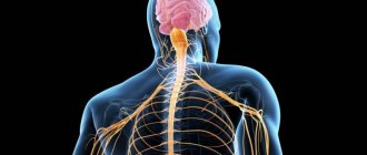
In any textbook that has a section on the nervous system, you can see how the nervous system is structured and what sections it consists of.
This is explained in both anatomy textbooks and physiology textbooks... And this site also has similar materials: see, for example, the Spinal Cord section. But how does it all work?
That's the main question!
A video about the operation of a neuron demonstrates external events that reflect the operation of the neuron.
It is well known that nerve cells use electricity to operate and exchange electrical signals in the form of nerve impulses. And for starters, it would be nice to know where nerve cells get electricity for this!
This question - about the occurrence of electrical charges in nerve cells - is answered in section: _1 Resting membrane potential
Once we understand where electrical potentials come from on the membrane, we can try to understand what the work of nerve cells is. How do they use these electrical potentials and electrically charged particles - ions?
The nervous system is very complex. And the nerve cells that make it up are also complex formations!
And yet we can highlight the most important thing in their work. We will try to figure out what they do and what they do mainly.
What can nerve cells do?
When we say “nerve cells,” we usually mean neurons. This is despite the fact that nerve cells also include glial cells, or glia (see: Neurons and glia in the hippocampus), and cellular sensory receptors. But they should still be considered separately. So we, too, following the others, will call neurons “nerve cells,” using these terms as synonyms.
Definition of "neuron"
the N neuron is an electrochemical dynamic voltage converter with a threshold operating principle, having heterochronic electrochemical inputs and synchronous chemical outputs with individual plasticity. © 2021 Sazonov V.F. © 2021 kineziolog.su.
Main tasks of a neuron
If we formulate the tasks of a neuron briefly and figuratively, we get only 3 main tasks.
1. Perception of arousal.
There is a perception of influence from other neurons or sensory receptors. Impact is the chemical release of mediators and their binding to the membrane receptors of the neuron. The neuron must perceive suitable (i.e. adequate) chemical signals (mediators) for it with its perceiving molecular membrane receptors. As a rule, these receptors are concentrated in the receptive sections of its synapses - on the postsynaptic (sometimes also called subsynaptic) sections of the membrane. The receptive part of the neuron is dendrites with synapses, or more precisely, with postsynaptic membranes. The result of the perception of excitation is the appearance of a local electrical potential on the membrane of the perceiving neuron.
2. Carrying out stimulation.
Conduction of excitation is the movement of excitation from the place of its origin to the place of transmission, i.e. from the presynaptic end of the dendrite to the postsynaptic end of the axon. This requires the birth of a nerve impulse based on a local excitatory potential and the movement of this nerve impulse along the neuron membrane to the postsynaptic end of the axon, where the chemical release of the transmitter will occur. Thus, the effect on the neuron occurs at one part of it (usually at the presynaptic end of the dendrite), but due to conduction, this excitation appears at the other end of the neuron - at the axon terminals.
3. Transfer of excitation.
Transmission of excitation is the chemical release of a transmitter from the axon terminals. Due to the release of the mediator, one neuron influences another neuron (or other cells). A neuron must exert a chemical effect on its targets: these are other neurons, muscle cells or glandular cells. To do this, it releases chemical control substances (mediators or modulators) from the endings of its transmitting process - the axon. The transmitting endings end in presynaptic membranes, through which the neuron releases its control substances.
So, a neuron must perceive chemical influences on its receiving part, conduct excitation in the form of a nerve impulse, and exert a chemical effect with its control substances on its targets using its transmitting part. To communicate between different parts of a neuron, nerve impulses are used that run from the receiving part of the neuron (dendritic endings) to its transmitting part (axon endings).
Thus, the neuron must release its control substances at the right time, in the right place, in the right quantity and in the right quality . And the neuron itself, in turn, must perceive control influences directed at it. If any of the above does not work out, then the desired effect from such a neuron will either not occur at all, or it will not be effective.
Nerve cells have a certain set of key reactions:
1. Formation and maintenance of the resting membrane potential . This ensures the excitability of neurons.
2. Formation of local potentials . This ensures subthreshold excitation of neurons.
3. Generation of action potential and nerve impulse . This ensures the transition from analog to digital coding of information by neurons.
4. Secretion of neurotransmitters (mediators and modulators). This ensures the transfer of excitation to other cells.
5. Changes in its intracellular metabolism and biosynthesis. This ensures the plasticity and adaptation of neurons.
6. Changing your cellular structures and properties. This ensures the plasticity of neurons.
7 Formation of new structures, for example, new synapses. This ensures the plasticity of nerve connections .
8. Movement of a nerve impulse along the membrane of a neuron. This ensures the conduction of excitation by neurons.
- Resting membrane potential, i.e. The difference in electrical potential between the inner and outer sides of the membrane is maintained by neurons at an average level of -70 mV and they spend up to 75% of all their energy received from nutrition on this.
- Subthreshold change in the electrical state of its membrane (local potential). This manifests itself in an increase in excitability or, conversely, in a decrease in excitability (inhibition).
- Generation of a nerve impulse (action potential). See: Action potential and nerve impulse
- The release of a special chemical that affects the condition of other nerve cells. These substances are called neurotransmitters (neurotransmitters and neuromodulators). See: Mediators and modulators. Perhaps this is the main function of the neuron - to release control substances to influence their targets.
- A change in metabolism in a given cell in response to a received chemical signal.
- A long-term change in the properties of this cell or its individual structures. This is a very important ability of living structures, distinguishing them from technical devices! Neurons can modify the molecules of their working proteins using their phosphorylation, i.e. addition of phosphorus residues to them. They can also synthesize new molecular receptors and integrate them into their membrane.
This phenomenon is called “plastic rearrangements,” and the ability for such rearrangements itself is
“plasticity . It is the plasticity of neurons and, in particular, the plasticity of synapses that provides the nervous system with such important properties as memory, learning, and the formation of conditioned reflexes. - An important property of a neuron is its ability to form new large structures in the process of life. Unlike mechanical devices, neurons grow new processes, form and remove spines, and form new synaptic contacts. These abilities of neurons also relate to their property of plasticity.
- Conducting excitation is receiving excitation at one end and transmitting it to the other. This is generally considered the most important function of a neuron. But, as you and I realized, it would be too easy!
Depolarization is a decrease in the electronegativity of a nerve cell.
We can say that the electrical potential of the membrane “creeps up” towards zero.
Depolarization means the cell is excited; a depolarized cell is more excitable, i.e. sensitive to excitement.
As a rule, depolarization and improvement of excitability of a nerve cell is produced by sodium entering it (positively charged sodium ions). If you want to cause depolarization, let sodium into the cell!
It is important to note that the term “depolarization” refers specifically to the loss of polarization. In this case, the electrical charge of the membrane decreases and tends to zero. A cell with “zero” charge is not polarized. If the charge becomes positive instead of negative, then this also means polarization of the cell. Only already positive. Unfortunately, textbooks can often erroneously conclude that depolarization continues even above zero, acquiring a positive value. This is not true!
Hyperpolarization is an increase in the electronegativity of a nerve cell.
The term "hyperpolarization" means "over-polarization." In this case, the electric charge of the membrane “creeps down”, i.e. moves away from zero.
Hyperpolarization means inhibition of the cell, it becomes less excitable, i.e. weakly sensitive to stimulation.
Generally, hyperpolarization can be induced in two ways:
1) release negatively charged chlorine ions into the nerve cell,
2) release an additional amount of positively charged potassium ions from the nerve cell.
If you want to cause hyperpolarization and worsen the excitability of a nerve cell, put chlorine into it or release potassium from it!
When the depolarization of the membrane reaches a certain critical level, a sharp jump in electrical voltage occurs from negative to positive and back to negative. This process involves membrane ion channels and the movement of sodium and potassium ions.
It is important to note that after this process has begun in one place, it, like a fire, will engulf the entire membrane of the neuron and run through all its processes. This is a nerve impulse —a spreading excitation. The term “action potential” is often used as a synonym for nerve impulse. However, it should be remembered that the concept of “action potential” refers only to the electrical process, i.e. reflects only part of a more general concept such as “nerve impulse”.
A nerve impulse is a wave of interconnected structural, chemical and electrical processes running along the membrane of a neuron.
The action potential is a sharp abrupt change in the electrical charge of the membrane from negative to positive and back to negative.
While living their cellular life, neurons constantly maintain themselves in an electrically charged state. How they do this is discussed in section: 2_1 Resting membrane potential. And at the same time, from time to time they change their charges, and also from time to time they release biologically active substances - transmitters (mediators and modulators).
The final
Image source: https://vk.com/doc183608877_438205454?hash=7699673c7e6de140a8&dl=511f143…
So, against the background of maintaining their constant electrical charge (resting potential), neurons undergo depolarization and hyperpolarization, generate nerve impulses (action potentials), release neurotransmitters (mediators and modulators), change their metabolism (metabolism), or change their properties using its ability to be plastic.
This is their cellular work for the benefit of the whole organism.
And now I will tell you the most important secret about the work of neurons...
If previously it was believed that the main job of neurons is the production of nerve impulses, now the emphasis has shifted.
The main job of neurons is the production and release of chemical control substances - neurotransmitters (mediators and modulators). It is with the help of their neurotransmitters that neurons control each other and the entire body!
And electrochemical nerve impulses only coordinate this chemical activity of neurons in time.
This is the great secret of how nerve cells work together with me.
Development and growth of neurons
Modern scientists are still debating the topic of nerve cell division, because There is currently no consensus on this issue in the field of anatomy. Many specialists in this field pay more attention to the properties rather than the structure of neurons, which is a more important and pressing issue for modern science.
The most common version is that the development of a neuron occurs from a cell, the division of which stops even before the release of processes. The axon develops first, followed by dendrites.
Depending on the main functionality, location and degree of activity, nerve cells develop differently. Their sizes vary significantly depending on their location and functions.
Structure
The general structure of a neuron is as follows: there is a body (soma), which contains a nucleus and other organelles, and processes - an axon and dendrites:
- axon
- this is the process through which the nerve impulse travels from a given cell to others. In other words, the axon is a signal output channel. - Dendrites
, accordingly, are channels for signal input, and there can be either a lot of them or very few. The number of dendrites depends on the type of neuron, and we will talk about this later.
Axons and dendrites
Axons are processes that can reach a length of more than a meter. To prevent the signal from “scattering” along the way from one cell to another, most axons in the body are covered with a myelin sheath, consisting of neuroglia cells (a general designation for the supporting cells of nervous tissue). The sheath provides insulation of one axon from the others and does not allow the electrical impulse to dissipate. Thanks to the myelin sheath, impulses travel along the axon more quickly. Dendrites are shorter and not covered with myelin.
Endoplasmic reticulum (ER) and Golgi complex
The most important organelles, in addition to the nucleus, are the rough ER, which has ribosomes and carries out protein synthesis, and the Golgi apparatus, which synthesizes various organic substances and “packs” them into membrane vesicles. Why these systems are so important for the functional activity of a neuron will be clear later.
Conducting excitation in the nervous system
Arousal as a response to irritation
. Phenomena associated with excitation have long been studied using an isolated neuromuscular preparation of a frog, to obtain which, most often, the gastrocnemius muscle is cut out from the hind leg along with the sciatic nerve corresponding to it. When a nerve is irritated, excitement occurs in it. It runs like a wave along the nerve fibers, passes to the muscle and causes its contraction, which is easy to register on a special device - a kymograph. The wave of excitation, or impulse, spreads along the nerves at different speeds: in motor nerves - up to 120 m/sec, and in sympathetic nerves - only a few meters per second. Excitation can be detected not only by muscle contraction, but also by the changes that occur in the nerve itself. The first, and, moreover, obligatory sign of excitation, wherever it arises, is an electrical reaction.
Rhythmic nature of excitation
. A single wave of excitation is easy to obtain under artificial experimental conditions. Under natural conditions, as a rule, each, even short-term, stimulation of receptors causes not one wave, but a series of waves following each other with a certain frequency. In other words, excitation is rhythmic in nature. Rhythmic excitation can also be obtained in experiments on a neuromuscular preparation of a frog. Electric current is usually used as a stimulus. Excitation occurs whenever the current is turned on and off, as well as when its direction changes. For rhythmic stimulation, intermittent direct current or induction current is used. A new wave of excitation can arise only after the previous wave has ceased. In human motor nerve fibers, the excitation wave lasts about 0.001 fractions of a second. Therefore, in one second, up to 1000 waves could pass along the nerve. However, under natural conditions, excitation waves, or impulses, travel along the nerves at a low frequency - usually 10-30 impulses per second.
Conduction of excitation in the central nervous system
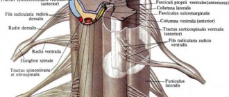
. An axon, i.e., a long process of one neuron, branches, approaches the body or dendrites of another neuron, forming small plaques or thickenings on its surface. Contacts between neurons are called synapses (Fig. 28). Excitation is transmitted through synapses from the axon of one neuron to the dendrites or body of another neuron. Chemical substances formed at the axon endings participate in the transmission of excitation. The body and dendrites of each neuron are approached by axons from many other neurons. In turn, the axon forms branches that connect to different neurons, often located far from each other.
Rice. 28. Diagram of synapses: 1 - nerve cell body; 2 - its axon; 3 - its dendrites; 4 - axon of another nerve cell; 5 - synapses
Many groups of nerve cells located in different parts of the central nervous system are interconnected bilaterally: excitation that arises in one of them is transmitted to the other. Of particular importance is a kind of circular connection: along the axon branch, the impulse directly or through intermediate neurons returns to the same nerve cell (Fig. 29). Such a ring connection can maintain the working state of the nerve cell: more and more new impulses arise in it.
Rice. 29. Ring connection between neurons
Inhibition of nerve cells
. Impulses entering the brain could spread through numerous intermediate neurons throughout all its parts and cause general excitation of the body. Under normal conditions, impulses travel along only a few of many possible paths. This is explained by the occurrence of a state of inhibition in nerve cells, in which they temporarily lose the ability to be excited, and thereby transmit impulses to other cells. Inhibition can occur in one or another neuron. Depending on which neurons are currently in a state of inhibition, the impulses will travel along one or another, but always a specific path (Fig. 30). That is why responses to the same irritation can be very different.
Rice. 30. Scheme of the passage of impulses from one neuron to another 1 - irritated section of the nerve; 2 - excitation does not transfer from the cell body to the dendrite; 3 — transition of excitation to the next neuron or muscle; 4 - braking
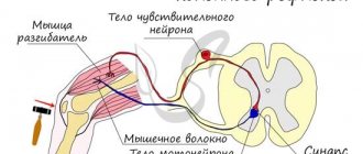
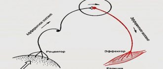
Reflex arcs
. The simplest reflex arcs consist of two or three neurons (color table IX). An example of a two-neuron arc is the knee reflex arc. If you sit a person on a chair, ask him to cross one leg over the other, and then hit the tendon below the kneecap with the edge of his palm, or better yet, with a light hammer, the leg will jump. The tendon that has been hit bends and pulls the muscle that extends the leg at the knee. The muscle is stretched, which causes irritation of the receptors located in it. The resulting flow of impulses along afferent neurons reaches the spinal cord, and from there along efferent neurons returns to the same muscle, causing its response shortening (color table IX).
Table IX. Reflex diagram: A - reflex arcs, consisting of two and three neurons; B - diagram of the knee reflex; centrifugal neurons are depicted in red, centripetal neurons from the skin - green, from muscles and tendons - blue, intermediate - black; arrows show the path of the reflex; the shaded neuron denotes inhibition of the muscle center of the opposite action
The bodies of afferent neurons are located in the dorsal root of the spinal nerve. They have two long processes: one conducts impulses from the muscle receptors to the cell body, and the other (axon) - from the cell body to the spinal cord. In the spinal cord, this process branches: one branch goes along the white matter to the underlying parts of the spinal cord, and the other goes up. Both of these branches give off lateral branches that enter the gray matter and end here. Efferent neurons have one long process (axon) and several short ones (dendrites). The cell body of the neuron is located in the anterior projections, or horns, of the gray matter. From here a long process passes through the anterior root, and then, as part of the spinal nerve, reaches the muscle.
In a two-neuron arc, the axon branches of the afferent neuron, approaching the anterior horns of the gray matter, come into contact with the efferent neuron. In the three-neuron arch there is one more neuron: it is called an intermediate or intercalary one. However, in the vast majority of cases, excitation passes through a large number of neurons to various parts of the brain.