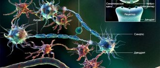
Amygdala
Admin
09.01.2019
Uncategorized
Amygdala
(amygdala, amygdala complex, (Latin) corpus amygdaloideum, from Greek αμυγδαλή, amygdalē - “almond”) - a paired almond-shaped group of nuclei located deep in each medial temporal lobe (MT) of the brain of complex vertebrates, including humans [1] . As numerous studies show, the amygdala plays a key role in processing and remembering emotional reactions. It is considered an integral part of the limbic system[2]. Moreover, anatomically, the amygdala[3], especially its central and medial nuclei,[4] are often considered part of the basal ganglia.
Internal structure of the amygdala
The amygdolar complex includes several different substructures that have different functional purposes. There are several options for their classification, with varying degrees of detail. Among the substructures of the amygdala, at least the basolateral complex of nuclei (or simply basolateral nuclei), the central, medial and cortical nuclei (or preamygdala cortex) are usually distinguished. The basolateral complex of the amygdala, in turn, is divided into the basolateral (lateral), basomedial (middle, or additional) and basoventral (lower) nuclei (see figure) [2][5][6].
The location and main nuclei of the human amygdolar complex (amygdala) using the example of a coronal section approximately through its middle. Designations of the amygdala nuclei in the figure: La
- Lateral nucleus,
BL
- Basolateral nucleus,
BM
- Basomedial nucleus,
BV
- Basoventral nucleus,
Ce
- Central nucleus,
Me
- Medial nucleus,
VCo
- ventral cortical nucleus (or preamygdala cortex).
In addition, around the amygdala are marked: Cl
- septum of the brain,
Hi
- Hippocampus
Ent
- Entorial cortex,
F
- endorchial cortical fold,
NbM
- basal ganglia of Meynert,
TrO
- optic tract,
V
- lateral cerebral ventricle.
Rat amygdala nuclei
We also present a diagram of the location of the nuclei of the (right) amygdala complex of the rat brain [7]. In this figure, all nuclei are divided into three groups: the central and medial nuclei are colored orange, the basolateral nuclei are colored green, and the cortical nuclei are colored brown. In addition, the nuclei called intercalary masses ( In
).
Designations in the figure: CEl
, central nucleus, lateral section;
CEm
, central nucleus, medial division;
CEc
, central nucleus, capsular compartment;
COa
, cortical nucleus, anterior region;
Cop
, cortical nucleus, posterior division;
BOT
, subject to the core of the olafactory tract;
Bi
, basal nuclei, intermedial section;
Bpc
, basal nucleus, parvocellular division;
BMmc
, basomedial nucleus, magnocellular division;
BMpc
, basomedial nucleus, parvocellular compartment;
Ld
, lateral nucleus, dorsal division;
Lvm
, lateral nucleus, ventromedial division;
Lvl
, lateral nucleus, ventrolateral division;
Md
, medial nucleus, dorsal division;
Mv
, medial nucleus, ventral division;
In
, intercalary masses (core);
Pir
, pear-shaped (piriform) bark. To simplify this diagram, the magnocellular region of the nucleus basalis, which is located more rostrally (anteriorly), is not shown, as is the rostral region of the medial nucleus, as well as the amygdalohippocampal region, which is located more caudally (posteriorly).
Pathologies
Excessive activity of the structures of the amygdala, which is located in the temporal zone, where the area between the putamen and the temporal pole is located, leads to disruption of the functioning of many body systems.
Effect on the heart
Cardiac dysfunction is associated with dysfunction of the brain's tonsils. Increased activity of the tonsil structures provokes the development of heart failure.
The pathogenetic mechanism is based on the participation of the amygdala in the formation of affects such as fear or rage. When a person is exposed to stress, the amygdala perceives the situation as a threat and activates the activity of the bone marrow, which intensively produces white blood cells (leukocytes). As a result, an inflammatory reaction develops in the blood vessels, which, as it progresses, can provoke angina pectoris, heart attack, and stroke.
Urbach-Wiethe disease
A rare, genetically determined pathology characterized by destruction of the amygdala. Patients with Urbach-Wiethe disease experience virtually no feeling of fear while simultaneously having the instinct of self-preservation.
Research shows that in order to scare patients, it is necessary to use unusual methods, for example, inhalation of air with a high (about 35%) concentration of carbon dioxide. Inhaling such air can cause a panic attack in patients. The clinical picture of the disease is variable and is represented by neurological deficits (epileptic seizures) and dermatological symptoms (scars, papules on the skin).
Main external relations
The lateral amygdala nucleus, which then sends impulses to the rest of the basolateral complex and the centromedial nuclei, receives signals from multiple sensory systems, including the neocortex, and is considered the main (multisensory) “input to the amygdala” [8].
The cortical nucleus of the amygdala (or preamygdala cortex) is involved in processing odors and responding to pheromones. Therefore, it receives signals from the Olafactor flask and Olafactor cortex.
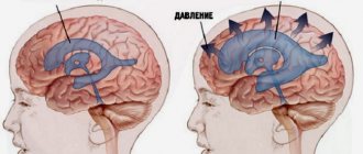
Neuroanatomical structures associated with panic disorder, their connections and functions.
The Amygdala sends its impulses to the Hypothalamus to activate the sympathetic nervous system, to the reticular nucleus of the thalamus to accelerate reflexes, to the nuclei of the trigeminal nerve and facial nerve, as well as to the ventral tagmental area, locus coeruleus, and to the laterodorsal tagmental area to activate the flow of dopamine, norepinephrine and epinephrine.[5]
The diagram on the left shows the central role of the amygdala in panic personality disorder. [9]
Amygdala
Amygdala (amygdala) of the brain
The tonsil of the brain, amygdala or amygdala (lat. Corpus amygdoloideum) is a subcortical structure of the limbic system, located deep in the temporal lobe of the brain.
“Wrong” tonsils - pharyngeal tonsils
The amygdala, as a brain formation, should not be confused with other tonsils - the pharyngeal tonsils!
Tonsils of the mouth (lat. tonsillae) are accumulations of lymphoid tissue located in the area of the nasopharynx and oral cavity. They perform protective and hematopoietic functions, participate in the development of immunity - they are a first-line protective mechanism against inhaled and ingested foreign harmful substances and antigens. The full immunological role of the tonsils still remains unclear. The commonly known term “tonsils” refers only to the palatine tonsils.
Both types of tonsils - cerebral and pharyngeal - act completely independently of each other and each in its own area, and the only thing they have in common is the same name.
And if your pharyngeal tonsils (tonsils) are suddenly removed, then do not be afraid that your brain activity will be disrupted in the same manner as in the unfortunate monkeys in experiments where their cerebral tonsils (amygdalae) were removed!
“Those same” tonsils - brain ones
So, the cerebral amygdala is an almond-shaped accumulation of gray matter in the depths of the temporal lobe of the brain, with an average size of 10x8x5 mm.
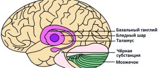
The amygdala belongs to the basal ganglia of the brain and is part of the limbic system, which controls emotions.
There are two tonsils in total - one in each hemisphere. The neurons of the amygdala are diverse in form, function and neurochemical processes in them.
Functions of the amygdala
The functions of the amygdala are associated with the provision of defensive behavior, autonomic, motor, emotional reactions, and the motivation of conditioned reflex behavior.
Moreover, the main thing, apparently, is motivation, i.e. encouragement to action.
The cerebral cortex allows you to create sensory (sensory) images, i.e. see, hear or feel something. The hippocampus (part of the limbic system, which “manages” memory) makes it possible to store a sensory image and remember it after some time. But the amygdala determines exactly what emotional feelings we experience for a given sensory image.
The amygdala is actually several separately functioning nuclei, which anatomists unite together due to their proximity to each other. Among these nuclei, the key ones are: the basal-lateral complex, the central medial nuclei and the corticomedial nuclei. The basal-lateral complex, necessary for the development of a conditioned fear reflex in rats, receives input signals from sensory systems. The central medial nuclei are the main output for the basal lateral complex and are involved in emotional arousal in rats and cats. The amygdala is connected to the rest of the nervous system and is strategically located, so it acts as a center for regulating emotions. It receives all the signals coming from the motor cortex, the primary sensory cortex, part of the association cortex, and the parietal and occipital lobes of your brain. In other words, from almost every available source. if you destroy it and look at the vegetative functions, nothing changes. But if it is irritated, a disruption occurs in the functioning of internal organs.
Axons emerging from the amygdala are concentrated in the reticular nuclei of the thalamus, which process signals from the sensory organs. Therefore, the amygdala can influence the work of the thalamus with sensory information: giving some information increased significance, and making others insignificant.
The amygdala reacts with many of its nuclei to visual, auditory, interoceptive, olfactory, and skin irritations, and all these irritations cause a change in the activity of any of the amygdala nuclei, i.e., the amygdala nuclei are polysensory. The reaction of the nucleus to external stimulation lasts, as a rule, up to 85 ms, i.e., significantly less than the reaction to similar stimulation of the neocortex. The neurons of the amygdala have pronounced spontaneous activity, which can be enhanced or inhibited by sensory stimulation. Many neurons are multimodal and multisensory and fire synchronously with the theta rhythm. If you destroy the amygdala and look at the autonomic functions, nothing changes. But if it is irritated, a disruption occurs in the functioning of internal organs. Irritation of the nuclei of the amygdala creates a pronounced parasympathetic effect on the activity of the cardiovascular and respiratory systems, leads to a decrease (rarely to an increase) in blood pressure, a decrease in heart rate, disruption of the conduction of excitation through the conduction system of the heart, the occurrence of arrhythmias and extrasystoles. In this case, vascular tone may not change. The slowdown in the rhythm of heart contractions when affecting the tonsils has a long latent period and has a long aftereffect. Irritation of the tonsil nuclei causes respiratory depression and sometimes a cough reaction. With artificial activation of the tonsil, reactions of sniffing, licking, chewing, swallowing, salivation, and changes in small intestinal motility appear, and the effects occur with a long latent period (up to 30-45 s after irritation). Stimulation of the tonsils against the background of active contractions of the stomach or intestines inhibits these contractions. The various effects of irritation of the tonsils are due to their connection with the hypothalamus, which regulates the functioning of internal organs. The amygdala provides emotional support for autonomic reactions. During an indicative reaction, when something new has arisen, as a rule, such a reaction is accompanied by a change in autonomic functions, such as a change in heart function, increased breathing, and a change in blood pressure. If the amygdala is destroyed, then this emotional accompaniment is absent, an indicative reaction occurs, but the autonomic nervous system does not turn on, and the autonomic reactions do not change. If you destroy the amygdala of a dominant male, then his career is over. The amygdala is responsible for recognizing a person by face. If scalerosis occurs in the temporal region, and the amygdala is located there, this especially often occurs with epilepsy, the disease prosopagnosia occurs, Prosop - face, agnosia - forget. As a result of this disease, a person does not even recognize himself in the mirror.
The amygdala has a low convulsive threshold; if an injury occurs in the amygdala, the focus of epilepsy very often occurs, the source of pathological impulses. The person develops post-traumatic amygdalar epilepsy, which is not related to glutamate or GABA. In the amygdala, pathological impulses arise that go to the cerebral cortex, where increased excitability occurs from there to the motor neurons of the spinal cord, and severe motor convulsions occur. This is often a birth injury. Damage to the amygdala in animals reduces the adequate preparation of the autonomic nervous system for the organization and implementation of behavioral reactions, leading to hypersexuality, the disappearance of fear, calmness, and inability to rage and aggression. Animals become gullible. For example, monkeys with a damaged amygdala calmly approach a viper that previously caused them horror and flight. Apparently, in case of damage to the amygdala, some innate unconditioned reflexes that implement the memory of danger disappear. In humans and other animals, this subcortical brain structure is involved in the formation of both negative (fear) and positive emotions (pleasure). Its size is positively correlated with aggressive behavior. In humans, this is the most sexually dimorphic brain structure—in men, it shrinks by more than 30% after castration. Conditions such as anxiety, autism, depression, post-traumatic shock and phobias are thought to be associated with abnormal functioning of the amygdala.
Diagram of the action of the amygdala ↙ ↘ With an intact amygdala With a damaged amygdala Monkey + fire = fear, flight Monkey + fire = indifference
Fence
The fence (Claustrum) is an elongated plate up to 2 mm thick, the front part of which thickens. The medial edge of the plate is smooth, and along the lateral edge there are small protrusions of gray matter. Located under the cerebral cortex, deep in the white matter. The deep localization and small size of the fence present certain difficulties for its physiological study. This structure contains polymorphic neurons of different types. It forms connections primarily with the cerebral cortex. Stimulation of the fence causes an indicative reaction, turning the head in the direction of irritation, chewing, swallowing, and sometimes vomiting movements. Irritation from the fence inhibits the conditioned reflex to light and has little effect on the conditioned reflex to sound. Stimulation of the fence during eating inhibits the process of eating food. It is known that the thickness of the fence of the left hemisphere in humans is somewhat greater than that of the right; When the right hemisphere fence is damaged, speech disorders are observed.
In the studies of E.N. Panakhova (2006) found that the role of the amygdala is not limited to its regulation of perceptual and cognitive processes - it takes part in the control of the conduction of integrated information along the entire visual pathway of both channels of specific signals entering the visual cortex of the brain - retinogeniculocortical and retinocolliculogeniculocortical. By the nature of their influence on the structures of the visual system, two phylogenetically heterogeneous sections of the amygdala are in an opposing relationship and have a phasic effect of the opposite direction on these structures. It has been established that the basolateral amygdala (BLA) leads to the actualization of the visual signal, and the more ancient phylogenetically, the corticomedial amygdala (CMA), has an inhibitory effect on the transmission of visual information to the cortex along the main retinogeniculocortical pathway.
Basic internal communications
Basic connections of the Amygdala nuclei. The power of the arrows corresponds to the power of the connections.
We will present the general scheme of internal connections of the amygdala complex of primates according to the work [8]. As already noted, the lateral nucleus (its dorsal part) receives many connections from the sensory cortex and sends its connections to almost all other nuclei of the amygdala, although the latter differ in their power. The strongest projections of the lateral nucleus go to all sections of the basal ganglia and preamygdaloid cortex. It is important to note that its projections into the central nucleus of the amygdala are relatively weak relative to, for example, connections with the basal ganglia.
Connections of the amygdala nuclei
Note again that, in general, the lateral nucleus is the main input to the amygdala. There, information is processed and transmitted basally - medially and further to the central nucleus, which can be called the main output instance of the amygdaloid complex. At the moment, intraamygdaloid connections have been studied in detail for rats, and there is also data for cats and primates.
Let us give as an example a diagram [7] of the main internal and internuclear connections for the amygdala of rats (see Fig., designations as before) obtained from anatomical studies. Most of these connections are glutamatergic. In [7], intranuclear connections are discussed in detail using the example of the lateral, basal and central nuclei of the amygdala. However, it is noted that the neurons of the basal and lateral nuclei have extensive dendritic trees located in neighboring sections of the same nuclei. The psychophysiological significance of such connections remains unclear.
Characteristic
The amygdala is a cluster of nuclei, a paired structure located in the limbic system within the brain. The area of brain tissue is responsible for the formation of emotions, such as fear and pleasure. It has been experimentally proven that stimulation of the right amygdala provokes the appearance of negative emotions (anxiety, sadness, worry), the left - more often positive (joy, happiness), less often negative.
According to anatomy, the amygdala in men is larger on average by 0.3 cm3 than in women. However, given that men's brains are on average 10% larger than women's (due to the larger size of the torso and skull), the increased volume of male tonsils is a predictable and logical result. The brain structure develops faster in women.
The left amygdala matures faster than the right, which is associated with the repeated occurrence of adequate reactions to dangerous situations in childhood. The larger the volume of the amygdala, the greater the number of connections formed with other parts of the brain. Scientists associate the number of synaptic connections formed by the amygdala with an individual’s communication activity.
The more social contacts there are, the more actively the brain region interacts with other brain systems. Research shows the relationship between the volume of the amygdala and such individual abilities as recognizing people's appearance and determining the nature of the emotions they show. The amygdala is a part of the brain that is responsible for the occurrence of fear, which allowed scientists to suggest the influence of increased activity of the structure on the development of anxiety-phobic disorders.
Stimuli that are associated with negative experiences can activate the activity of the amygdala, which provokes the body’s preparation for stress and to face a dangerous situation. Some scientists suggest a similar pathogenetic mechanism for the occurrence of panic attacks. Increased activity of the amygdala complex is detected in conjunction with post-traumatic neurotic, bipolar, and panic disorders.
The studies revealed that in patients diagnosed with depression of various origins, emotionally significant verbal stimuli (words, phrases) provoked prolonged activation of the tonsils. Similar physiological reactions are observed in patients who are presented with visual stimuli associated with feelings of sadness and grief.
After a course of treatment with antidepressants (Venlafaxine), the increased activity of the amygdala regresses, and the structure begins to function normally. Aggression is not an independent neurotic disorder. Aggressive behavior can be a consequence of a normal reaction to an external stimulus or a manifestation of mental pathology. The implementation of aggressive behavior patterns occurs with the direct participation of the amygdala.
Damage to the brain matter in the temporal lobe is often accompanied by a loss of control over manifestations of aggression. Research results provide an example of this. In 20-28% of patients with temporal lobe epilepsy and atrophic changes in the substance of the amygdala complex, interictal (between attacks) aggressive behavior is observed.
The amygdala cannot be called a gland because it does not produce specific substances. This department is involved in the process of detecting threats, expressing emotions of fear, and enhancing emotional memory. The amygdala complex is a structure that monitors danger and provides a response to facial expressions of threat and rage.