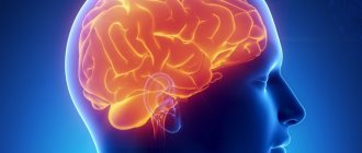
One of the most inexplicable things in the universe is the brain. Almost nothing is known about it with regard to its operating principles. From a physiological point of view, this organ has been well studied, but most people have more than a superficial understanding of its structure.
The majority of educated people know that the brain is two hemispheres, covered with a cortex and convolutions; it consists of several sections and somewhere there is gray and white matter. We will talk about all this in special topics, and today we will look at what the basal ganglia of the brain are, which few have heard of and know about.
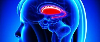
Structure and location
The basal ganglia of the brain is a collection of gray matter in the white matter, located at the base of the brain and part of its anterior lobe. As we can see, the gray matter not only forms the hemispheres, but is also located in the form of separate clusters called ganglia. They have a close connection with the white matter and cortex of both hemispheres.
The structure of this region is based on a slice of the brain. It includes:
- amygdala;
- striatum (composed of the caudate nucleus, globus pallidus, putamen);
- fence;
- lenticular nucleus.
Between the lenticular nucleus and the thalamus there is a white substance called the internal capsule, and between the insula and the fence is the external capsule. Recently, a slightly different structure of the subcortical nuclei of the brain has been proposed:
- striatum;
- several nuclei of the midbrain and diencephalon (subthalamic, pedunculopontine and substantia nigra).
Together they are responsible for motor activity, motor coordination and motivation in human behavior. This is all that can be said for sure about the function of the subcortical nuclei. Otherwise, they, like the brain as a whole, are poorly understood. Absolutely nothing is known about the purpose of the fence.
What are the basal ganglia?
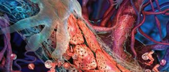
The basal or subcortical ganglia in the brain are small collections of gray matter. The gray mass consists of nerve ganglia (axons, dendrites and auxiliary nervous tissue), which, when connected to each other, form a kind of chain.
The basal ganglia lie in deep brain structures and act as a link for transmitting information between the right and left hemispheres. This connection allows the brain to function as a single mechanism.
Long processes extending from the nerve ganglia (axons) ensure the reception and transmission of impulses to other parts of the central nervous system. The basal ganglia, or “subcortex,” interact closely with the motor control systems (corticospinal and cerebellar).
Inclusions of the gray substance are located in the thickness of the white matter. At the stage of embryogenesis, the basal ganglia are formed from a temporary structure - the ganglion tubercle. It acts as a source of neurons for the formation of mature structures of the central nervous system.
The nuclei are located near the base of the brain, slightly to the side of the optic thalamus. The basal ganglia are anatomically a structural unit of the forebrain.
The functionality of the “subcortex” is combined into two systems:
- Striopallidal - acts as the main transmitter of information between the trunk of the central nervous system and the cortex.
- Limbic - provides communication between structures that are located between the thalamic region.
These systems include different structural units of the “subcortex”.
Physiology
All subcortical nuclei are again conventionally combined into two systems. The first is called the striopallid system, which includes:
- pale globe;
- caudate nucleus of the brain;
- shell.
The last two structures consist of many layers, which is why they are grouped under the name striatum. Ballus pallidus is a brighter, lighter color and is not laminated.
The lenticular nucleus is formed by the globus pallidus (located inside) and the shell, which forms its outer layer. The amygdala and the amygdala are components of the limbic system of the brain.
Let's take a closer look at what these brain nuclei are.
Caudate nucleus
Paired component of the brain related to the striatum. The location is in front of the thalamus. They are separated by a strip of white matter called the internal capsule. Its anterior part has a more massive thickened structure, the head of the structure is adjacent to the lenticular core.
Structurally, it consists of Golgi neurons and has the following characteristics:
- their axon is very thin, and dendrites (processes) are short;
- nerve cells have reduced physical dimensions compared to normal ones.
The caudate nucleus has close connections with many other distinct brain structures and forms a very wide network of neurons. Through them, the globus pallidus and thalamus interact with sensory areas, creating pathways with closed circuits. The ganglion also interacts with other parts of the brain, and not all of them lie in its vicinity.
Experts do not have a consensus on what the function of the caudate nucleus is. This once again confirms the scientifically unfounded theory that the brain is a single structure, any of its functions can be easily performed by any part. And this has been repeatedly proven in studies of people injured due to accidents, other emergencies and diseases.
It is certainly known that it takes part in autonomic functions and plays an important role in the development of cognitive abilities, coordination and stimulation of motor activity.
The striatal nucleus consists of layers of white and gray matter alternating in a vertical plane.
Black substance
The component of the system that is most involved in the coordination of movements and motor skills, maintaining muscle tone and controlling postures. Participates in many autonomic functions, such as breathing, cardiac activity, and maintaining vascular tone.
Physically, the substance is a continuous strip, as was believed for decades, but anatomical sections have shown that it consists of two parts. One of them is a receiver that sends dopamine to the striatum, the second - a transmitter - serves as a transport artery for transmitting signals from the basal ganglia to other parts of the brain, of which there are more than a dozen.
Lenticular body
Its location is between the caudate nucleus and the thalamus, which, as stated, are separated by the external capsule. In front of the structure, it merges with the head of the caudate nucleus, which is why its frontal section has a wedge-shaped shape.
This nucleus consists of sections separated by a thin film of white matter:
- shell – darker outer part;
- pale ball.
The latter is very different in structure from the shell and consists of type I Golgi cells, which predominate in the human nervous system, and are larger in size than their type II. According to neurophysiologists, it is a more archaic brain structure than other components of the brain nucleus.
Other nodes
The fence is the thinnest layer of gray matter between the shell and the island, around which there is a white substance.
The basal ganglia are also represented by the amygdala, located under the shell in the temporal region of the head. It is believed, but not known for sure, that this part belongs to the olfactory system. It is also where the nerve fibers coming from the olfactory lobe end.
Functions of the basal ganglia
The main functions of the basal ganglia are to regulate and maintain the functioning of all life support systems and organs. But since the basal ganglia also interact with other brain structures, as well as various systems, the functional orientation is complemented by a regulatory set.
General functionality of the basal ganglia in the brain:
- Control and regulation of motor skills, including fine motor skills and complex postures;
- Regulation of the autonomic nervous system;
- Participation in the processes of higher nervous activity;
- Activation of neurotransmitters during concentrated work (impulse transmission).
The basal ganglia in the brain have one feature - when a person is in a state of complete rest (sleep), they suspend their work. Some scientists correlate this ability as a connection with consciousness, but there has been no confirmation so far.
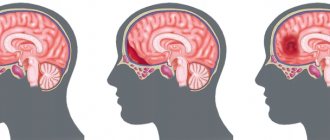
Symptoms of basal ganglia dysfunction
The subcortical ganglia, along with other brain structures, are subject to various primary and secondary pathologies. And since their functionality mainly consists of maintaining the autonomic nervous system, the general condition of a person directly depends on the healthy functioning of the nerve connections.
Symptoms of the pathology increase gradually and quite often patients do not pay attention to them at the initial stage. Calcification of the basal ganglia is considered a sluggish process, since a long period of time is required for the accumulation of calcifications on the surface of the nucleus.
Progressive pathologies include corticobasal degeneration. The clinical picture increases gradually, with an increase in the number of self-destruction of nerve cells.
All clinical manifestations of pathology of the basal ganglia are divided into two groups: hyper or hypokinetic disorders.
General symptoms:
- Various movement disorders - tremor, impaired sense of balance, atony or hypertonicity of muscles, erratic movements, pathological postures;
- Changes in the emotional sphere - apathy or depression;
- Feeling tired (broken);
- Disturbances in the functioning of vital centers (blood circulation, breathing);
- Speech center disorder;
- Poor facial expressions;
- Inadequate assessment of one's behavior.
Symptoms can also be supplemented by general cerebral manifestations, since the basal ganglia of the brain function in conjunction with other structures and systems.
Pathological states of nuclei
The causes of nuclear pathology in the brain are numerous infections and traumatic brain injuries, and congenital anomalies cannot be ruled out.
In patients with pathologies of the basal ganglia of the brain, there are signs of irritation - the damaged nervous tissue does not respond to various stimuli (external or internal), as a result of which spontaneous outbreaks of irritation occur in the cerebral cortex without an obvious source.
The most common diseases:
- Cortical paralysis - the striopallidal system is affected, resulting in convulsive syndrome. A characteristic feature is the proboscis symptom and involuntary head movements;
- Parkinson's disease is a progressive degenerative disease where neurons die and dopamine production stops. The muscles become rigid, as a result of which the patient suffers from various movement disorders;
- Hethington's disease is a genetic disease that, in addition to muscular manifestations, is combined with mental personality disorders.
To treat diseases, complex therapy is selected, which also includes correctional work with a psychologist and speech therapist. Thanks to complex work, the irritation of the nervous system is restored and the correct physiological reaction is formed.
Diagnosis and prognosis of pathology
An initial examination based on the patient’s complaints is carried out by a neurologist. To differentiate the pathology, the patient is sent for functional diagnostics:
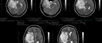
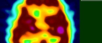
- CT or MRI of brain structures.
- Angiography or ultrasound examination of the vessels of the brain and neck.
- EEG.
The prognosis for life with pathologies of the basal ganglia depends on many factors (gender, concomitant diseases, age), including the compensatory capabilities of the body, which are individual.
For positive dynamics during rehabilitation, it is important not only the timeliness of the diagnosis, but also the characteristics of the disease. Most pathologies, unfortunately, cannot be treated and it is only possible to carry out a course of maintenance therapy.
According to statistics, depending on the type of pathology, patients have a 50/50 chance, that is, half of the cases have an unfavorable prognosis for life, the other part of the patients has a chance of returning to normal life after long-term rehabilitation.
Consequences of basal ganglia pathologies
Manifestations of pathology, even with successful treatment, will accompany the patient throughout his life and can cause disability. The development of the disease is most often corrected by taking medications, physiotherapeutic procedures, physical exercise, and strengthening the nervous system.
As you know, the adaptive forces of the body are great. But at the same time, the sick person and his relatives need to be patient and follow all the specialists’ instructions: the effectiveness of rehabilitation measures and future adaptation in society depend on this.
Pathological states of nuclei
Pathologies of the basal ganglia are expressed by a number of diseases, since the vital activity of the body depends on their functioning. The degree of their manifestations may vary.
- Functional deficiency. The first signs of pathology appear at an early age. It is usually a consequence of genetic abnormalities and is inherited. In adults, the deviation can lead to the development of Parkinson's disease or paralysis.
- Neoplasms and cysts. Like any other structure of the brain, cells of the basal ganglia are capable of mutating into atypical ones and forming tumor-like neoplasms. Their localization may vary. The impetus for the development of a tumor is a metabolic disorder in cells, atrophy and necrosis of brain tissue. The appearance of neoplasms can occur both in utero and after the birth of a child, as he grows up. For example, some experts associate cerebral palsy with damage to the basal ganglia in the second half of pregnancy. Some types of pathologies can be triggered by a difficult course of labor, head injuries, and infectious diseases in the first year of a child’s life. An obvious manifestation of damage to the basal ganglia of the brain are neurological abnormalities in which excessive irritation (excitation) of formations occurs: hyperactivity, attention deficit disorder. There are also asymptomatic small cysts that may disappear over time.
- Calcification of the basal ganglia. A striking example of pathology is Idiopathic calcification of the basal ganglia or Fahr syndrome. It is characterized by the appearance of calcium accumulations (calcification) on the surface of the ganglia. The causes of the pathology are unknown, but there is an opinion that it can develop as a result of a chromosomal malfunction. The patient experiences degradation of motor functions, dementia, convulsions, headache, fatigue, dysarthria, and muscle spasms. Signs of parkinsonism may also appear - tremors, muscle rigidity, shuffling gait, “rolling” movements of the fingers. In the final stages, mental disorders develop.
- Corticobasal degeneration. Refers to progressive pathologies of the central nervous system. When it occurs, self-destruction of ganglion cells occurs due to disruption of the metabolic processes of the brain. The manifestation of pathology depends on the functioning (to one degree or another) of the part of the brain to which the affected area belongs. For example, the first symptom is often a feeling of numbness or awkwardness in a limb, a disorder of its sensitivity. Then other symptoms appear: various forms of muscular dystonia, myoclonus, postural tremor, etc.
Treatment of pathologies of the basal ganglia should be comprehensive. A psychotherapist, speech therapist and some other specialists, depending on the manifestations of the disease, must take part in this.