What it is
Perivascular spaces are located between the walls of blood vessels and the white matter of the brain. These formations are also called criblures or Virchow-Robin spaces. They are filled with cerebrospinal fluid and regulate the outflow of cerebrospinal fluid.
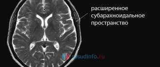
Normally, criblures are so small that they are not visible on an MRI image. However, there are cases when examination reveals expanded perivascular spaces. What does this diagnostic result mean? This suggests that criblures are visualized during an MRI examination. They look like white spots in the photo.
Interesting Facts
The perivascular spaces are named after the two scientists. However, as often happens, the area was first discovered by another person. This was done in 1843 by Durand Fardel.
Only 10 years later, the German scientist Rudolf Virchow described in detail the structure of this area. This fact is surprising, given that a conventional microscope was used for the study.
A few years later, his French colleague clarified that this is not just a gap, but a channel inside which a cerebral vessel passes.
Causes
Enlarged perivascular Robin-Virchow spaces are not always a sign of pathology. This diagnostic result is also observed in completely healthy people. Most often, expansion of criblures is observed in elderly patients and is associated with age-related changes in the brain.
However, in some cases, dilated perivascular spaces may be a sign of the following diseases and conditions:
- brain atrophy;
- leukoaraiosis;
- cerebral ischemia (including cerebral infarction);
- disseminated encephalomyelitis.
In elderly people, expansion of criblures is often observed with hypertension, atherosclerosis, and dementia. These pathologies are usually accompanied by memory impairment and other cognitive impairment.
Additional diagnostic methods
What to do if the MRI results indicate that you have dilated perivascular Virchow-Robin spaces? It is necessary to show the transcript of the study to a neurologist. Only a specialist can determine whether this is a variant of the norm, an age-related feature, or a sign of pathology.
There are cases when MRI does not reveal any changes in the brain, but the image shows dilated perivascular Virchow-Robin spaces. What does this mean? As a rule, such a sign does not indicate pathology. Doctors consider an increase in criblures only in combination with other changes identified during an MRI examination.
If necessary, the doctor may prescribe additional tests:
- multislice computed tomography;
- vascular angiography;
- Dopplerography;
- cerebrospinal fluid examination.
Let's take a closer look at the most common diseases and conditions that can lead to expansion of criblures.
Expanded spaces in pathology
The localization and intensity of the MRI signal will help to distinguish pathology or age-related norms.
If criblure is visualized in an atypical place and there is an asymmetrical picture, then most likely we are talking about a disease.
Several reasons can lead to true expansion.
Cryptococcosis
This is a fungal disease that occurs in people with immunodeficiency. Most often occurs in HIV-infected people. In this case, fungal spores can be localized inside the perivascular spaces, causing their expansion. Such accumulations are called gelatinous pseudocysts. Their difference from normal dilated spaces is the preservation of hyperdensity in FLAIR mode.
An MRI picture of concomitant meningitis, hydrocephalus and the presence of foci of cryptococcosis in the brain substance also helps to identify the disease.
Treatment is carried out with antifungal drugs.
Foci of cryptococcosis on MRI
Mucopolysaccharidosis
This is an inborn metabolic disease in which the decomposition of glycosaminoglycans is impaired. Excess substances accumulate, forming criblures. In the pictures they look like latticework. Hyperintense white matter is also visualized, which helps distinguish pathology from normality.
Since the disease is based on enzyme deficiency, the goal of therapy is their synthetic replacement: Aldurazyme, Elaprase.
Brain atrophy
If the patient’s perivascular spaces are expanded and the volume of the brain is reduced, then doctors talk about organ atrophy. Most often this is a sign of the following diseases:
- senile dementia;
- atherosclerosis;
- Alzheimer's disease.
In these diseases, neurons die. This is accompanied by memory impairment, mental impairment, and mental disorders. Typically, such diseases occur in elderly patients.
In some cases, dilated perivascular Virchow-Robin spaces are detected in newborns. This may be a sign of serious genetic diseases accompanied by the death of neurons.
How to treat such pathologies? After all, it is no longer possible to restore lost neurons. You can only slow down the process of death of nerve cells. Patients are prescribed the following drugs for symptomatic therapy:
- nootropics: Piracetam, Cavinton, Nootropil;
- sedatives: “Phenazepam”, “Phenibut”;
- antidepressants: Valdoxan, Amitriptyline.
The prognosis of such pathologies is usually unfavorable, as brain atrophy and neuronal death progress.
Focal changes in the brain: development, types, symptoms, dangerous or not, how to treat
Any violations in this department negatively affect the functioning of the entire vital system.
In the case of multiple lesions, the functioning of the nervous system is completely disrupted, the functioning of all parts of the brain suffers, leading the person to complete helplessness.
What does the disease lead to:
- Severe surges in blood pressure.
- Encephalitis of the brain.
- Multiple sclerosis.
- Poor circulation in all organs.
- Complete damage to the central nervous system.
At the first symptoms of this disease, it is necessary to consult a doctor and examine the brain to identify such disorders. There are techniques to reduce the progression of gliosis.
For newborn children, a diagnosis such as gliosis is practically a death sentence. As a result of genetic mutations, pathological processes in the brain begin to occur in the fetus at the age of 5 months, which leads to severe gliosis. Children suffering from this disease rarely survive to the age of 4 years, although in the first months of their life everything seems to be fine, and the disease does not make itself felt.
Important Basal ganglia structure, location and functions
Leukoaraiosis
Doctors call leukoaraiosis a thinning of the white matter of the brain. Due to structural changes in the nervous tissue, patients have enlarged perivascular spaces. This is also a sign of diseases characteristic of older people:
- hypertension;
- atherosclerosis;
- senile dementia.
Changes in the white matter of the brain cause cognitive impairment. Patients are treated symptomatically with nootropic drugs. These drugs improve the nutrition of neurons and stop their death. For atherosclerosis, statins are indicated. For high blood pressure, antihypertensive drugs are prescribed.
Perivascular spaces are expanded along the penetrating vessels
subarachnoid) space. Currently, there is no common understanding of PVR. It is believed that there is no PVR around the cerebral veins. At the same time, other authors believe that the PVR is surrounded by both arteries and veins and venules. Some authors describe the PVR as a space located between the vessel wall and the nervous tissue of the brain.
One of the most important functions of the VRP is the regulation of circulation (drainage) of cerebrospinal fluid and the exchange of soluble factors between the cerebrospinal fluid and tissue fluid. There is a "tide hypothesis" which posits that cardiac contractions create and maintain pressure waves to modulate flow from the subarachnoid space to the VRP and back.
The second function of the VRP is that these spaces are an important part of the blood-brain barrier (BBB). The third important function of VRP is their participation in immunoregulation, since they contain immunocompetent cells
Macrophages constantly migrate from the blood and do not pass through the membrane formed by the legs of glial cells.
PVRs contain vasoactive neuropeptides that regulate blood pressure and heart rate, the activity of microgliocytes, are involved in signal transmission, and serve to prevent the development of inflammation (by activating the enzyme adenylate cyclase, which then produces cAMP, which is involved in the modulation of autoreactive and regulatory T cells).
Ischemic conditions
With ischemia, the blood supply to the brain deteriorates. This is usually a consequence of atherosclerotic changes in the vessels. The patient periodically experiences dizziness, double vision, motor coordination disorders, speech and memory disorders. Due to changes in the vessels, the spaces around their walls also expand.
Patients are prescribed nootropic drugs (Piracetam, Cerebrolysin, Actovegin), as well as drugs that normalize metabolism in brain cells (Cortexin, Ceraxon). In this case, it is very important to carry out etiotropic treatment of atherosclerosis with statins. The drugs Lovastatin, Atorvastatin, and Simvastatin are prescribed. This therapy eliminates the cause of ischemia.
Treatment
The method of treating brain damage depends on its type, the degree of pathological changes, and the severity of the general condition. Typically, treatment for traumatic brain injury and organ disease differs.
Traumatic brain injury
Immediately after receiving a traumatic brain injury, it is important to provide proper first aid, which will help alleviate the condition and improve the prognosis. If there is no breathing or pulse, perform artificial respiration and cardiac massage. If these are not changed, the victim must be laid on his side, which avoids disruption of respiratory function due to vomiting
If these are not changed, the victim must be laid on his side, which avoids disruption of respiratory function due to vomiting
If there is no breathing or pulse, perform artificial respiration and cardiac massage. If these are not changed, the victim must be placed on his side, which avoids disruption of respiratory function due to vomiting.
If there is a closed injury, a cold compress is applied to the injury site to reduce pain and swelling. If there is bleeding from a wound on the skin, it is covered with a piece of gauze, and then the head is bandaged.
It is not recommended to independently remove bone fragments and other elements protruding from the wound before the ambulance arrives, because in this case the bleeding will only intensify. In addition, you can get an infection.
To correct post-traumatic disorders, the following is prescribed:
neuropsychological treatment to restore memory, attention, and emotional state; taking medications to normalize blood flow in the brain; conducting speech therapy sessions to restore speech; psychotherapeutic treatment to correct the emotional background; a diet that includes foods that normalize brain function
Damage of a different etiology
If brain damage is caused by an infectious agent, antibacterial drugs that are sensitive to the pathogen are prescribed. For example, for viral diseases, antiviral agents are used, for bacterial diseases, antibacterial agents are used. In combination, immunomodulators are prescribed to increase the body's protective function.
If a hemorrhagic stroke occurs, the hematoma is surgically removed. In the ischemic form of the pathology, the use of decongestants, nootropic, and anticoagulant drugs is indicated.
Mental disorders are corrected with medications (nootropics, tranquilizers, antidepressants) and non-drug (psychotherapy, etc.) methods. In most cases, they are combined.
It is worth noting that patients with acute brain tumors often have decreased memory, so they forget to take medications prescribed by the doctor. For this reason, this responsibility falls on the shoulders of relatives: they need to monitor the implementation of medical recommendations every day.
Brain infarction
Often the perivascular spaces are dilated in patients who have suffered a cerebral infarction. This disease is a consequence of prolonged ischemia. In some cases, cerebral infarction is asymptomatic and goes unnoticed by the patient. Its consequences can only be seen in an MRI scan.
It is important to remember that if the patient has risk factors (high blood pressure, atherosclerosis, diabetes), then the heart attack may recur in severe form. To prevent a recurrent attack of acute ischemia, patients are prescribed antihypertensive medications, hypoglycemic agents, and blood thinners.
Disseminated encephalomyelitis
Disseminated encephalomyelitis (DEM) is an acute pathology of the central nervous system. In this disease, the myelin sheath of nerve fibers is destroyed. The perivascular Virchow-Robin spaces are dilated due to damage to the white and gray matter. The MRI image shows foci of demyelination.
This pathology is of autoimmune origin. The clinical picture of the disease resembles signs of multiple sclerosis. Patients experience gait and movement disturbances, speech disorders, dizziness, and inflammation of the optic nerve.
Unlike many other demyelinating diseases, REM is treatable. Patients are prescribed corticosteroids to suppress the autoimmune reaction:
- "Prednisolone";
- "Dexamethasone";
- "Metypred."
After a course of therapy, 70% of patients experience complete recovery. In advanced cases, patients may still experience the consequences of the disease: impaired sensitivity of the limbs, gait disturbances, and visual disturbances.
Symptoms
Pathologies of the central nervous system, which are characterized by the presence of lesions, exhibit a complex of similar symptoms:
- Cephalgia, or headache. It is observed in most cases, is permanent and intensifies as the disease worsens.
- Fatigue, lethargy, deterioration in concentration, loss of memory and intelligence.
- Lack of emotions, apathy. The sick person ceases to enjoy previous sources of pleasure, and gradually loses interest in life.
- The sleep-wake process is disrupted.
- In the presence of foci of excitation, epileptic seizures are observed.
Depending on the location of the pathological area, the patient may experience:
- Lack of self-control and self-criticism (with destruction of the frontal part of the cerebral hemispheres);
- Violation of social norms (foci are located deep in the organ);
- Irritability, anger appear, behavior goes beyond the normal: the patient behaves defiantly, strangely, impulsively.
As the disease worsens, the manifestations of damage to the central nervous system structures intensify.
The limbic system of the brain is important, what it is and the principle of functioning
Prevention
How to prevent the above pathologies? It can be concluded that older patients are more susceptible to such diseases. Therefore, all people over 60 years of age need to regularly visit a neurologist and undergo an MRI examination of the brain.
It is also important to constantly monitor blood cholesterol levels and blood pressure. After all, diseases accompanied by pathological changes in the white matter most often develop against the background of atherosclerosis and hypertension.
Sites of Alzheimer's disease
MRI can be used to diagnose and monitor the progress of Alzheimer's disease. Focal formations in this disease are not white, but almost black. This is due to atrophic processes occurring in the organ, which begins to decrease in size.
The affected areas do not respond well to the radio signal sent to them, so they are called low signal intensity areas. Dystrophy of the posterior parts of the brain is especially well visualized.
Magnetic resonance imaging reveals structural abnormalities in the brain. Therefore, this research method is useful in diagnosing diseases that cause changes in the structure of the organ and the blood vessels that penetrate it. Anyone can distinguish an image of a healthy brain from an image with pathological foci. But only a doctor can make a diagnosis after a long study of the MRI results.