Veins of the brain
The cerebral veins (vv. cerebri) are divided into superficial, forming in the cortex of the cerebral hemispheres, and deep, starting from the central parts of the cerebral hemispheres.
The following belong to the superficial veins of the brain (Fig. 416).
416. Veins of the brain. 1 - sinus signoideus; 2 - sinus transversus; 3 - confluence sinuum; 4 - sinus rectus; 5 - sinus sagittalis superior; 6 - vv. cerebri superiores
1. The superior veins of the cerebrum (vv. cerebri superiores) collect blood from the cortex of the dorsolateral surface of the cerebral hemispheres, forming a network of veins in the choroid. Large venous vessels are located mainly in the cortical grooves. Vv. cerebri superiores pierce the arachnoid membrane and flow into the sinus sagittalis superior.
2. The superficial middle vein of the cerebrum (v. cerebri media superficialisis) is a paired large vein that runs in the central sulcus and connects the sinus sagittalis superior and sinus cavernosus.
3. The anterior cerebral vein (v. cerebri anterior) originates on the medial surface of the cerebral hemisphere, extends to the base of the brain and connects the great cerebral vein with the inferior sagittal sinus.
4. The inferior veins of the cerebrum (vv. cerebri inferiores) originate in the basal cortex of the brain, flow into the sinus caroticus, intersigmoideus et. sphenoparietalis.
5. The basal vein (v. basalis) is formed in the area of the substantia perforata anterior, then accompanies the optic nerve tract. Having gone around the cerebral peduncles, it flows over the pineal gland into v. cerebri magna.
6. The superior veins of the cerebellum (vv. serebelli superiores) begin on the upper surface of the cerebellar hemispheres, flow into the sinus rectus and v. cerebri magna.
7. The inferior veins of the cerebellum (vv. serebelli inferiores) are located on the lower surface of the cerebellum and anastomose with the previous ones. They flow into sinus transversus and sinus petrosus inferior.
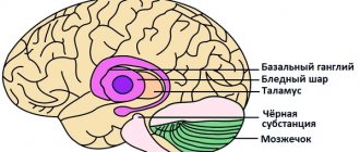
The internal veins of the cerebral hemispheres begin in the basal ganglia and white matter. They are represented by the following trunks.
1. The internal veins of the cerebrum (vv. cerebri internae) collect blood from the white matter of the cerebral hemispheres, the walls of the ventricles, the optic thalamus and the basal ganglia. In the transverse fissure of the brain near the quadrigeminal, all branches of the veins merge into the great vein of the brain (v. cerebri magna).
2. The vein of the choroid plexus (v. choroidea) is formed from the veins of the choroid plexus of the lateral ventricle, penetrating through the for. interventriculare into the central part of the lateral ventricle. It flows into v. cerebri magna.
3. The veins of the transparent septum (vv. septi pellucidi) begin in the substance of the brain, forming the anterior horn of the lateral ventricle. They flow into v. choroidea.
Great vein of the brain
The great vein of the brain (v. cerebri magna) is single, represents a short trunk 0.5-1 cm long. It is formed by the fusion of the above branches of the deep veins of the cerebral hemispheres. In the transverse sulcus of the brain above the superior colliculus, the midbrain flows into the sinus rectus.
Veins of the spongy substance of the bones of the cranial vault
Diploic veins (vv. diploicae) are located in the spongy substance of the bones of the cranial vault. They are oriented towards the large openings of the outer plate of the skull bones, through which the so-called exhaust veins (vv. emissariae) pass. The graduates anastomose with the saphenous veins of the skull, and the diploic veins with the venous sinuses. Due to the absence of valves in the veins of the spongy substance, blood flow through them is possible in two directions. The following diploic veins are distinguished.
1. The frontal diploic vein (v.diploica frontalis) is located in the scales of the frontal bone. Connects the supraorbital vein with the superior sagittal venous sinus.
2. The anterior temporal diploic vein (v.diploica temporalis anterior) is located in the parietal bone and the squama of the temporal bone. Connects the deep temporal veins and sinus sphenoparietalis, anastomoses with the frontal diploic vein.
3. The posterior temporal diploic vein (v.diploica temporalis posterior) connects the parietal outlet with the mastoid outlet and flows into the posterior auricular vein.
4. The occipital diploic vein (v. diploica occipitalis) begins in the squama of the occipital bone and flows into the occipital outlet.
Emissary veins of the skull
Emissary veins (vv. emissariae) are located in various parts of the skull.
1. The parietal emissary vein (v. emissaria parietalis) is a pair, connects the superficial temporal vein with the superior sagittal sinus.
2. The mastoid emissary vein (v. emissaria mastoidea) establishes an anastomosis between the sinus sigmoideus occipital and posterior temporal veins.
3. The condylar emissary vein (v. emissaria condylaris) connects the sinus sigmoideus with the venous plexuses of the spinal column and the deep vein of the neck.
4. The occipital emissary vein (v. emissaria occipitalis) is located on the external occipital eminence, reports vv. occipitales with the transverse sinus or the confluence of the sinuses.
Venous plexuses in the skull
The venous plexuses (plexus venosus) surround the contents of the foramen ovale, carotid and sublingual canals.
1. The venous plexus of the foramen ovale (plexus venosus foraminis ovalis) is located in the foramen ovale and connects the cavernous sinus with the pterygoid venous plexus.
2. The venous plexus of the carotid canal (plexus venosus canalis carotid) surrounds the internal carotid artery in the cranial canal of the same name, collects blood from the mucous membrane of the tympanic cavity and establishes a connection between the cavernous sinus and the pterygoid plexus.
3. The venous plexus of the hypoglossal canal (plexus venosus canalis hypoglossi) connects the occipital sinus with the sinus petrosus inferior and the internal vertebral plexus.
Veins of the orbit
From the contents of the orbit, the frontal region, and partly the upper jaw, small veins originate, which are the sources of the upper and lower ophthalmic veins, which flow into the cavernous sinus and veins of the head.
1. The nasofrontal vein (v. nasofrontalis) originates in the forehead and external nose. In the medial corner of the orbit it connects with v. angularis, representing the beginning of the facial vein.
2. Ethmoid veins (vv. ethmoidales) collect blood from the mucous membrane of the cells of the ethmoid bone, exit through the openings of the same name into the orbit.
3. The lacrimal vein (v. lacrimalis) originates in the lacrimal gland.
4. Veins of the eyelids (vv. palpebrales) and veins of the connective tissue membrane (vv. conjunctivales), vorticose veins (vv. vorticosae), ciliary veins (vv. ciliares), central retinal vein (v. centralis retinae), suprascleral veins (vv. episclerales) are formed in formations of the same name.
All of the listed orbital veins gather on the upper surface of the eyeball and merge into the superior ophthalmic vein (v. ophthalmic superior).
The superior ophthalmic vein has no valves and is characterized by a well-developed muscle layer. Initially, the vein is located in the medial corner between the superior and medial walls of the orbit, then it goes to the outer wall of the orbit, crossing the optic nerve under the superior rectus muscle of the eye. It leaves the orbit through the superior orbital fissure, flowing into the cavernous sinus.
The inferior ophthalmic vein (v. ophthalmica inferior) is formed from small veins of the lacrimal sac, medial, inferior rectus and oblique muscles of the eye. From the medial corner of the orbit, the vein passes to its lower wall and accompanies m. rectus inferior. In the upper part of the orbit, the vein divides into two branches: one of them flows into the sinus cavernosus or the superior ophthalmic vein, the other, passing through the inferior orbital fissure, connects with the deep facial vein. Anastomoses with the venous pterygoid plexus and the infraorbital vein. There are no valves in the system of these veins, so blood can pass both from the veins of the face to the cavernous sinus and back. This creates conditions for inflammation, when it is possible for infection to spread from the upper jaw, orbit and nasal cavity into the venous sinuses of the dura mater.
Veins of the meninges
Veins of the dura mater (vv. meningeae) originate in the dura mater and accompany the arteries. Veins from the shell of the calvarium flow into the sinus sagittalis superior, from the shell of the base of the skull into the venous sinuses of the base of the skull. Among the veins of the hard shell of the base of the skull, the middle vein (v. meningea media) is distinguished, accompanying a. meningea media. The vein flows into the sinus sphenoidalis and anastomoses in the area of the foramen ovale with the plexus of the same name and the pterygoid plexus.
What is the cerebral venous system
The vascular structure makes up 2/3 of the total volume of the cerebral bed.
The veins of the brain are a whole complex of sinuses, an anastomosis arranged in tiers inside the skull. It differs from the arterial one in that it has a much larger reservoir. Includes:
- capillaries;
- sinuses (large collectors);
- diploic, emissary;
- veins of the orbit, eyeball;
- jugular - a paired organ located on the neck, forming an upper hollow structure.
Basically, cerebrospinal fluid accumulates in the sinus sinuses with walls made of hard shell lined with endothelium.
A special feature of the structure is the presence of sinus canals that prevent compression of the cortex, although the tubes do not have a rigid frame. Therefore, the outflow of lymph can become difficult if the pressure of the cerebrospinal fluid begins to rise rapidly.
There are paired and unpaired sinuses.
The pair group includes:
- cavernous with location on both sides of the skull, starting from the Turkish ganglion
- sphenoparietal, extending downward from the main ossicle in the parietal lobe
- superior petrosal with origin at the cavernous sinus, extending along the edges of the pyramid of the temporal bone, flowing into the sigmoid region
- transverse, originating from the lateral confluence of sinuses
- sigmoid as a continuation of the transverse section with an S-shaped bend.
Unpaired:
- upper longitudinal, located at the site of attachment of the falciform process, collecting lymph from the bones of the calvarium and superficial components;
- lying below, collecting cerebrospinal fluid from the inner lobe of the convolutions of the gray matter and the corpus callosum;
- straight, passing along the line of junction of the tentorium of the cerebellum with the falciform process;
- occipital, originating from the occipital bone (internal tubercle), passing along the line of attachment of the falciform process.
Anatomy of the internal veins of the cerebral hemispheres
Internal veins form in the interventricular space, begin to bend around the shallow edge of the thalamus and bend back between the leaves of the ventricle.
The internal cavity is equipped with many inflows.
Basic:
- Thalamostriatal, passing between the thalamus, caudate nucleus in the intermediate sulcus for collecting cerebrospinal fluid, entering the area of the anteroposterior septum. As it moves back, it begins to join with the villous, into which the small capillaries of the vestibule flow.
- Lateral.
- Veins of the corpus callosum.
- Dorsal, posterior.
- The villous vein is one of the powerful ones, flowing into the large cerebral vein with the drainage of cerebrospinal fluid from the plexus of tubules of the lateral ventricles.
The deep areas of the brain stem include veins:
- midbrain;
- oblong;
- lateral, flowing into the large cavity and into the internal structures.
What are diploic veins and their characteristics
Diploic - wide tubes, but with thin walls and no valves. They lie in diploe channels. Located towards the base of the skull.
Diploic and meningeal veins are constantly in close contact with each other.
These veins include smaller branches:
- Frontal with location in the thickness of the frontal bone, flowing into the sagittal region. The functions include the supply of lymph to the supraorbital and superficial part of the sagittal canal.
- Anterior temporal, feeding the sphenoparietal sinus with cerebrospinal fluid.
- Occipital and temporal, flowing into emissary capillaries.
- Posterior temporal, flowing into the auricular vessel and the transverse sinusoid recess with a deviation back.
The functions include the collection of venous lymph from the temporal and parietal bones in the area of the mastoid emissary tube.
Diagnostic methods
In addition to general clinical tests, the patient is also prescribed:
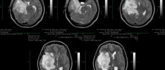
- computed tomography (CT);
- magnetic resonance imaging (MRI);
- angiogram.
One of the most informative methods for vascular malformation is angiography. This is one of the x-ray methods that involves intravenous administration of a contrast agent with further visualization of the result in the image. The popularity of the method also lies in its simplicity and accessibility.
Anatomy of the superior veins
The superior veins are located on the surface of the cortex of the gray matter convolutions.
The composition includes varieties of smaller branches:
- medium – large, paired with passage in the central groove;
- the inferior veins of the cerebrum with their origin from the base, located in the region of the cerebellum below. This is a whole network of branches in the gyral cortex with the collection of cerebrospinal fluid, starting from the dorsolateral component;
- anterior, originating from the medial lobe of the gyri, extending to the base of the cephalic cortex, joining the large semi-tube with the sagittal canal from below;
- shallow, extending from the surface of the cerebellum;
- basal, located at the foot of the medulla, responsible for the functionality of the optic nerve.
Arteriovenous malformations
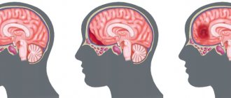
An arteriovenous aneurysm (malformation) is a congenital malformation of cerebral vessels in which arterial blood is directly discharged into the venous bed, bypassing the capillary network. Such aneurysms consist of a dilated adductor arterial vessel, a tangle of vessels forming an arteriovenous shunt, and one or more sharply dilated efferent veins. The walls of the tangle of vessels of arteriovenous malformation are thinned, and it is almost impossible to determine their arterial or venous origin.
Since in arteriovenous malformation there is no capillary network, part of the blood entering the cerebral hemispheres does not take part in tissue metabolism, so-called hemorrhagic stealing of the brain occurs. This causes persistent cerebral ischemia and causes psychopathological disorders, as well as progressive brain atrophy.
The clinical picture of arteriovenous malformation is varied and depends on its location. Often, arteriovenous malformations are clinically manifested by epileptic seizures or spontaneous intracranial hemorrhages.
The prehemorrhagic period may be asymptomatic or manifest itself as epileptic seizures
Functions of veins
The system includes shallow and deep veins of the head.
The functions of the former include:
- outflow of cerebrospinal fluid, metabolic products and gray matter of the convolutions into the straight vein;
- outflow into the sinuses and then into the upper hollow, brachiocephalic region.
Through the deep veins of the brain, according to anatomy, blood is carried away to the external and internal jugular components, starting from the subcortical formations.
Well-developed baro-chemoreceptor structures are responsible for supplying information to the cerebral cerebellum regarding any changes in the composition of the cerebrospinal fluid and pressure.
Thanks to the many anastomoses located inside the medulla, unity of blood circulation and structural integrity are created in the area of superficial and deep capillaries.