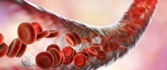
Causes of subarachnoid hemorrhage
In most cases, this condition is caused by non-traumatic factors:
- aneurysm rupture;
- rupture of a vessel in the brain against the background of arterial hypertension, atherosclerosis and other vascular diseases.
Subarachnoid hemorrhage in the brain can also be caused by traumatic factors:
- traumatic brain injury;
- brain contusion;
- fracture of the skull bones.
Main services of Dr. Zavalishin’s clinic:
- consultation with a neurosurgeon
- treatment of spinal hernia
- brain surgery
- spine surgery
Treatment of cerebral hemorrhage in hypertension
Surgical removal of clotted blood in the acute stage of intracerebral hemorrhage is indicated for patients only in rare cases. However, by removing blood clots from a hematoma lying above the tentorium of the cerebellum (supratentorial), it is possible to prevent herniation of the temporal lobe of the brain in comatose patients with still preserved reflex eye movements. Surgical removal of a hematoma from the focus of acute hemorrhage in the cerebellum is usually the method of choice, as it often saves the patient’s life and gives an excellent prognosis in terms of restoration of impaired functions.
If the patient has a clear consciousness and there are no symptoms of focal damage to the brain stem, then if he has a small cerebellar hematoma, the neurosurgeon may refuse immediate surgical intervention to remove it. However, it is necessary to remember the likelihood of rapid deterioration of the clinical condition when the hematoma is localized in the cerebellum. Therefore, in such patients there should always be the possibility of urgent surgery if clinical need arises.
To reduce cerebral edema around intracerebral hemorrhage, mannitol and other osmotic diuretics are prescribed. The activity of steroids in intracerebral hematoma is insignificant. Monitoring intracranial pressure can help evaluate the effectiveness of drug therapy and avoid significant deviations in the direction of arterial hypo- and hypertension. A sharp decrease in blood pressure in order to “stop bleeding” during intracerebral hemorrhage is ineffective, since most often stopping the bleeding that has occurred during intracranial hemorrhages occurs spontaneously even before examining patients.
In conditions such as toxicosis of pregnancy and malignant arterial hypertension, early diagnosis and prudent treatment are especially necessary. This is necessary in order to avoid an excessive or sudden decrease in blood pressure in the patient.
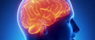
Clinical picture of subarachnoid hemorrhage
This condition develops rapidly against the background of a person’s normal well-being and manifests itself:
- sharp acute headache, worsening with the slightest physical activity;
- nausea, vomiting;
- convulsions;
- psycho-emotional disorders (fear, agitation, drowsiness);
- increased body temperature (up to febrile and subfebrile levels);
- disorders of consciousness (from stupor to fainting, coma);
- symptoms of damage to the oculomotor nerve (gaze paresis, drooping eyelid), hemorrhage in the eyeball.
In half of the cases, this condition leads to death. The main complications and consequences of subarachnoid hemorrhage:
- spasm of cerebral vessels caused by ischemia;
- recurrence of an aneurysm episode (may occur early or several weeks after the first episode);
- hydrocephalus (in the initial stages or in the late period);
- pathologies caused by subarachnoid hemorrhage in the brain (myocardial infarction, pulmonary edema, bleeding from a stomach or duodenal ulcer).
Among the long-term consequences of subarachnoid hemorrhage in the brain: memory disorders, attention, psycho-emotional disorders (depression, insomnia, agitation). Also, a standard complaint of patients who have suffered this condition is headache. Somewhat less frequently, hormonal disorders develop in the hypothalamic and pituitary gland systems.
Diagnosis and treatment of subarachnoid cerebral hemorrhage
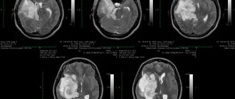
The doctor clarifies the victim’s medical history and conducts an external examination. The diagnosis is established on the basis of CT data, which makes it possible to determine the extent of the process, the presence of edema, and assess the condition of the cerebrospinal fluid system. If the results are negative, a lumbar puncture is performed.
MRI is a sensitive tool for diagnosing pathology after a few days. Vascular angiography is used to determine the source of bleeding in the brain.
Therapy for subarachnoid hemorrhage takes place in a neurological hospital and depends on the severity of the patient’s condition. It is aimed at solving the following problems:
- stabilization of the patient's condition;
- prevention of recurrent episode;
- normalization of homeostasis;
- treatment and prevention of ischemia, vascular spasm;
- therapy for the disease that caused the hemorrhage.
The aneurysm is clipped or endovascular occlusion is performed. Vasospasm and ischemia are treated with medication. In case of local spasm of a vessel, a vasodilator drug is injected directly into it or balloon angioplasty is performed.
In 50% of cases, subarachnoid hemorrhage leads to death. The majority develop significant impairments in the functional activity of the brain.
You can get help with this pathology at the Department of Neurosurgery of the City Clinical Hospital named after. A.K. Eramishantseva. The clinic has modern diagnostic equipment and advanced equipment for performing complex operations.
Hemorrhagic stroke
The first and most important rule is to start treating hemorrhagic stroke with stem cells right away. Rehabilitation therapy after a stroke should be carried out in an “ambulance” mode - this is a guarantee of the patient’s return to normal life and “biological insurance”. That's why you need to have your own stem cell bank just in case!
Experience has shown that stem cells administered intravenously can penetrate the brain, replacing damaged neurons (brain cells) at the site where the hematoma occurred, and thus treat hemorrhagic stroke.
Whether a person has suffered a mini-stroke or a major stroke, stem cell treatment can return him to normal life!
In addition, stem cells synthesize substances that activate regeneration processes, resulting in the appearance of new blood vessels and nerve cells, which leads to the restoration of brain function, and this, in turn, eliminates the neurological symptoms of the disease.
In a word, treatment of stroke with stem cells is one of the most effective methods of rehabilitation. The clinic has helped a huge number of people recover. And this is the main evidence that stem cells provide effective treatment for ischemic stroke, hemorrhagic stroke and their consequences.
But treating a disease is always more difficult than preventing it. If your plans do not include hemorrhagic stroke, there should be only one prevention - lead a healthy lifestyle, and, above all, avoid stress.
And if you already have cardiovascular diseases - hypertension, atherosclerosis - or simply high cholesterol levels in the blood, you simply need to undergo a course of cell therapy on time!
Therapeutic measures for stroke should begin as early as possible, preferably within the “therapeutic window” - in the first 3-6 hours from the moment of development of the disease. Their adequacy to the patient’s condition and intensity largely determine the further course and outcome of the disease. Patients are advised to be hospitalized in a neurological or neurovascular hospital, or in the case of a major stroke - in the intensive care unit. Given the high frequency of a combination of vascular lesions of the brain and heart, most patients require consultation with a cardiologist. If possible, the question of the need and possibility of neurosurgical treatment should be resolved as early as possible. Hospitalization of patients in a state of deep coma with disorders of vital functions, severe organic dementia, and incurable oncological diseases is inappropriate.
Patients with PNMK require bed rest until the end of the acute period and stabilization of the condition. Inpatient treatment is indicated in the case of acute hypertensive encephalopathy, severe hypertensive crisis, and repeated TIAs. Indications for hospitalization are also the lack of effect from outpatient therapy and exacerbation of concomitant diseases, in particular coronary artery disease.
There are two main directions of treatment - differentiated, depending on the nature of the stroke (hemorrhagic or ischemic) and undifferentiated (basic), aimed at maintaining vital functions and correcting homeostasis.
Undifferentiated treatment. Correction of the cardiovascular system is primarily aimed at controlling blood pressure. Its numbers should be 15–25 mm Hg. Art. exceed what is usual for the patient. Rare decreases in blood pressure should be avoided to avoid the development of steal syndrome. Antihypertensive therapy includes the use of beta blockers (anaprilin, atenolol), calcium channel blockers (both short-acting - nifedipine and long-acting - amlodipine), diuretics (furosemide), and, if necessary, ACE inhibitors (captopril, enalapril). If oral administration is impossible or ineffective, drugs are administered intravenously under blood pressure control. If arterial hypotension develops, cardiotonic drugs (mesaton, cordiamine) are prescribed; if there is no effect, intravenous corticosteroids (hydrocortisone, dexamethasone) are prescribed. If indicated, correction of coronary circulatory disorders, acute cardiac arrhythmia and conduction disturbances, and heart failure is carried out.
Monitoring the function of the respiratory system includes ensuring patency of the airways, toileting the oral cavity and nose, removing secretions and vomit from the upper respiratory tract using suction. Intubation and transfer of the patient to artificial ventilation are possible. With the development of pulmonary edema, the administration of cardiac glycosides (corglycone, strophanthin) and diuretics is required. In case of a severe stroke, broad-spectrum antibiotics (synthetic penicillins, cephalosporins) should be started from the first day to prevent pneumonia. In order to prevent congestion in the lungs, it is necessary to begin active and passive (including turning from side to side) breathing exercises as early as possible.
To maintain homeostasis, it is necessary to administer an adequate amount of saline solutions (2000-3000 ml per day in 2-3 doses): Ringer-Locke, isotonic sodium chloride solution, 5% glucose solution, while it is necessary to control diuresis and expiratory fluid losses. Considering that patients with stroke often develop acidosis, the use of 4-5% sodium bicarbonate solution, 3.6% trisamine solution is indicated (under the control of CBS indicators). If necessary, the content of potassium and chlorine ions in the blood is adjusted. In the acute period of stroke, patients should receive a diet rich in vitamins and proteins, low in glucose and animal fats. For swallowing problems, food is administered through a nasogastric tube.
The fight against cerebral edema includes the use of corticosteroids, primarily dexazone (16–24 mg per day, 4 injections) or prednisolone (60–90 mg per day). Contraindications to their use are intractable arterial hypertension, hemorrhagic complications, severe forms of diabetes mellitus. Glycerol perosa is also indicated for intravenous drip administration of osmotic diuretics (15% mannitol solution, reogluman) or saluretics (furosemide).
Control of autonomic functions includes regulation of intestinal activity (a diet rich in fiber and lactic acid products, if necessary, the use of laxatives, cleansing enemas) and urination. If necessary, catheterization of the bladder is carried out, and uroseptics are prescribed to prevent ascending urinary tract infections. From the first day, regular treatment of the skin with antiseptic drugs is required to prevent bedsores; it is advisable to use functional anti-bedsore mattresses. For hyperthermia, use antipyretics
Differentiated treatment. The main directions of differentiated therapy for acute cerebrovascular accidents are restoration of adequate perfusion in the ischemic penumbra zone and limiting the size of the ischemic focus, normalization of rheological and coagulation properties of blood, protection of neurons from the damaging effects of ischemia and stimulation of reparative processes in nervous tissue.
One of the most effective methods of treatment is hemodilution - the administration of drugs that reduce the level of hematocrit (up to 30–35%). For this purpose, rheopolyglucin (reomacrodex) is used, the daily volume and rate of administration of which is determined by both hematocrit indicators and blood pressure levels and the presence of signs of heart failure. For low blood pressure, it is possible to use polyglucin or isotonic saline solutions. At the same time, solutions of aminophylline, pentoxifylline (Trental), and nicergoline (Sermion) are prescribed intravenously. In the absence of heart rhythm disturbances, vinpocetic (Cavinton) is used. As the patient's condition stabilizes, intravenous administration of drugs is replaced by oral administration. The most effective are acetylsalicylic acid (1–2 mg/kg body weight), it is advisable to use forms of the drug. having a minimal negative effect on the gastric mucosa (thromboass): pentoxifylline, cinnarizine, prodectin (anginin).
In the case of increasing thrombosis of the cerebral arteries, with a progressive course of stroke, cardiogenic embolism, the use of anticoagulants is indicated. Heparin is administered intravenously in a daily dose of 10–24 thousand units or subcutaneously at 2.5 thousand units 4-6 times a day. When using heparin, mandatory monitoring of the coagulogram and bleeding time is necessary. Contraindications to its use, as well as thrombolytics, are the presence of sources of bleeding of various locations (peptic ulcer, hemorrhoids), persistent intractable hypertension (systolic pressure above 180 mm Hg), severe disorders of consciousness. With the development of DIC syndrome, due to a decrease in the level of antithrombin III, administration of native or fresh frozen blood plasma is indicated. After stopping the administration of heparin, indirect anticoagulants (phenyline, syncumar) are prescribed with monitoring of blood coagulation parameters.
The established nature of the thrombotic stroke allows the use of thrombolytics (urokinase, streptase, streptokinase) in the first hours of the disease. Due to the fact that with the intravenous administration of these drugs there is a high risk of hemorrhagic complications, the most effective method is targeted thrombolysis, in which the drug is injected directly into the thrombotic zone under X-ray control. Recombinant tissue plasminogen activator has a powerful fibrinolytic effect, the administration of which is also advisable only in the first hours of the disease.
In the complex treatment of patients with acute cerebrovascular accidents, the use of drugs that have antiplatelet and vasoactive effects is indicated: calcium channel blockers (nimotop, flunarizine), vasobral, tanakan. The use of angioprotectors is justified: prodectin (anginin). The use of these drugs is advisable after the acute phase of the disease has passed, as well as in patients with TIA.
In order to prevent hemorrhage in the ischemic zone in case of extensive infarctions, dicinone (sodium etamsylate) is prescribed intravenously or intramuscularly.
The use of drugs that have a neurotrophic and neuroprotective effect on brain tissue is extremely important. For this purpose, use nootropil (up to 10–12 g per day), glycine (1 g per day sublingually), aplegin (5.0 ml in 200.0 ml of isotonic sodium chloride solution intravenously 1–2 times a day), Semax (6–9 mg 2 times a day intranasally), Cerebrolysin (10.0–20.0 ml per day intravenously). The use of these drugs contributes to a more complete and rapid restoration of impaired functions. In some cases, in particular with global cerebral ischemia, it is possible to use barbiturates (sodium thiopental) to reduce the energy needs of the brain under ischemic conditions. The widespread use of this method is limited by the pronounced cardiodepressive and hypotensive effects of the drug and depression of the respiratory center. A certain effect is achieved by drugs that inhibit lipid peroxidation processes: unithiol, vitamin E, aevit.
Differentiated conservative treatment for hemorrhagic stroke. The main direction is to reduce the permeability of the vascular wall and prevent the lysis of a formed thrombus. To inhibit fibrinolysis and activate thromboplastin production, epsilon-aminocaproic acid is used. Over 3–5 days, 50.0–100.0 ml of a 5% solution of the drug is administered intravenously 1 or 2 times a day. Inhibitors of proteolytic enzymes are used: trasylol (contrical, gordox) in an initial dose of 400–500 thousand units per day, then 100 thousand units 3–4 times a day intravenously. An effective hemostatic drug with a low risk of thrombosis is dicinone (sodium etamsylate). To prevent vasospasm, which complicates the course of subarachnoid hemorrhage, patients are prescribed Nimotop.
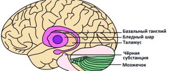
Surgical treatment for hemorrhagic stroke. Removal of medial hematomas, typical for hemorrhagic stroke, localized in the subcortical ganglia, internal capsule, and thalamus, as a rule, does not lead to an improvement in the condition of patients and does not significantly change the prognosis. Only sometimes indications for surgery may arise in relatively young patients with an increase in cerebral and focal symptoms after a period of relative stabilization of the condition. In contrast, removal of hematomas localized in the white matter of the cerebral hemispheres lateral to the internal capsule, as a rule, leads to a significant improvement in the patient’s condition and regression of dislocation symptoms, and therefore surgical intervention for these hematomas should be considered absolutely indicated.
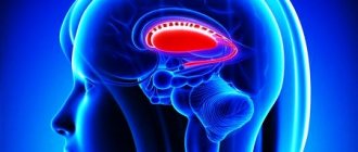
The main method of surgical treatment to remove intracerebral hematomas is craniotomy. If the hematoma is located laterally and extends to the insula, the least traumatic approach to the hematoma is through the lateral (Sylvian) fissure, with trepanation performed in the frontotemporal region. Hematomas localized in the area of the visual thalamus can be removed through an incision in the corpus callosum. For atypical hemorrhages, the surgical approach is determined by the location of the hematoma in the brain.
To remove deep-seated hematomas, stereotactic aspiration can be used. Based on the results of a CT study, the coordinates of the hematoma are determined. Using a stereotactic apparatus fixed on the patient's head, a special cannula connected to an aspirator is inserted through the burr hole. In the lumen of the cannula there is a so-called Archimedes screw, the rotation of which leads to the destruction and removal of the hematoma. The advantage of this method is its minimal trauma.
Hemorrhage into the cerebellum can cause life-threatening compression of the brainstem, making surgery necessary. Resection trepanation of the posterior cranial fossa is performed above the location of the hematoma. The dura mater is subsequently opened and the cerebellar tissue is dissected, and the accumulated blood is removed by aspiration and washing the wound.