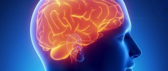
According to statistics, such a problem as cerebral aneurysm occurs in 5% of the population. Often such an anomaly occurs covertly and is discovered only through the efforts of a pathologist. In half of the cases, aneurysms, which are small in size, do not rupture. Moreover, such a condition is extremely dangerous, since if a gap in education nevertheless occurs, the person runs the risk of becoming disabled, or even the matter may even end in death.
What is a ruptured brain aneurysm and who can it threaten?
The essence of the problem
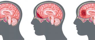
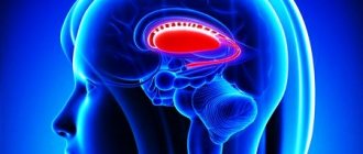
An aneurysm is an enlargement of a section of the lumen of an artery. A rupture is called a serious complication when, due to damage to the formation, blood begins to flow into the brain, leading to irreversible changes in it. Experts say that in a third of cases, a breakup ends in the death of the person.
From a physiological point of view, the process is as follows: in the area where the aneurysm has formed, the vessels become inelastic, weaken and do not resist blood flow. It is because of this that a protrusion is formed, i.e. a small sac literally filled with blood. If the education is small, a person may not know about it. But the problem is that it can increase in size over time, and this leads to a violation of the integrity of the blood vessels. The weak point of an aneurysm is called the apex - this is where it ruptures most often.
Bleeding in the damaged area lasts literally seconds, but this is enough for serious disorders to begin in the brain. At first, the body tries to cope on its own, reflexively spasming the artery so that a blood clot forms, which will stop the release of blood and save life. But it also happens that this process is delayed. And in such a situation the prognosis is unfavorable.
What kind of unique artificial vessels were created in Russia? More details
Causes of the disease
An aneurysm is a dangerous condition that primarily requires surgical treatment. Otherwise, complications will not be avoided. Experts believe that the disease is a consequence of developmental abnormalities that disrupt the structure of the vascular wall. Often, a cerebral aneurysm is combined with connective tissue dysplasia, polycystic kidney disease, and vascular disorders. The acquired form of the disease can be a consequence of traumatic brain injury. Also, a cerebral aneurysm occurs against the background of atherosclerosis and hypertension.
The vascular wall defect itself occurs gradually. Against the background of degenerative processes, tissue damage or underdevelopment, the area loses its former elasticity. Under the pressure of blood flow, the vascular wall begins to bulge. This is how a brain aneurysm forms. Most often, the formation occurs in the place where the arteries branch and there is the strongest pressure on the vessel wall.
Symptoms of a rupture
As doctors note, a ruptured aneurysm has a clear clinical picture. All signs appear suddenly. A sharp headache occurs, which many characterize as unbearable. The situation may also be accompanied by vomiting, and in particularly severe cases, loss of consciousness and coma follow.
A breakup can occur against the backdrop of some emotional events and upheavals, or it can happen spontaneously. There is a risk of re-bleeding. So, after the first rupture, a blockage of the wall may occur, but after 2-3 weeks the situation can repeat. Doctors note that after repeated hemorrhage, the prognosis becomes unfavorable. After the person returns to consciousness, he or she experiences weakness, dizziness, and poor orientation in space. Rapid breathing and increased heart rate may also occur. In addition, stiff neck muscles, problems with oculomotor functions, loss of speech, and paralysis may occur.
The general condition of a person in such a situation is regarded as serious - he needs emergency medical care.
Symptoms
If a vessel in the head bursts, signs of intracerebral bleeding appear - sudden pain in the head area, intensifying after exercise, nausea, which is often accompanied by vomiting. In elderly patients, pain in the head is sometimes absent or moderate.
In cases where the hemorrhage focus is large, neurological symptoms may be supplemented by confusion. A typical symptom is focal seizures (affecting specific areas of the body) or generalized seizures (distributed throughout the body).
Sometimes, before a vessel in the head bursts, precursor symptoms appear, which include a feeling of heat, visual dysfunction, and increasing pain in the head. More often, a stroke develops rapidly without previous signs. Typically, rupture of the vascular wall occurs during the daytime and is caused by physical or emotional stress.
If a vessel bursts in the head, the condition is accompanied by specific symptoms, which manifest themselves acutely with a tendency to increase. The clinical picture develops quickly. Confusion, stupor, and fainting are often observed. In severe cases, stunning and coma occur. Focal neurological symptoms depend on the location of the hemorrhage:
- Hemispheres. The pathology is accompanied by hemiparesis (paresis in one half of the body).
- Posterior fossa of the skull. Symptoms of damage to the structures of the brainstem and cerebellum are observed - paralysis of the eye muscles, narrowing of the pupils to a pinpoint state, stertorous breathing (noisy breathing, accompanied by inspiratory and expiratory wheezing, characteristic of obstruction, blockage of the airways).
- Ventricular system. The most life-threatening form, accompanied by meningeal (stiff neck, Kernig and Brudzinski symptoms, changes in reflexes) and brain stem (tachypnea - rapid, shallow breathing, tachycardia, stupor or stupor, increased blood pressure, increased tone of skeletal muscles) symptoms, hyperthermia (accumulation of excess heat in the body), hormetonic-type convulsions, rapid depression of consciousness.
When blood enters the ventricular system, hydrocephalus of the occlusive type may develop, occurring in an acute form. The focus of hemorrhage localized in the frontal lobe leads to a pronounced impairment of cognitive abilities (memory, mental activity, speech).
A supratentorial (located in the cerebral hemispheres) hematoma, accompanied by cerebral edema, can lead to transtentorial (the temporal lobe is wedged through the foramen of the cerebellar tentorium and under it) herniation, which in turn causes compression of the trunk and the appearance of secondary foci of hemorrhage in the area of the midbrain and pons .
Hematomas localized in the cerebellum, when enlarged, can block the 4th ventricle, which leads to the development of hydrocephalus, which occurs in an acute form. Foci of hemorrhage located in the cerebellar area with a diameter exceeding 3 cm can provoke dislocation of the median structures. The described conditions often lead to severe depression of consciousness, the development of coma and death of the patient.
Extensive intracerebral bleeding usually (70% of cases) ends in death. In surviving patients, neurological symptoms gradually regress in parallel with the process of resorption of blood poured into the cranial cavity. Hemorrhages cause less damage to brain structures than infarctions, so neurological deficits are often quickly corrected.
If a small vessel ruptures with the subsequent formation of a small focus of hemorrhage, focal neurological symptoms usually develop without depression of consciousness. In this case, the headache is moderate. Nausea may not be a clinical sign.
Risk factors
The question of why some aneurysms are asymptomatic and do not lead to serious consequences, while others rupture, worries many doctors and scientists. Based on a number of studies, risk factors have been identified that increase the likelihood of rupture. Among them:
- age point - risks increase for people over 50 years of age;
- location of the formation - if the aneurysm is located in the middle or anterior cerebral artery;
- size of the formation - aneurysms larger than 5 mm become dangerous.
If a person has such risks, it is important to begin treatment as quickly as possible in order to prevent a potential disaster.
Dried apricots will cleanse blood vessels, and raisins will calm the nerves. 6 dried fruits instead of tablets Read more
Diagnostics
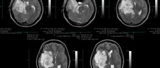
The first choice methods for making a differential diagnosis are MRI and CT studies. Differentiation is made in relation to ischemic stroke and subarachnoid (intrathecal) hemorrhage. When making a diagnosis, it should be taken into account that acute neurological deficit can develop as a result of an epileptic seizure or hypoglycemia (a critical decrease in blood glucose levels). A blood test shows your glucose level. Computed tomography allows you to establish:
- Exact localization of the source of hemorrhage.
- Volume and prevalence of the pathological process.
- The presence and severity of cerebral edema.
- The presence and severity of dislocation of brain structures.
If MRI and CT examinations are not possible, a cerebrospinal fluid sample is taken as an alternative. Blood is detected in the cerebrospinal fluid if the focus of hemorrhage is in the area of the subarachnoid space or the posterior fossa of the skull. The attending physician will tell you what to do if a blood vessel in your head bursts, taking into account the results of the examination.
Why does the gap occur?
There are several reasons why a cerebral aneurysm begins to rupture. One of them is genetics. Here we are talking about the fact that weakness of the artery wall is inherited. Also on the list of causes that provoke the problem are kidney pathologies, injuries, cancer, and atherosclerosis.
The following factors can damage the integrity of the vessel wall::
- increased physical activity;
- a history of arterial hypertension;
- emotional tension;
- drinking alcohol;
- development of infections accompanied by high fever.
Definition
If a vessel in the head bursts, the pathology is called intracerebral hemorrhage, which is reflected in the IBC-10 as section 161. Hemorrhagic stroke ranks second in prevalence after stroke developing of the ischemic type. Pathology is more often detected in patients aged 45-60 years. Statistics show that most patients are representatives of the Negroid and Mongoloid races.
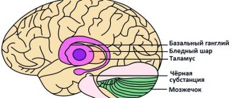
The most common localization of hemorrhage is the cerebral hemispheres, basal ganglia, cerebellum, pons, and trunk. Hemorrhages that occur against the background of arterial hypertension are usually acute and extensive. Intracranial hematomas are space-occupying formations, which often tend to expand due to the growth of the pathological focus.
If a vessel in the head bursts, the consequences arise as a result of the mechanical effect of the space-occupying formation on the adjacent brain structures. An intracerebral hematoma compresses surrounding tissues, causing neurological symptoms.
How are they treated?
First you need to provide first aid to the person. Naturally, urgent hospitalization will be required, but it is impossible to leave the situation unattended until the ambulance arrives. The patient should be laid so that the head is elevated - this will ensure the necessary outflow of blood and prevent the development of brain swelling. You should also worry about the flow of oxygen; accordingly, you need to relieve the neck - unbutton your shirt, loosen your tie, etc. If you lose consciousness, you should protect the person by clearing the airways - remove dentures, turn your head to the side. It is also worth applying cold objects to the head to reduce the risks of swelling and hemorrhage.
Aneurysm treatment is predominantly treated surgically. The method is selected depending on a number of parameters - the location of the formation, the person’s condition, the severity of the violations and the time that has passed since the moment of rupture.
Cleaning of blood vessels. How to get rid of cholesterol plaques Read more
The following treatment options are used for therapy::
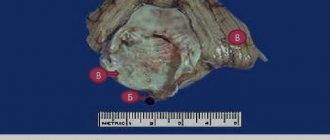
- Clipping is a microsurgical method that involves placing a clip on the base or body of the aneurysm: this will shut it off from the bloodstream.
- Endovascular method - here an intervention is performed by inserting a catheter through the femoral artery.
- Combined method - first a blood clot is injected into the aneurysm, and then it is clipped.
There is such a nuance that it is important to perform the operation in the first 72 hours after the start of bleeding, since the risk of recurrent hemorrhage is high. If you are late, spasm begins to increase, ischemia develops, and the operation will not make sense.
Treatment methods
Treatment of conditions that develop due to a burst blood vessel in the brain is often symptomatic. Regulated risk factors are monitored. In cases where intracranial bleeding threatens serious complications, neurosurgical intervention is performed. Typically, surgery is prescribed if the diameter of the volumetric focus of hemorrhage exceeds 3 cm.
If the patient has been treated with anticoagulants, the drugs are discontinued and medications are prescribed that neutralize the effect. The following events are taking place in parallel:
- Monitoring blood pressure indicators.
- Anticonvulsant therapy (if seizures are present).
- IVL (artificial pulmonary ventilation).
- Correction of hypoglycemia and hyperglycemia.
Early neurosurgical removal of large lobar (blood does not extend beyond the white matter) hematomas improves survival. However, surgical intervention is associated with the risk of relapse - repeated hemorrhage. The operation is not performed if the focus of hemorrhage is located in the deep parts of the brain.
Consequences of a breakup
Of the most common consequences if a person remains alive, the following points:
- paralysis and paresis;
- problems with speech if the hemorrhage is in the left hemisphere;
- problems urinating or constipation if damage occurs in the corresponding brain structures;
- mental problems - aggression, rage, apathy, depression may appear;
- cognitive disorders - memory problems, inability to remember new information;
- epilepsy.
So you should worry about your health in advance in order to prevent the appearance and, especially, rupture of a defect.
Rehabilitation after surgical treatment of an aneurysm
The rehabilitation period takes place under the supervision of specialists. Professionals do everything necessary to prevent the occurrence of postoperative complications. The patient remains in the intensive care unit for several days under the constant supervision of specialists. Afterwards, the patient is transferred to a general ward, where strict bed rest must be observed for some time. The return to a normal lifestyle occurs gradually.
If the patient's well-being worsens after surgery, transcranial Doppler ultrasound may be prescribed. Rehabilitation is aimed primarily at preventing vasospasm and increased blood pressure. Specialists also monitor the general condition of the cardiovascular system and, if necessary, prescribe osmodiuretics to eliminate swelling of the brain tissue. Additionally, anti-inflammatory therapy is carried out.
Rehabilitation after surgical treatment of an aneurysm necessarily includes a program to restore impaired body functions and further socialize the patient. The recovery period lasts at least 6 months. With the help of high-quality rehabilitation, it is possible, among other things, to eliminate the adverse consequences of surgical treatment.
Rehabilitation measures include physiotherapy, massage, and the implementation of an individually selected rehabilitation program. After endoscopic clipping, the patient can return to normal life within a few weeks.
Prevention of vascular fragility
To prevent vascular rupture, you need to pay attention to doing eye exercises. It is also very effective to drop warm black tea into your eyes from time to time. An excellent way to prevent rupture of blood vessels in the eyes are special vitamin complexes and eye drops. For example, vitamin complexes, including blueberry extract, as well as beta-carotene, have an excellent strengthening effect. Such prevention is especially necessary in winter, when there is a shortage of fresh berries, vegetables and fruits.
You also definitely need vitamins of groups A, B and C, so when choosing a complex of vitamins, be sure to pay attention to whether these elements are included in its composition. Strengthening eye drops, such as Katachrom and Quinax, are also very useful. Due to their composition, they are an effective prevention of many diseases of the organ of vision. The same cannot be said about the well-known Visine, which helps to avoid rupture of blood vessels only if it is applied in a timely manner, for example, after a tiring day at work.
For contact lens users, eye drops that imitate natural tears are ideal. They are especially useful when the eyes need natural hydration.
In the medical department, everyone can undergo examination using the most modern diagnostic equipment, and based on the results, receive advice from a highly qualified specialist. The clinic is open seven days a week and operates daily from 9 a.m. to 9 p.m. Our specialists will help identify the cause of vision loss and provide competent treatment for identified pathologies.
You can make an appointment at the Moscow Eye Clinic by calling 8 8 (499) 322-36-36 (daily from 9:00 to 21:00) or using the online registration form.
Dagaev Adam Huseinovich
How can I help you
You can determine that a person has a hemorrhagic stroke and a burst blood vessel in the brain by inappropriate behavior and a sharp change in his well-being.
Necessary:
- Ask him to smile. With a stroke, the smile will be one-sided, since one half of the body does not work.
- Ask to say your first and last name. The patient's speech will be slurred, slow, and hesitant.
- Suggest raising both hands up. A person either will not be able to do this, or the arm on the damaged part of the body will be lower than the other.
- Ask to stick out your tongue as far as possible. During a stroke, the tongue becomes bent or retracts to one side.
The further fate of the patient is in the hands of the people around him at the time of the attack. The first thing you need to do is call an ambulance. And then, while waiting for the doctors:
- Lay the victim so that the head and shoulders rise above the body. It is important not to allow him to stand up or move around.
- Loosen the straps, unbutton the cuffs and collar of the shirt.
- If you have dentures, you need to get them.
- Turn the patient's head to the side.
- If vomiting, ensure that the mouth is cleaned with a handkerchief or piece of cloth.
- Apply a cold compress to your forehead.
- Rub your palms and feet to improve blood circulation.
You cannot give any medications to the patient yourself. Any erroneous and illiterate action can be fatal.
What types of strokes are there?
The brain consumes more energy than other organs and systems. Blood flows to it through four large arteries - two carotid and two vertebral. Even if blood flow is disrupted in one of the vessels, the other three take over its function. There have been cases in medical practice that three out of four arteries were blocked by cholesterol plaques and only one provided the brain with nutrients and oxygen.
Blood circulation in the brain should not be allowed to be disrupted, otherwise hemorrhage into the nervous tissue may occur. 80% of all cases are ischemic strokes. In this case, the blood does not pour into the brain, forming a large hematoma, but the artery becomes blocked and a certain area of the brain becomes bloodless. This leads to the death of nerve tissue.
A stroke or acute cerebrovascular accident does not begin suddenly. It is usually preceded by unpleasant and painful symptoms that need to be paid attention to and taken action.
Ischemic strokes are divided into subtypes:
- Lacunar (25% of all cases). Small vessels are affected; there may be no dangerous consequences.
- Atherosclerotic (atherothrombotic) - blood supply is disrupted in large vessels and a large area of the brain is affected. The number of cases is also 25%.
- Cardioembolic stroke, in which a blood clot forms in the heart or aorta. It ends up in the brain and clogs the blood flow.
- Strokes of rare etiology, in which the destruction of arterial walls occurs or are caused by some congenital pathologies.
Hemorrhagic strokes are considered the most dangerous. However, it is difficult to predict the severity and extent of the consequences. The cause of hemorrhage is high blood pressure.
There are also subtypes here:
- Intracerebral, if a vessel in the head bursts and blood spills into the brain matter.
- Intraventricular - rupture occurs in the ventricles of the brain.
- Subarachnoid hemorrhage - a hematoma forms between the skull and the brain.
The doctor urgently needs to find out where the capillary in the head has burst (or a large artery is blocked) in order to begin treatment correctly. Most people with cerebrovascular accidents arrive in the intensive care unit in an unconscious state.
Stroke is the most common cause of death. Only 8% of patients return to normal life. Most die or become disabled for the rest of their lives.