Author
Eremin Dmitry Sergeevich
Radiologist (CT)
Doctor
Radiologist
until October 31
Nightly 20% discount on computed tomography and MRI More details All promotions
MRI
(magnetic resonance imaging) is a modern type of diagnostics that uses a magnetic field and radio waves (rather than ionized radiation, which is used in X-ray machines and computed tomography). Therefore, MRI is safe and can be performed as often as necessary.
Magnetic resonance imaging is a highly informative diagnostic method, most revealing in the study of soft tissues, which allows obtaining images in the form of tissue sections of a particular organ.
What you need to know about the principle of MRI and the design of a magnetic resonance imaging scanner
The patient is placed on a movable table that moves through a tunnel-shaped magnet. The magnet creates a powerful magnetic field; Radiofrequency pulses are sent to the examined area of the patient in a magnetic field. As a result of this radio wave exposure, hydrogen atoms resonate in the tissues of the body.
Our body is mostly composed of water and fat, and these substances, in turn, are characterized by a high hydrogen content. The amount of hydrogen varies in different tissues; including in tissues affected by pathological processes, it differs from what is characteristic of healthy tissue of a given organ.
Information about atomic resonance is read by special sensors (coils) and processed using a computer program that reconstructs an image in the form of a slice of the organ under study.
Indications for magnetic resonance imaging
- Pathologies and diseases of cerebral vessels.
- Head injuries and bruises, which are accompanied by internal bleeding.
- Speech impairment and hearing loss.
- Tumors of the brain, cerebellopontine angle.
- Paroxysmal states.
- Infectious diseases of the central nervous system (abscess, meningitis, HIV infection).
- Pituitary adenoma.
- Abnormal development of the blood vessels of the head (thrombosis, aneurysms).
- Epilepsy.
- Systematic headaches of unknown origin.
- Sinusitis.
- Multiple sclerosis.
- Neurodegenerative diseases.
- Pathology of the base of the skull.
Types of MRI studies
The most popular types of MRI studies are:
- MRI of the spine
. Allows you to assess the condition of the spinal cord, cartilage, ligaments and back muscles. Circulatory disorders, consequences of injuries, developmental anomalies, changes in intervertebral discs, etc. are identified. An MRI examination of a specific section or the entire spine may be performed. - MRI of joints
. A specific joint is examined: knee, shoulder, hip. MRI allows you to study in detail the structure of the articular joint, visualize intra-articular (menisci, joint fluid) and periarticular formations (ligaments, muscles). Developmental anomalies, inflammatory and degenerative changes in the joint, and pathologies of periarticular tissues are diagnosed. - MRI of the brain
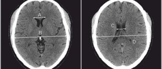
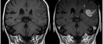
. MRI examination of the brain is highly sensitive and allows you to visualize both hemispheres of the brain, its stem part, the ventricular system and other structures. Using MRI of the brain, vascular abnormalities, vasodilation, hemorrhages, tumors, foci of inflammation and degeneration, fluid accumulations, etc. can be identified. - MRI of the pituitary gland
. MRI shows the condition of the pituitary gland itself and the sella turcica (anatomical area in which the pituitary gland is located). MRI can detect adenomas and other pituitary lesions. - MRI angiography of the brain
. MRI provides an opportunity to assess the condition of cerebral vessels without the introduction of a contrast agent. This is possible because the method allows one to distinguish a substance in motion (blood) from stationary structures (vascular walls). - MRI cholangiography
– study of the patency of the bile ducts. The intrahepatic ducts, cystic duct and common bile duct are examined, as well as (partial) liver and pancreatic tissue. Allows you to identify stones, polyps, tumors and narrowing of the bile ducts. - MRI of the prostate
. MRI allows you to evaluate in detail the structure of the prostate gland, identify prostate adenoma (benign hyperplasia), foci of inflammation and prostate tumors. - MRI of the pelvic organs
(uterus and ovaries). MRI can detect changes in tissue structure, endometriosis, adhesions, fibroids, polyps, tumors, and helps determine the type of ovarian formation.
Indications for MRI and CT procedures
Computed tomography is used for:
- skull injuries (brain scan);
- ruptures of internal organs;
- insufficient information content of x-ray images;
- suspected tumor (any location);
- monitoring the condition of tissues after operations.
MRI is prescribed for research:
- vessels of any organ (mainly brain);
- joints;
- brain and spinal cord;
- spine;
- abdominal and pelvic organs.
Harmlessness of MRI
To date, there have been no cases where the magnetic field or radio waves used in MRI scans have caused harm to a patient. MRI is not done in the first trimester of pregnancy, but this is just a precaution; the facts when an MRI would cause any harm to the fetus are also unknown to medicine.
contraindications for MRI
.
First of all, they are determined by the presence of electronic devices implanted into the body (hearing aids, artificial heart pacemakers), as well as any metal structures and fragments (joint endoprostheses, metal plates, knitting needles, consequences of metal osteosynthesis and gunshot wounds, etc.). Dental implants, vascular stents and umbrella filters produced in the last 5-7 years are usually made from MRI-ready materials. Therefore, for such patients, an MRI examination can be performed after presenting a certificate or confirmation from the medical institution that performed the installation.
MRI is not done if you have a pacemaker (an absolute contraindication).
An MRI cannot be performed if the patient is afraid of confined spaces (suffers from claustrophobia).
Sometimes during the study, a burning sensation or irritation of the skin occurs due to the applied cream, ointment, or certain types of tattoos, in these cases the study must be stopped and, if the cause cannot be eliminated, the study may be stopped.
Breastfeeding, menstruation and the presence of an intrauterine device are not an obstacle to undergoing an MRI.
Contraindications for procedures
What is more harmful and what is safer, CT or MRI, will show a list of contraindications to the procedures. CT cannot be done if:
- pregnancy;
- large body weight (the device is designed for 150 kg);
- allergies to contrast agent;
- renal failure;
- serious condition of the patient;
- pathologies of the thyroid gland;
- multiple myeloma.
MRI cannot be done if:
- wearing a pacemaker (the magnetic field will disable it);
- installed metal implant in any organ, dentures and braces;
- Ilizarov apparatus;
- pregnancy (first trimester);
- presence of tattoos containing ink with metallic impurities;
- the patient's serious condition;
- mental illness of the patient (risk of claustrophobia, violence, epilepsy attack).
Preparing for an MRI
No special preparation is required for MRI. The only exception is MRI of the liver and gall bladder, which are performed strictly on an empty stomach, preferably in the morning.
The patient should be prepared for the fact that he will have to spend quite a significant amount of time (from 15 minutes to almost an hour, depending on the type of study) inside the tomograph tunnel. At the same time, the installation produces noticeable noise (this is an inevitable consequence of the technology used). Clothing in which you can undergo an MRI must not contain metal parts (zippers, fasteners, buttons). All accessories (watches, hairpins, hairpins, jewelry, etc.) will need to be removed. It is also necessary to leave electronic magnetic cards and any other magnetic media (flash drives, memory cards, etc.) in a special booth, otherwise all information on them will be erased in the magnetic field.
When to Apply Contrast
Magnetic resonance diagnostics can provide a very high degree of clarity of the resulting images. In most cases, the use of contrast is not required.
But when it comes to diagnosing tumors and small anatomical structures, a contrast agent can still be used.
The staining agents are made from the rare earth metal gadolinium and are injected intravenously into the patient during an MRI.
MRI contrast agents are much better tolerated than CT contrast agents. This makes the use of the dye safe even for patients with kidney pathology and does not require a preliminary test for creatinine, which is necessary for CT diagnostics with contrast.
MRI with contrast is used in the following cases:
- suspicion of a neoplasm;
- the need for differential diagnosis of a malignant tumor;
- study of the pituitary gland;
- the need to diagnose demyelinating diseases.
The use of contrast allows one to obtain a comprehensive picture of the disease, its course and the effectiveness of the therapy used.
Advantages of MRI at Family Doctor
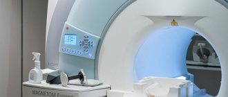
The “Family Doctor” uses a Brivo MR355 Inspire tomograph, manufactured by GE Healthcare (the medical division of the American company General Electric), to conduct magnetic resonance imaging. Brivo MR355 Inspire is a world-class MR scanner with a magnetic field of 1.5 Tesla (the higher this figure, the more detailed the resulting image can be). This tomograph belongs to the class of equipment with high magnetic field power. Thanks to modern technologies, Brivo MR355 Inspire provides high-quality visualization to help make the diagnosis as accurate as possible.
MRI is performed at the Diagnostic Department of the Hospital Center (Baumanskaya metro station). Research is carried out on the orders of Family Doctor doctors, as well as third-party medical organizations.
Sign up for diagnostics Do not self-medicate. Contact our specialists who will correctly diagnose and prescribe treatment.
MRI: “magnetic eye” monitors health
"Full body scan!" - the doctor orders from the television screen, faced with a complex diagnostic case. What would have seemed fantastic when our parents were young is now a completely ordinary procedure. Why feel and tap a person's body in an attempt to find the source of the problem, if you can use one of the magical technologies that allows you to easily look inside the body?
Previously, only the rays of an X-ray machine and ultrasound waves were capable of this, but today nuclear magnetic resonance rules the roost, thanks to which the abbreviation MRI has firmly entered the lexicon of modern doctors. Moreover, each of us can satisfy our curiosity even without a referral from a doctor: fortunately, in large cities, MRI centers are now almost more common than state clinics. If there was money... well, there would be reasons to worry.
How does an MRI scanner work?
Even at the previous stage of development of medical imaging - with the invention of computed tomography (CT) - scientists learned to obtain an image similar to anatomical illustrations from an atlas, allowing for a detailed examination of each section of the body in the area of medical interest. But despite all the advantages of CT, this method is based on the properties of x-ray radiation - and therefore is accompanied by potentially harmful effects of radiation on the body.
Contraindications
Given the properties of a magnet to attract and move objects containing metal, persons with metal implants are not allowed to undergo an MRI procedure. People with electronic devices embedded in their bodies should also not enter the MRI room. Therefore, patients who have:
- cardiac defibrillators and pacemakers not compatible with MRI;
- almost all types of ferromagnetic heart valves;
- ear implants;
- clamps, clips, pins, prostheses, spirals, intravascular catheters made of ferromagnetic materials;
- electronic devices and medication ports that are not compatible with MRI.
It should be remembered that not all implants are a contraindication for MRI examination.
In each specific case, you should consult a diagnostician. Based on the device data sheet, he will be able to tell whether the materials from which they are made are compatible with tomographic scanning or not. It is also necessary to warn the doctor about the presence of dentures, braces, tattoos, which are safe, but can reduce the quality of the images obtained by the tomograph. The radiologist will either change the machine settings or suggest a different type of examination. Pregnant women and patients with serious renal impairment should not undergo MRI diagnostics if the MRI is performed with contrast. Author: Belik Ekaterina Mikhailovna
Radiologist with 19 years of experience
How is an MRI of the brain performed?
- An MRI machine is a cylinder with holes at both ends. The patient sits on a movable table that is placed inside the device during the MRI procedure.
- The length of the tunnel depends on the specific type of apparatus. Some devices only partially surround the table. Tomographs can also be open on the sides. They are used for patients with severe claustrophobia. In closed-type devices, the table slides in completely.
- During the examination, the patient is secured with belts. The less it moves, the more accurate the diagnostic results.
- During magnetic resonance imaging of the brain, devices with wires that receive and send radio waves are placed around the head.
- Patients are often given earplugs. This helps protect your hearing from the loud noise that comes from the MRI machine when it is in operation.
- The table is placed inside the tomograph, and the specialist moves to an adjacent office, which houses a computer system that processes the data. The device takes a series of pictures. Each one takes a few minutes to shoot. As a rule, an MRI of the brain without contrast takes about 20–30 minutes, with a contrast agent – approximately 45–50 minutes.
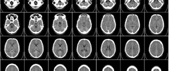
- An MRI machine takes layer-by-layer images of tissue. During a head scan, data is collected from approximately 20 levels, each 4–5 mm thick. The higher the magnetic field strength of the tomograph and the thinner the sections, the more accurate the research result.
What is MRI?
MRI is a non-invasive, that is, without instrumental internal intervention, medical examination. To obtain detailed images of internal organs, bones, soft tissues - essentially all the internal structures of the body, a magnetic resonance imaging scanner uses a powerful magnetic field, pulsed radio frequency and a computer. Detailed images obtained using the MR method allow doctors to examine in detail the condition of the body part being examined and determine the presence, extent and severity of the pathological process. The result of the examination is a series of tomograms that show internal bone and soft tissue structures with high anatomical accuracy in 3D format. These images can be viewed on a monitor, transmitted electronically, printed or copied to a CD, or uploaded to a digital server in the cloud. Tomography data recorded on electronic media is understandable and readable for a doctor in any part of the world and can be used:
- to make a final diagnosis;
- prescribing a course of therapy;
- assessing treatment progress;
- determining the surgical intervention strategy.
Improvements in recent years have brought the MRI diagnostic method to a new level, since modern tomographs have high power, which makes it possible to diagnose most diseases, including cancer of internal organs, degenerative changes in the central nervous system, inflammatory diseases, autoimmune pathologies, at a very early stage of development.