Hippel-Lindau disease (Cerebroretinal angiomatosis)
The onset of neurological manifestations usually occurs in the 3rd-4th decades of life. In childhood, Hippel-Lindau disease is characterized by the appearance of neurological symptoms against the background of pre-existing visual disorders. In some cases, the disease in children manifests itself with subarachnoid hemorrhage.
CNS damage
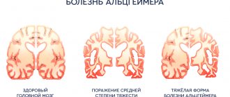
. The most common source of primary symptoms is cerebellar cysts (cerebellar cysts). They manifest with general cerebral symptoms (diffuse headaches, nausea not associated with food intake, vomiting, tinnitus) caused by increased intracranial pressure. The first signs also include epileptic seizures; they can be generalized or focal. Over time, signs of cerebellar damage appear, forming the symptom complex of cerebellar ataxia: static and dynamic discoordination, adiadochokinesis, hypermetria and asynergia, intention tremor, myodystonia. As the cerebellar tumor grows, displacement and compression of the brain stem occurs, accompanied by brain stem symptoms, primarily swallowing disorder, diplopia, and dysarthria. Spinal tumors (usually angioreticulomas) are manifested by radicular syndromes, loss of deep types of sensitivity, and absence of tendon reflexes. In 80% of cases of spinal pathology, a clinical picture similar to syringomyelia is observed. A picture of complete damage to the diameter of the spinal cord is possible.
Eye damage
in the early stages they are diagnosed only by ophthalmoscopy. After 8 years, complaints about image hazyness and distortion (metamorphopsia) appear. In half of the patients, both eyes are affected. Retinal angiomas that increase over time lead to circulatory disorders in its vessels, ischemia and cystic degeneration. In the late stage, uveitis, cataracts, retinal detachment, glaucoma, and hemophthalmos are possible.
Kidney damage
in 60-90% of cases it is represented by cysts, in 45% of cases - renal cell carcinoma. As a rule, renal carcinoma clinically debuts between the ages of 40 and 50 years in patients who have previously been treated for neoplasms. In half of the cases, at the time of diagnosing carcinoma, its metastases are detected. The combination of polycystic kidney disease with retinal angiomatosis is more typical than its combination with cerebral angiomas. In 35% of patients with Hippel-Lindau disease, polycystic disease is diagnosed posthumously. In childhood, with a familial type of the disease, polycystic kidney disease is often its only manifestation.
Pheochromocytoma
in almost half of the cases it is bilateral. May be the only clinical manifestation of the disease. In combination with renal carcinoma it is quite rare.
Pancreatic damage
from 30 to 72% are cysts. Pancreatic cysts are benign and rarely lead to clinically significant pancreatic enzyme deficiency. Although there are known cases of complete replacement of normal gland tissue by a cyst with the development of diabetes mellitus.
Causes
Hippel-Lindau disease is an autosomal dominant disorder resulting from a deletion or mutation of the VHL gene, located on the short arm of chromosome 3. Each child of a person with the syndrome has a 50% risk of inheriting an altered copy of the VHL gene.
The normal VHL gene acts as a tumor suppressor gene with the function of preventing tumor formation. The gene acts as a key regulator of cellular hypoxia signaling through its product, the VHL protein (pVHL). pVHL, through the HIF (hypoxia-inducible factor) complex, is indirectly responsible for elevated levels of growth factors, including vascular endothelial factor, platelet-derived growth factor, and transforming growth factor alpha.
In the case of a nonfunctioning gene, such as in Hippel-Lindau disease, regulation of the HIF complex does not occur. As a result, levels of various growth factors increase, which promotes increased blood vessel growth (angiogenesis) and tumor formation. This is the same process as other more common forms of cancer, such as kidney, breast, pancreatic, and adrenal cancer.
The researchers believe that interfering with growth factor and/or HIF activity will prove to be an effective treatment for Hippel-Lindau syndrome and other forms of cancer.
Symptoms and treatment
There are no symptoms that are generally characteristic of von Hippel-Lindau disease. The symptoms that occur depend rather on the individual tumors in the various organs. Therefore, diagnosis and treatment depend on individual components and may vary in each individual case.
If we talk about the treatment of Hippel-Lindau disease, we can mention magnetic resonance imaging, atraumatic neurosurgery, endoscopic surgery, and ultrasound examination.
However, many problems remain unresolved. The main issue is the correction of the genetic disorder. They are trying to find the answer in drug treatment, but suitable therapy still does not exist.
Pheochromocytoma
When the adrenal glands are damaged, Hippel-Lindau disease is manifested by the formation of pheochromocytoma. This neoplasm, originating from the substance of the adrenal medulla, is usually benign in nature. It is diagnosed in most cases at the age of about 30 years and is characterized by bilaterality, multiple nodes and the ability to transfer to tissues located nearby.
Pheochromocytoma manifests itself with the following symptoms:
- Arterial hypertension resistant to antihypertensive therapy.
- Blood pressure-related cranialgia.
- Pale skin.
- Tendency to hyperhidrosis.
- Tachycardia.
Criteria used to make a diagnosis
The diagnosis of von Hippel-Lindau disease can be made if at least one of the symptoms presented in the list below is detected. But this is possible provided that at least one of the patient’s family members has a history of hemangioblastoma of the central nervous system, retinal hemangioblastoma, cystic renal formations or adenocarcinoma:
- hemangioblastoma, which is localized in the tissue of the cerebellum, as well as in other parts of the central nervous system;
- spinal cord hemangioblastoma;
- hemangioblastoma localized in the bone marrow;
- hemangioblastoma on the retina;
- cystic lesion of kidney tissue;
- renal adenocarcinoma;
- cystic formations of the pancreas;
- cystadenocarcinoma;
- pheochromocytoma;
- epididymal adenoma.
It should be noted that the medical specialties whose representatives deal with the problem of Hippel-Lindau syndrome are neurology, ophthalmology, urology, and therapy.
Description
The first descriptions of von Hippel-Lindau disease come from the mid-19th century. Although the disease itself is much older. The disease got its name not from the person who discovered it, but from two researchers. Eugen von Hippel was a German ophthalmologist. Arvid Lindau was a pathologist in Sweden.
Von Hippel-Lindau disease is a rare genetic cancer syndrome. It is based on a mutation of the so-called von Hippel-Lindau gene. This violation of the hereditary gene can lead, first of all, to cell proliferation, that is, a tumor, on the other hand, to the formation of cysts, which causes fluid to fill cavities in various organs. Typical of Hippel-Lindau disease is a multiplicity of tumors that affect paired organs (for example, the eyes).
This disease can develop in various organs of the body. The severity and organs affected can vary significantly even among close relatives of the same family. Tumors can be bilateral.
Mechanism of disease development
Hippel Lindau syndrome is transmitted in an autosomal dominant manner. This means that if one parent has the defective gene, the child has a 50% risk of developing the disease.
This syndrome appears in people of different ages and has different manifestations even in close relatives.
Approximately 90% of people with this disease develop at least one symptom by age 65.
Until methods for early diagnosis were developed, patients usually did not live past the age of fifty.
In recent years, the prognosis of the disease has improved markedly, since there are methods for early detection of pathology and new technologies for its treatment.
Retinal angiomatosis
For ophthalmological localization of the pathological process, a characteristic feature is a triad of signs:
- presence of angioma;
- vasodilation in the fundus;
- the presence of subretinal exudation (accumulation of fluid - a product of inflammation - under the cornea), which can cause the development of exudative retinal detachment at an advanced stage of the disease.
The initial stages of the disease are characterized by decreased pigmentation in the fundus and increased tortuosity of blood vessels. When pressing on the eyeball, pulsation of both arteries and veins is noted.
At later stages, vasodilation and vascular tortuosity progress, and the formation of aneurysms and angiomas, which have the characteristic appearance of rounded glomeruli, is possible - this is a specific sign of Hippel-Lindau disease.
Types of disease
Traditionally, the types of Hippel-Lindau disease differ depending on the occurrence of pheochromocytoma. This medical term refers to a tumor with good blood circulation, surrounded by a specific capsule. Previously, only the presence or absence of a tumor was considered a criterion. This issue has now been reviewed. The dominant occurrence of pheochromocytoma is also taken into account.
- Type 1 Hippel-Lindau disease: families with significant absence of pheochromocytoma;
- Type 2: families with a dominant presence of pheochromocytoma;
- Type 2A: families as type 2, but without renal carcinomas;
- Type 2B: families as type 2, but with frequent renal carcinoma;
- Type 2C: families with a dominant presence of pheochromacytoma, but without manifestation of the disease in other organs.
Occurrence of the disease, gender and age of patients
Hippel-Lindau disease runs in families. The inheritance of this disease is autosomal dominant. This means that both sexes can be affected and no generation will be skipped. The mutation can be detected already at birth. But tumors appear only with age. The earliest recorded case of tumor appearance was 5 years. Most tumors appear between 15-35 years of age. However, there are no significant clinical differences between men and women.
Hippel-Lindau disease has an onset in every family. In every family there is a person who gets it for the first time.
It is often unclear who is the first carrier of such a gene, or who was the carrier. In such a person, the mutation occurs for the first time. Today, new mutations are frequently observed.