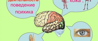
Like any other organ of the brain, the thalamus has an extremely important and irreplaceable function for the body. It’s hard to imagine, but this relatively small organ is responsible for all mental functions: perception and understanding, memory and thinking, because thanks to it we see, understand, feel the world and perceive everything that surrounds us. Thanks to its work, we navigate in space and time, feel pain, this “sensitivity collector” perceives and processes information received from all receptors, except the sense of smell, and transmits the necessary signal to the desired part of the cerebral cortex. As a result, the body gives the correct reaction, displays the correct behavior patterns to the corresponding stimulus or signal.
General information
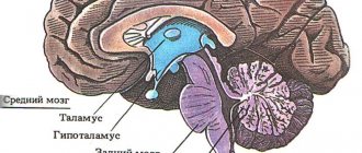
The diencephalon is located under the corpus callosum and consists of: the thalamus (thalamic brain) and the hypothalamus.
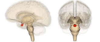
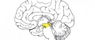
The thalamus (aka: visual thalamus, sensitivity collector, body informant) is a section of the diencephalon located in its upper part, above the brain stem. Sensory signals and impulses from various parts of the body and from all receptors (except smell) flow here. Here they are processed, the organ evaluates how important the incoming impulses are for a person and sends the information further to the central nervous system (central nervous system) or to the cerebral cortex. This painstaking and vital process occurs thanks to the components of the thalamus - 120 multifunctional nuclei that are responsible for receiving signals, impulses and sending processed information to the corresponding section of the cerebral cortex.
Thanks to its complex structure, the “visual thalamus” is capable of not only receiving and processing signals, but also analyzing them.
Ready information about the state of the body and its problems reaches the cerebral cortex, which, in turn, develops a strategy for solving and eliminating the problem, a strategy for further actions and behavior.
If something breaks
As you may have noticed, the thalamus has a complex structure and its functions are varied, therefore, if one of its parts begins to work incorrectly, completely different symptoms may appear. And if changes occur in the functioning of the thalamus, this can affect the functioning of the entire organism as a whole. After all, he plays such an important role as a redistributor. For example, anterograde amnesia may begin, in which a person forgets events that occurred after the onset of the disease. At the same time, the memory of what preceded the onset of symptoms remains intact. Another rare disorder affecting the thalamus was first described in 1979. This is "fatal familial insomnia." If a certain mutation has occurred in the PRPN gene, amyloid plaques begin to accumulate in the area of the thalamus that regulates sleep. Due to the improper functioning of this department, a person stops sleeping. A mutation in a gene is transmitted through the pedigree, which is why the name contains the word “family.” Known in only about 40 families worldwide and owned by 100 people. There is another type, this is “sporadic fatal insomnia,” which also has no special treatment, and the cause of which is also the malfunction of the thalamus.
To treat some diseases that affect the thalamus, electrodes are used that are implanted in the brain and can stimulate a certain part of it. For example, it is used to relieve symptoms of Parkinson's disease. The method is invasive and changes electrical activity, therefore magnetic resonance imaging is contraindicated for patients with such stimulators. But stimulation can be stopped at any time and the electrodes can be removed. A more drastic solution is surgical intervention, when certain areas of the thalamus are deliberately destroyed - thalamotomy. It is used to treat tremors in Parkinson's disease.
Text: Nadezhda Potapova
Read materials from our website on Facebook , VKontakte , Yandex-Zen and the Telegram channel , and also follow the new pictures of the day on Instagram .
Structure
The thalamus is a paired ovoid formation consisting of nerve cells that are united into nuclei, thanks to which the perception and processing of signals and impulses coming from different sense organs occurs. The thalamus occupies the bulk of the diencephalon (approximately 80%). Consists of 120 multifunctional gray matter nuclei. It is shaped like a small chicken egg.
Based on the structure and location of individual parts, the thalamic brain can be divided into: metathalamus, epithalamus and subthalamus.
Metathalamus (subcortical auditory and visual center) - consists of medial and lateral geniculate bodies. The auditory lemniscus ends in the nucleus of the medial geniculate body, and the visual tracts end in the lateral geniculate nucleus.
The medial geniculate bodies constitute the auditory center. In the medial part of the metathalamus, from the subcortical auditory center, cell axons are directed to the cortical end of the auditory analyzer (superior temporal gyrus). Dysfunction of this part of the metathalamus can lead to hearing loss or deafness.
The lateral geniculate bodies constitute the subcortical visual center. This is where the optic tracts end. The axons of the cells form the optic radiation, along which visual impulses reach the cortical end of the visual analyzer (occipital lobe). Dysfunction of this center can lead to vision problems, and severe damage can lead to blindness.
Epithalamus (suprathalamus) - the upper posterior part of the thalamus, which rises above it: includes the pineal gland, which is the supracerebral endocrine gland (pineal gland). The pineal gland is in a suspended state, as it is located on leashes. It is responsible for the production of hormones: during the day it produces the hormone serotonin (the hormone of joy), and at night it produces melatonin (a regulator of the daily routine and the hormone responsible for the color of the skin and eyes). The epithalamus plays a role in the regulation of life cycles, regulates the onset of puberty, sleep and wakefulness patterns, and inhibits the aging process.
Lesions of the epithalamus lead to disruption of life cycles, including insomnia, as well as sexual dysfunction.
The subthalamus (subthalamus) or prethalamus is a small-volume brain substance. It consists mainly of the subthalamic nucleus and has connections with the globus pallidus. The subthalamus controls muscle responses and is responsible for action selection. Damage to the subthalamus leads to motor disturbances, tremors, and paralysis.
In addition to all of the above, the thalamus has connections with the spinal cord, with the hypothalamus, subcortical nuclei and, naturally, with the cerebral cortex.
Each department of this unique organ has a specific function and is responsible for vital processes, without which the normal functioning of the body is impossible.
Functions of the thalamus
The “sensitivity collector” receives, filters, processes, integrates and sends information to the brain that comes from all receptors (except smell). We can say that in its centers the formation of perception, sensation, and understanding occurs, after which the processed information or signal enters the cerebral cortex.
The main functions of the body are:
- processing of information received from all organs (receptors of vision, hearing, taste and touch) senses (except smell);
- managing emotional reactions;
- regulation of involuntary motor activity and muscle tone;
- maintaining a certain level of activity and excitability of the brain, which is necessary for the perception of information, signals, impulses and irritations coming from the outside, from the environment;
- responsible for the intensity and feeling of pain.
As we have already said, each lobe of the thalamus consists of 120 nuclei, which, based on functionality, can be divided into 4 main groups:
- reticular;
- lateral (lateral);
- medial (middle);
- associative.
Reticular group of nuclei (responsible for balance) – responsible for ensuring balance when walking and balance in the body.
The lateral group (vision center) is responsible for visual perception, receives and transmits impulses to the parietal, occipital part of the cerebral cortex - the visual zone.
The medial group (hearing center) is responsible for auditory perception, receives and transmits impulses to the temporal part of the cortex - the auditory zone.
Associative group (tactile sensations) - receives and transmits tactile information to the cerebral cortex, that is, signals emanating from receptors of the skin and mucous membranes: pain, itching, shock, touch, irritation, etc.
Also, from a functional point of view, nuclei can be divided into: specific and nonspecific.
Specific nuclei receive signals from all receptors (except smell). They provide a person’s emotional reaction and are responsible for the occurrence of pain.
Specific kernels, in turn, are:
- external - receive impulses from the corresponding receptors and send information to specific areas of the cortex. Through these impulses feelings and sensations arise;
- internal - do not have direct connections with receptors. They receive information already processed by the relay cores. From them, impulses go to the cerebral cortex to the associative zones. Thanks to these impulses, primitive sensations arise and the relationship between sensory areas and the cerebral cortex is ensured.
Nonspecific nuclei support the general activity of the cerebral cortex, sending nonspecific impulses and stimulating brain activity. Having no direct connection with the cortex, the nonspecific nuclei of the thalamus transmit their signals to subcortical structures.
Thalamus
Rhythmic activity of the thalamus
Numerous studies have shown that the sources of electrical phenomena reflected on the surface of the scalp in the form of the EEG alpha rhythm are located in the neocortex.
At the same time, the location of the generators that trigger these events is less obvious. The idea that the thalamus is involved in the generation of alpha rhythm was first proposed by Berger in the 1930s. To date, a huge evidence base has been accumulated confirming the involvement of the thalamus in the formation of alpha activity. The most obvious evidence is the results of experiments on local damage to the thalamus. Such damage leads to disorganization or complete suppression of alpha activity on the EEG. In the 1960s P. Andersen and S. Andersson (Andersen R. A., Andersson S., 1968), having performed a series of elegant experiments, showed for the first time that the thalamus is the primary generator of all types of rhythmic fusiform activity recorded in the cortex. As further evidence of the role of the thalamus, the results of studies performed by Lopes da Silva et al. (1973) are usually cited. During a period of quiet wakefulness with eyes closed, dogs exhibit a pronounced alpha frequency rhythm with maximum amplitude in the posterior parts of the scalp, which is analogous to the dominant occipital alpha rhythm in humans. Alpha activity in dogs was recorded synchronously in the visual part of the thalamus - in the lateral geniculate nucleus (LGN) and visual cortex. The thalamus is the structure in which the primary processing and integration of almost all signals entering the cerebral cortex occurs, for which it has received the name “gate to the cortex.” The basis of specific thalamic nuclei are “relay” neurons that have direct axonal projections to a strictly defined area of the cortex. Relay nuclei receive afferent stimulation directly from the receptor zones of the corresponding analyzers. The function of relay nuclei is to switch information seeking in the cerebral cortex from the receptors of the corresponding analyzer. The first-order relay nuclei for the visual analyzer are the lateral geniculate bodies.
Neurons of the relay nuclei of the thalamus have pacemaker activity: the electrical potential of the neuronal membrane spontaneously decreases to a critical level, which causes the rhythmic generation of action potentials. These cells are capable of generating single or double discharges of action potentials. Through thalamocortical fibers, impulses enter the corresponding zones of the cortex, forming rhythmic activity in the form of so-called “cortical twins”. As a result, the frequency of spike activity of thalamic neurons coincides in frequency with the cortical alpha wave.
Thus, studies of the mechanisms of generation of bioelectrical activity of the human brain have now identified the “thalamic” theory of alpha rhythm generation as dominant. This hypothesis implies the presence of a key neuronal mechanism: volley pacemaker activity in thalamocortical neurons of the relay nuclei, primarily the lateral geniculate nucleus, and active interaction between neurons of the thalamus and cortex, forming the so-called thalamocortical system.
Thalamocorticothalamic loop
Rhythmic excitation of pacemaker neurons along corticopetal projections forms EPSPs on the postsynaptic membrane of cortical pyramidal cells. The frequency of spontaneous depolarization of pacemakers can vary widely and ranges from 3-4 to 15-20 Hz. Why is the main rhythm in the EEG the alpha rhythm - cortical activity with a frequency of about 10 Hz?
Frequency stabilization is achieved by a return braking mechanism. The reticular nucleus of the ventral thalamus, which belongs to the nonspecific nuclei, plays a key role in regulating the frequency of rhythmic activity. Taking some assumptions, the overall process can be described as a thalamo-cortico-thalamic loop of recurrent inhibition.
Neurons of the relay nucleus of the thalamus, through thalamocortical fibers, cause activation of neurons in the corresponding cortical zone. Excitation along associative fibers through a network of interneurons reaches the frontal cortex and “returns” to the thalamus along descending corticofugal projections. One of the main descending pathways is the corticothalamic pathway through the neurons of Meynert's nucleus, the cholinergic projections of which enter the reticular nucleus of the ventral thalamus. When excited, reticular neurons release the inhibitory transmitter GABA, causing hyperpolarization of the pacemaker neuron membrane. The refractory phase begins - spontaneous depolarization of the pacemaker becomes impossible for some time. As a result, the frequency of spontaneous depolarization decreases, and a stable frequency of rhythmic activity in the cortex is maintained at about 10 Hz, which corresponds to the alpha frequency range.
Thus, spontaneous excitation of thalamic pacemaker neurons leads to excitation of neurons in the projection zone of the cortex and is transmitted to neurons of the frontal cortex, which in turn activate neurons of the reticular nucleus of the thalamus. The reticular nucleus includes GABAergic neurons, which, when excited, release the inhibitory transmitter GABA into the synaptic cleft and inhibit the activity of pacemakers. This closes the thalamo-corticothalamic loop and implements the mechanism of recurrent inhibition.
The reticular nucleus has a topographic organization of corticofugal projections. As a result, the spatial organization of recurrent inhibition is ensured: the reticular nucleus provides selective regulation of the thalamocortical activity of the nuclear structures of the thalamus. Therefore, by regulating the cortical input, the reticular nuclei regulate the distribution of rhythmic activity over the surface of the cerebral cortex. This mechanism underlies the correct zonal distribution of the alpha rhythm. Normally, the alpha rhythm dominates in amplitude in the occipital leads and is practically absent in the frontal and temporal regions.
Thus, in the mechanisms of alpha rhythm generation, in particular in maintaining a stable frequency of oscillations, a key role is played by reciprocal inhibition of the activity of thalamic pacemakers from the frontal cortex.
Literary sources
- Aleksandrov M.V., Ivanov L.B., Lytaev S.A. [etc.]. Electroencephalography: manual / ed. M. V. Alexandrova. — 3rd ed., revised. and additional - St. Petersburg: SpetsLit, 2021. - 224 p.
- Aleksandrov M.V., Ivanov L.B., Lytaev S.A. [etc.]. General electroencephalography / ed. M. V. Alexandrova. - St. Petersburg: Strategy of the Future, 2021. - 128 p.
- Brezhe M. Electrical activity of the nervous system: trans. from English - M.: Mir, 1979. - 264 p.
- Gnezditsky V.V. Inverse EEG task and clinical electroencephalography. - M.: MEDpress-inform, 2004. - 624 p.
- Rusinov V.S., Mayorgik V.E., Grindel O.M. |et al.]. Clinical electroencephalography / ed. V. S. Rusinova. - M.: Medicine, 1973. - 339 p.
Separately about the visual thalamus
Previously, it was believed that the thalamus processed only visual impulses, and then the organ received the name - visual thalamus.
Now this name is considered obsolete, since the organ processes almost the entire range of afferent systems (except for smell). The system that provides visual perception is one of the most interesting. The main external organ of vision is the eye, a receptor that has a retina and is equipped with special cells (cones, rods) that transform the light beam and electrical signal. The electrical signal, in turn, passing through the nerve cells enters the lateral center of the thalamus, which sends the processed signal to the central part of the cerebral cortex. Here the final analysis of the signal occurs, thanks to which what is seen is formed, that is, the picture.