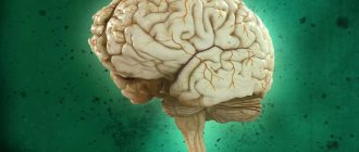
Good afternoon, dear readers! Today we have on our virtual anatomical table the diencephalon, that is, the diencephalon.
Before we get started, I would like to say a huge thank you to all those who leave comments here, on my VK page or on my super humble YAD channel. I’ve had quite a difficult time in the last few months (the session was disgusting), and your messages to me and your gratitude helped me get through this period. I couldn’t answer you for quite a long time because I didn’t even have time to open my website and look at the counters. However, now I am armed with audio lectures from my teachers, atlases, textbooks and my modest knowledge, so we begin.
Diencephalon - general knowledge
The diencephalon is a continuation of the brain stem and, as it were, a transitional part between the brain stem and the cerebral hemispheres. I urge you, dear readers, to immediately remember the Latin terminology (which, in fact, is based on Greek in our case), according to which the diencephalon sounds like “diencephalon,” the midbrain is “mesencephalon,” and the medulla oblongata is “myelencephalon.” This will greatly help you in studying brain development and in working with English books and video guides.
An illustration from Sinelnikov’s atlas will help us understand the topography of the diencephalon:
The diencephalon in such illustrations always reminded me of the head of a bird, which is attached to a long and thick “neck”, that is, on the brain stem.
I will deliberately skip the description of the boundaries of the diencephalon, because this requires a good knowledge of the structures of the telencephalon, which we have not yet covered. It can only be noted that behind and below the diencephalon borders on the midbrain, being its direct continuation. Above and laterally, the diencephalon is surrounded by the cerebral hemispheres. What exactly are the structures of the cerebral hemispheres adjacent to the thalamus? We will look at this in guides about the large hemispheres themselves.
So, the diencephalon has this name for a reason - as we already know, it is literally hidden between the cerebral hemispheres. To understand why our body needs the diencephalon, we need to know what it consists of. The diencephalon consists of two main parts:
- Thalamic brain;
- Hypothalamic region.
The thalamic brain includes:
- Thalamus (you've probably heard the old school name for thalamus - "visual thalamus");
- Metathalamus;
- Epithalamus.
The hypothalamic region includes the nuclei of the hypothalamus, parts of the visual pathway, subcortical olfactory centers and the pituitary gland.
Diencephalon - functions
The diencephalon is responsible for the regulation of the most important processes and is involved in higher nervous activity. The above sections of the diencephalon perform specific functions that are determined by the nuclei of gray matter contained in their structure. They interact with each other, thanks to which the assigned tasks are carried out in a coordinated and correct manner.
Thalamus - functions
According to the structure and mechanism of operation, the visual hillocks are considered a mini-version of the cerebral hemispheres. The thalamus contains the neuronal nuclei of the diencephalon, which are classified into 3 types:
- specific;
- nonspecific;
- associative.
The function of specific nuclei is to transmit signals to the corresponding centers of the cerebral cortex. They receive data from receptors:
- skin;
- ears;
- muscles;
- eye;
- internal organs.
These nuclei are involved in thermoregulation processes, determine tactile and pain sensitivity, visual and auditory sensations. The nonspecific group is a continuation of the midbrain into the diencephalon; the thalamus is a “platform” for the formation of the reticular formation from them. They are necessary for the correct distribution of nerve impulses going to the cortex. The pulsation of nonspecific nuclei additionally supports the excitability of neurons responsible for the formation of consciousness.
Association nuclei are connected to the parietal, temporal and frontal lobes of the cortex. They play the role of a “filter” that selects the most important signals and transmits them to the correct data processing centers. The functions of the associative nuclei are to ensure the normal activity of the areas of the cerebral cortex responsible for:
- speech perception and generation;
- hearing;
- obtaining and analyzing visual information.
Hypothalamus - functions
The section in question also contains numerous nuclear centers of the diencephalon. These accumulations of gray matter determine what the hypothalamus is responsible for. According to a simplified classification, the nuclei are divided into anterior, middle and posterior groups. They closely interact with each other, exchanging information, and regulate the following processes in the body:
- heart contractions;
- eating behavior;
- motility of the stomach and intestines;
- arterial pressure;
- heat production and release;
- all types of metabolism;
- energy costs;
- pregnancy and lactation;
- reproductive behavior;
- acid-base balance;
- memorization;
- formation of emotions;
- formation of hormones (vasopressin and oxytocin);
- control of the pituitary gland.
Epithalamus - functions
The basic task of this area is to ensure a stable relationship between the limbic system and the basal ganglia with other parts of the brain. The epithalamus serves as a kind of bridge between the structures surrounding it. In this area, the diencephalon regulates:
- fluctuations in the intensity of various physiological processes depending on the time of day;
- secretion of hormones by the hypothalamus and pituitary gland;
- release of melatonin;
- motor functions;
- the occurrence of positive and negative emotions;
- cognitive abilities and memory.
Metathalamus - functions
This name is already considered obsolete and is rarely used in medicine. Modern science includes the metathalamus in the structure of the thalamus. It refers to its posterior part and is called the geniculate body. The main task of the described zone is the primary processing of sensory information, its analysis and integration, and subsequent redirection to the appropriate centers of the cerebral cortex.
Subthalamus - functions
A special feature of this part of the diencephalon is its direct connection with the telencephalon. The largest and most pronounced structure is the subthalamic nucleus, whose neurons are capable of stimulating the globus pallidus and substantia nigra. Thanks to this, the subthalamus is included in the extrapyramidal system of the brain - a set of departments that regulate:
- motor activity;
- equilibrium;
- holding the body in a certain position;
- stimulation of muscle tone;
- orientation in space.
Pituitary gland - functions
The pituitary gland is considered the main organ in the endocrine system. The pituitary gland is responsible for most important life processes; the functions and hormones it secretes regulate:
- metabolism;
- body growth;
- reproductive abilities (especially in women);
- adrenal gland function;
- mental condition.
The diencephalon is a structure of components interconnected with each other. The activity of the pituitary gland is directly dependent on the activity of the hypothalamus. Together they form a single functional system, where one element plays a regulatory role, and the second - an effector role. Hypothalamic neurons produce transmitters that stimulate or inhibit the production of hormones by the pituitary gland.
Thalamic brain
Many students make the mistake of saying that the diencephalon consists of the thalamus and hypothalamus. This is incorrect, because with such a classification we lose many important structures that are not the thalamus, but are part of the thalamic brain. Let's look at this in detail.
Thalamus
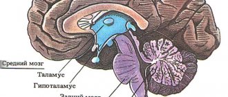
Wikipedia has a very good picture of the projection of the thalamus in relation to other parts of the brain:
Also, the thalamus has always reminded me in shape of the spaceship of the inhabitants of Cloud City on the planet Bespin. It was there that the most dramatic events of the 5th episode of Star Wars took place. So, the residents of Cloud City traveled on small spaceships that looked like two cabins connected by a small isthmus:
In this illustration from the atlas of Yu.L. Zolotko you can see both thalami and partially the caudate nucleus, which is located directly in front of the thalamus. Here the thalami are sagittally truncated by approximately half, but the absence of the cerebral hemispheres gives us the opportunity to see the thalami themselves and other components of the thalamic brain, about which we will see below.
So what is the thalamus? The thalamus is a paired egg-shaped formation with a pronounced sharpening in the front and a pronounced thickening in the back. The anterior point is called the anterior tubercle of the thalamus (tuberculum anterius thalami), and the posterior thickening is called the pulvinar thalami. The two thalami are connected to each other by a thin isthmus called the interthalamic fusion (adhesio interthalamica).
As I said before, the thalamus has the old school name “optic thalamus”. This is due to the fact that the thalamus actually processes and conducts visual signals, being one of the subcortical centers of vision. However, the thalamus processes not only visual signals, but in general all signals of all types of sensitivity.
According to the apt expression of Professor V.A. Dubynin, the thalamus is the “secretary” of the cerebral cortex. This is a very accurate definition, because the thalamus not only duplicates the functions of the cortex, it processes a huge amount of visual, auditory, tactile, proprioceptive information and decides which of these information flows to send to the cortex and what to slow down or completely block.
Indeed, the thalamus actively blocks unnecessary flows of information that tend to get into the cortex - you can appreciate this effect while observing a small, restless child who cannot concentrate on any one task and is constantly distracted. This is a consequence of imperfect development of the central nervous system, including the thalamus, which is only learning to stop numerous impulses and impulses that interfere with performing the same work, for example, eating.
To summarize, let's remember that the thalamus is the main subcortical center of almost all types of sensitivity. All sensory pathways going to the cortex are somehow connected to the thalamus.
Nuclei and pathways of the thalamus
In this lesson we will study the material a little differently than in previous neuroanatomy guides. Today we will deviate from our usual scheme, in which we studied the nuclei separately and the pathways separately (if you don’t understand what these are, look here). The thalamus has a lot of nuclei interspersed with relatively little white matter, so I decided to look at both nuclei and tracts at the same time.
In order to outline the approximate location of the thalamic nuclei on the diagram, we will need to draw the following marking in the form of something similar to a diverging lens:
Anterior nuclei of the thalamus
Having thus divided the thalamus into several fields, we can mark the nuclei. First of all, we will note a group of nuclei called the anterior nuclei of the thalamus (nuclei anteriores thalami). The subcortical sense of smell[/anchor] is located here.
The conducting path of the olfactory analyzer is interesting in that here the impulse first enters the cerebral cortex, and only after that - to the subcortical centers. In the case of other types of sensitivity, everything happens the other way around - subcortical centers process information and send it further to the cortex.
Based on this, we can schematically note the tracts that go to the anterior nuclei of the thalamus from the cortex and limbic system (I did not find the names of these tracts), as well as the tracts connecting the thalamus and the mammillary bodies that have not yet been studied by us. These tracts are called the mastoid-thalamic fascicle (fasciculus mammilothalamicus), I designated them as number 1.
While working with American, English and other foreign atlases, I was surprised to discover that Western neuroanatomists designate the functions of the anterior nuclei of the thalamus in a completely different way. It indicates that the anterior nuclei of the thalamus are responsible for learning, short-term memory and the ability to concentrate on long-term tasks. When compiling this article, I rely primarily on Professor Gaivoronsky’s textbook and Professor Izranov’s video lessons, so I present the structure of the thalamus in accordance with the Russian anatomical school.
Posterior nuclei of the thalamus
The posterior nuclei of the thalamus (nuclei posteriores thalami) are grouped in the rounded posterior end, which, as we know, is called the cushion. Fortunately, there is no confusion here and all sources indicate the main function of these nuclei - subcortical analysis of visual signals. The fibers of the optic tract (tractus opticus) enter the thalamic pad, where they switch to neurons of the posterior nucleus. The processes of these neurons go directly to the higher cortical center for processing visual information, that is, to the occipital lobe cortex.
Ventrolateral nuclei of the thalamus
The ventrolateral nuclei of the thalamus (nuclei ventrolaterales thalami) should be remembered better than all other nuclei by my readers. This is where the pathways that we have been teaching and drawing for so long end. First of all, this is the medial lemniscus (which at the level of the spinal cord consisted of the Gaulle and Burdach fascicles), as well as the spinothalamic tract, which (as I just learned) is also called the spinal lemniscus. I find it easier to remember these pathways by their full names - ganglio-bulbo-thalamo-cortical tract and ganglio-dorsal-thalamo-cortical tract, respectively.
Both pathways carry nonspecific sensation from the trunk and limbs to the central nervous system, with the medial lemniscus conveying proprioceptive sensation and the spinal lemniscus transmitting cutaneous, temperature, and pain sensation.
Median nuclei of the thalamus
If you look at the figure with which we marked the thalamus, you will see a fairly large field in the very center of the marking. This is the space for the median nuclei of the thalamus (nuclei mediani thalami). According to Gaivoronsky’s textbook, these nuclei are responsible for processing signals received from the auditory and vestibular analyzer, including the well-known lateral lemniscus (number 6). Axons of the median nuclei of the thalamus are sent to several areas at once, including both areas of higher analysis (the cortex of the temporal and frontal lobes of the cerebral hemispheres) and subcortical centers (red nuclei of the midbrain, posterior nuclei of the hypothalamus).
Medial nuclei of the thalamus
The medial nuclei of the thalamus (nuclei mediani thalami) in Russian anatomical literature are indicated as “integration centers of the extrapyramidal system.” This means that the auxiliary nuclei of the system of unconscious movements are located in the medial nuclei of the thalamus. In the diagram from Gaivoronsky’s textbook, the medial nuclei have processes from all other nuclei of the thalamus. I will draw as many lines as possible so as not to clutter our drawing, but I hope you will remember this feature of the medial nuclei.
Axons of the medial nuclei (number 7) connect the thalamus with several formations at once, such as the frontal cortex, limbic system and red nuclei.
Epithalamus
Many students confuse the metathalamus and epithalamus, however, it is not difficult to distinguish between them. One of the most important anatomical structures of the epithalamus is a gland called the pineal gland. You can simply remember the same prefix (epi), which will guide you.
So, the epithalamus includes the pineal gland (also known as the pineal gland), leashes, triangles of leashes and commissure of leashes. Let's figure out where it is.
Leashes
The thalamus is wrapped in broad white matter structures that separate the thalamus itself and parts of the cerebral hemispheres called the basal ganglia. I could not find complete information about exactly what signals these pathways carry. However, Sapin's textbook suggests that among these fibers there are fibers that conduct signals from the olfactory organ.
So, the medial part of this white matter is called the stria medullaris of the thalamus, and the lateral part is called the terminal stria (stria terminalis). On the posterior part of the thalamus, the medullary and terminal striae form triangular thickenings, which are called leash triangles (trigonum habenulae). Further, the leashes themselves (habenulae) extend from the triangles of the leashes, which are connected in a section called the commissure of the leashes (comissura habenularum).
In our illustration I have indicated:
- number 8 - triangles of leashes;
- number 9 - leashes;
- number 10 - soldering of leashes.
Pineal gland
The pineal gland looks like a very small spruce cone, so the second name for the pineal gland is the pineal gland (glandula pinealis). The pineal gland is located in the area of the thalamic cushion; it is, as it were, suspended on leashes. The quadrigeminal plate of the midbrain is also a good landmark, because the epiphysis literally hangs over it, and many people mistake it for the “fifth colliculus.” In our illustration, the pineal gland corresponds to the number 11:
Here I highlighted the leash triangles in green, and the pineal gland in lilac:
What is the pineal gland for? This small gland controls our body's circadian rhythms through the synthesis of the hormone melatonin. Melatonin is what helps us fall asleep when we close our eyes. Melatonin inhibits the production of stress hormones, lowers body temperature, and also increases the activity of cellular immunity.
By the way, some fish, amphibians and reptiles literally have a third eye - an unpaired light-sensitive organ that is located just above the forehead. Histologically, this organ, also called the parietal eye, is a greatly simplified traditional eye, with receptors capable of detecting only the difference between light and dark.
The parietal eye is directly or indirectly connected via conductive pathways to the pineal gland - this is how the synthesis of melatonin is triggered in response to the onset of darkness, so that the daytime animal falls asleep. In humans, the function of the parietal eye is performed by the traditional organs of vision. This is why physiologists advise turning off the lights a few minutes before bedtime - this creates optimal conditions for the production of melatonin and falling asleep.
Metathalamus
The metathalamus is the part of the thalamic brain that connects the diencephalon and midbrain. The metathalamus includes the medial and lateral geniculate bodies, as well as the superior and inferior arms. It's actually very simple.
Top and bottom handles
As you remember, the dorsal surface of the midbrain is formed by the quadrigeminal plate, in which the subcortical centers of vision (superior colliculi) and hearing (inferior colliculi) are localized. From each hillock towards the thalamus, bundles of pathways extend, which on the outside look like small cylindrical thickenings. These are the upper and lower arms (brachia colliculi superior et inferior).
There is a certain circumstance - in some sources the upper and lower arms of the quadrigeminal region are attributed to the midbrain, and in others - to the metathalamus. Maybe the whole point is that having a midbrain and arms and legs would sound too funny?
In my guide, I attributed the handles to the metathalamus (this is exactly what happened in Professor Izranov’s lecture), so let’s add them to our drawing as numbers 12 (upper) and 13 (lower):
geniculate bodies
The geniculate bodies are another subcortical centers of hearing and vision. Externally, the geniculate bodies appear as four small protuberances on the posterolateral part of the thalamic cushion. Unlike the colliculi and the manubrium, the geniculate bodies are divided into lateral and medial.
The lateral geniculate bodies (corpora geniculata lateralia) are the subcortical centers of vision, which are approached by the upper arms, starting from the superior colliculi. Accordingly, the medial geniculate bodies (corpora geniculata medialia) are responsible for subcortical sound processing and are approached by the lower arms, starting from the lower colliculi. The geniculate bodies, unlike the handles, are indicated in absolutely all sources as part of the thalamic brain. Let us also mark them - number 14 for the lateral geniculate bodies, and number 15 for the medial ones.
I found this illustration on the Internet, where we see the dorsal side of the brain stem. Here I have marked the upper arms and lateral geniculate bodies in yellow, and the lower arms and medal geniculate bodies in blue:
Without any secretions, you will also be able to identify these anatomical formations. And, I hope, you can easily find in this illustration the quadrigeminal plate, the pineal gland, the massive middle cerebellar peduncles, the rhomboid fossa and the medulla oblongata:
Features of the diencephalon
In Latin, the name of the described area is diencephalon. Translated, this means “located inside the head.” The intermediate section is formed during embryonic development. It is formed from the secondary medullary vesicle, combining from two parts (posterior and anterior) into one. This structure is small in size, but has a complex structure and performs vital tasks in the body.
Diencephalon - location
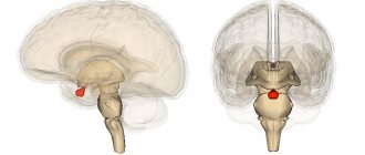
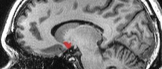
The area in question owes its name to its location. The diencephalon is a “bridge” between the cerebral hemispheres, with which it borders in front and above, and the middle section, with which it comes into contact below and behind. In relation to the third ventricle, the diencephalon is located laterally (on the sides). If you look at its localization in cross-section, the diencephalon is located in the depths and almost in the center of the skull. The location is clearly shown in the figure.
Diencephalon - structure
The diencephalon is simplified into two parts. The diencephalon consists of the thalamic and hypothalamic regions. Between them there is a small slit-like cavity called the third ventricle. The main structures of the diencephalon are further divided into several more segments. Thalamic part:
- Epithalamus.
The most superior posterior zone, closely connected with the basal ganglia and limbic system. The epithalamus consists of the pineal gland, habenula, leash triangle, commissure and subcommissural organ. - Thalamus.
An alternative name is optic bumps. It is a collection of gray matter. It is a pair structure consisting of two symmetrical halves. - Metathalamus.
Previously, this segment was considered a separate area; in modern anatomy it belongs to the posterior part of the thalamus called the “geniculate body”. - Subthalamus.
The “substrate” of the thalamus, formed from several gray matter nuclei and white matter structures.
The diencephalon in the hypothalamic region is divided into the following units:
- Hypothalamus.
The central link between the nervous and endocrine systems. The hypothalamus contains more than 40 pairs of nuclei and is connected to almost all parts of the brain, including the cortex. - Pituitary gland (posterior lobe).
Pituitary gland is another name. It is a rounded brain appendage located in the bony cavity (sella turcica). It is closely connected with the hypothalamus and “subordinates” to it.
Age-related features of the diencephalon
The sections of the described area develop heterochronously (not in parallel) with each other. The human diencephalon is formed in the 2nd month of intrauterine development in the form of a visual thalamus. By the 20th week of pregnancy, nerve fibers are formed that stretch to the cortex. At 6 months, reticular formations appear. The thalamus develops much faster. At the age of 4, increased growth is observed, but the sensory nuclei have been working since birth. The functional development of the thalamus ends by the age of 14.
The diencephalon in the hypothalamic zone progresses more slowly. Its structures are clearly visible only in the 8th month of intrauterine development. The onset of maturation occurs at 2-3 years of age. By the age of 5, most of the basic nuclei of the thalamus are formed, but they are finally activated only in adolescents. Full maturity of the hypothalamus is achieved by 16 years of age.