: 30 Oct 2021, The Fellowship of the Ring, volume 88, no. 3
Nowadays, one of the first places among the causes of mortality and disability of the population is occupied by congenital and acquired pathologies of the brain: from strokes and cancer to injuries and neuropsychiatric disorders. It is not surprising that maintaining brain health is one of the central problems of modern medicine. Therefore, such an important place is given to the development of methods for prediction, early diagnosis and monitoring of the effectiveness of treatment of neuropathologies of various origins. Among modern technologies for intravital non-invasive study of the state of the central nervous system, the leading methods are radiation diagnostics, while the palm belongs to magnetic resonance imaging, which offers the widest range of approaches to visualization of brain tissues and structures
The brain is one of the most well-supplied areas of the human body. Adequate functioning of all parts of the higher nervous system and related structures ensures sufficient arterial blood
and
venous
, as well as constant circulation
of cerebrospinal fluid
(
CSF
).
Violation of the speed, pressure, viscosity and other parameters of these biological fluids can cause severe pathology with a fatal outcome. Moreover, most of the works in this area are devoted to the study of the arterial part of cerebral hemodynamics, and studies of the role of the venous and cerebrospinal fluid systems are rare. And although the experience of recent years has significantly expanded our knowledge of liquor dynamics, there are still many unresolved and controversial issues. MRI IN THE LEADERS
Pathological processes in the human body are always based on disturbances in the movement of any biological fluid (blood, lymph, urine, bile, intra-articular, etc.), the main component of which is water. In different human organs, normally and in pathology, there are different conditions for the movement of such fluids, which is reflected in their linear and volumetric velocities, the nature of movement, interaction with the walls of conducting systems and other dynamic parameters. Diagnostic visualization and quantitative assessment of the speed characteristics of the movement of these substrates are considered the basis of clinical diagnostics in many medical fields: cardiology, neurology and neurosurgery, urology, gastroenterology, etc., however, technologies for intravital visualization of the movement of biofluids in the human body were invented and introduced into clinical practice only in the last 60–70 years. In this regard, MRI is unique, since it is the hydrogen atoms of water molecules and organic compounds that provide the “base” of the MRI signal. Other methods make it possible to assess the movement of fluid in the body only indirectly, by the movement of various tracers (radioactive isotopes, dyes, etc.), which cannot be called completely safe. In addition, the introduction of such foreign substances into a rather fragile system, which is any biofluid, can lead to uncontrolled changes in the parameters of its movement
Based on the information available today, we cannot create a holistic picture that describes the imbalance between the fluid media of the central nervous system in various pathologies. One of the reasons is the shortcomings of existing instrumental visualization methods, so the development of new approaches and original techniques is extremely promising.
An integrated approach to the morphological and functional assessment of brain tissue can be offered by modern methods of radiation diagnostics: magnetic resonance imaging
(MRI),
multislice computed tomography
(MSCT) and
ultrasound examination
(US).
However, today only MRI allows non-invasively and even without the use of contrast agents to visualize fluid flow and evaluate its quantitative parameters. Modifications of this method ( MR angiography
,
MR venography
,
MR myelography
) make it possible to obtain a large amount of additional information to assess the functional parameters of the flow of biological fluids, which opens up the possibility of early diagnosis of a wide range of diseases.
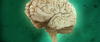
What is the brain, encephalon, its meaning?
The brain is the anterior part of the central nervous system. Brain
located in the cranial cavity, it interacts with the human body (men, women) with the external environment, integrating the functioning of all body systems.
The brain has the ability to assimilate, organize, store, and retrieve information about past experience. The brain is the material substrate of higher nervous activity. Phylogenetically, the brain is the anterior end of the neural tube.
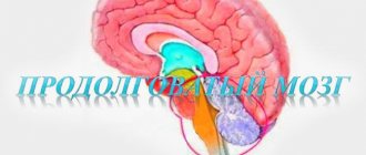
Ontogenetically, the brain is a derivative of the cerebral vesicles, from which parts of the brain are formed: the telencephalon, the diencephalon, the midbrain, the hindbrain, which is represented by formations such as the pons, cerebellum, and medulla oblongata. The cavities of the brain vesicles develop into the ventricles of the brain.
Descending Paths
Descending projection pathways are a group of tracts that send neural information from the telencephalon cortex and subcortical formations to brainstem structures. These include:
- Pyramid path. This tract connects the motor gyrus with the motor nuclei of the brainstem. So, with the help of this path, a person manages to control the muscles of the neck, head, eyes, face and torso.
- Extrapyramidal pathway. Thanks to this tract, people maintain their balance in space.
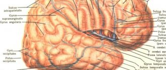
Brain structure
The cerebrum, or telencephalon, is represented by two hemispheres, which are connected to each other by the corpus callosum, corpus callosum. It consists of nerve fibers running transversely from one hemisphere to the other. The corpus callosum ensures the unity of functioning of both hemispheres. When the corpus callosum is cut, each hemisphere of the brain begins to function independently of each other. Under the corpus callosum is the fornix, fornix. Anterior to the pillars is the anterior commissure, comissura anterior. Between the anterior part of the columns of the fornix and the knee of the corpus callosum there is a thin vertical plate of brain tissue - the transparent septum. Between the plates of the septum there is a slit-like cavity that does not have an ependymal lining. A number of authors call it the 5th ventricle.
The surface of the hemispheres is covered with a layer of gray matter - this is the cerebral cortex. Underneath it is the white matter and subcortical nuclei: striopallidal system, extrapyramidal system.
If you make a horizontal section of the brain through the cerebral hemispheres at the level of the thalamus and subthalamic nuclei, you can see the following formations: pineal body, superior colliculus, thalamus, frenulum, posterior limb of the internal capsule, globus pallidus of the lenticular nucleus, putamen of the lenticular nucleus, lateral sulcus, fence , anterior part of the internal capsule, head of the caudate nucleus, column of the fornix, anterior horn of the lateral ventricle, genu of the corpus callosum, septum pellucidum, interthalamic commissure, lentiform nucleus, external capsule, extreme capsule, convolutions of the insula, lateral sulcus, internal capsule, subthalamic nucleus, tail caudate nucleus, lateral geniculate nucleus, red nucleus, gray matter of the superior colliculus, cerebellar vermis.
Sinuses and processes of the dura mater of the brain
In some areas, the hard shell is divided into 2 parts, while its inner plate extends deep into the convolutions, forming processes:
- Sickle cerebral hemispheres;
- Cerebellar tent;
- Falx cerebellum;
- Seat diaphragm.
In the areas of contact of the dura mater with the bones of the skull, as well as where it splits into two plates, triangular-shaped canals appear - the sinuses of the dura mater of the brain. They are part of the venous system of the central nervous system. The walls of the canals are formed by the petals of the membrane, which are covered inside with a layer of endothelium. Thanks to this, they became elastic.
On the periphery of the cerebral hemispheres the following sinuses are distinguished:
- Superior sagittal. It extends along the upper part of the falciform process, ending at the level of the internal occipital protrusion.
- Inferior sagittal. Located along the bottom of the sickle.
- Straight. Extends at the confluence of the falx and the cerebellar tent. Has a tetrahedral shape.
- Transverse. Pair structure. It runs in the transverse grooves of the bones of the skull.
- Occipital. Located in the wall of the cerebellar falx, it stretches along the length of the internal crest of the occiput to the foramen magnum.
- Cavernous. Pair education. Stretched along the lateral wall of the sphenoid bone. It is an irregular triangle.
- Intercavernous. Pair structure. It goes along the circumference of the sella turcica. They are the connecting link of the cavernous canals, forming a venous ring with them.
- Sphenoparietal. It is a pair structure. Its walls are adjacent to the sphenoid bone. Connects with the cavernous sinus.
- The upper one is rocky. It is a paired structure running from the cavernous sinus along the superior petrosal groove of the temporal bone.
- The lower one is rocky. A paired structure located in the inferior petrous gyrus of the occipital and temporal bones. It stretches from the posterior edge of the cavernous canal to the superior bulb of the jugular vein.
- Basilar plexus. Located in the area of the clivus of the occipital bone. Associated with the cavernous and petrosal sinuses, as well as with the venous plexuses of the spine.
Venous sinuses have become a kind of collectors in which waste blood is concentrated. Next, it enters the jugular veins and is transported to the lungs, where it is enriched with oxygen and again sent to the central nervous system.
Brain of a newborn, children, child, human: structure, anatomy
The brain of a newborn baby is shorter and wider than that of school-age children and adults. It is devoid of all tertiary and a number of secondary grooves. By the end of the child’s first year of life, the brain increases in size by 2–2.6 times. By 3 years it increases 3 times. From birth to adulthood, brain weight increases 4 times, and body weight increases 21 times.
The mass of the right hemisphere is most often greater than the mass of the left hemisphere. After birth, the parietal and frontal lobes develop most intensively. And because of this, the overall configuration of the brain changes. Unlike the adult brain, in a newborn the neurons of different layers are located closely next to each other, because of this, the radial striation of the cortex may be absent. Single neurons may be located in the subcortical white matter. In the substantia nigra of the brain stem, neurons do not yet have the pigment melanin, which usually appears by 3–4 years of age. Up to 3–6 months of extrauterine life, the outer embryonic layer, which is called “Obersteiner’s layer,” remains in the cerebellar cortex. Obersteiner's layer consists of medulloblasts and spongioblasts. The surface of the inferior olives of the fetal medulla oblongata is smooth. After the birth of a child, the olives acquire elevations and then noticeably increase in size with age. Almost always in newborns, immature cellular elements are found in the subependymal parts of the ventricular system of the lateral ventricles, the presence of which erroneously resembles manifestations of Virchow's local encephalitis. Immature cells are located in the subependymal layer diffusely or in the form of separate foci. Sometimes they can be traced along the blood vessels over a significant extent of the white matter. By 3–6 months of a child’s life, these cells gradually disappear. The presence of a large number of immature cells in the subependymal parts of the ventricular system is an additional morphological sign of fetal prematurity.
General information
The head and spinal regions of the central nervous system require additional protection. In addition to the bone frame, they are shrouded in meninges. There are only 3 of them:
- Solid (TMO). It differs from others in density and strength. Its outer side is rough, covered with a developed blood network, and loosely adheres to the bones of the skull, forming the epidural space in the cranial vault. Formed by fibrous tissue cells.
- Translucent arachnoid (arachnoid). Located under the hard layer. It looks like a thin web, which is why it got its name. Formed by cells of loose connective tissue. It consists of 2 layers, between which there are a large number of partitions.
- Soft. It is the lowest layer that fits tightly to the brain and spinal cord.
In some pathologies of the central nervous system, a subdural space appears between the hard and arachnoid membranes, which is filled with a small amount of cerebrospinal fluid. Normally it shouldn't be there.
How does a child's brain mass change with age?
If you monitor how the mass of a child’s brain changes depending on age, you will notice the following picture. If the child’s age is from 3 to 8 days, body length is 49 – 50 cm, then the brain weight will be 336 grams. At 1 month, the child’s height is 52 cm, brain weight is 360 grams. At 3 months, the child’s height is 56 cm, brain weight is 520 grams. At 6 months, with a height of 62 cm, GM weight is 670 g. At 9 months, with a height of 67 cm, GM weight is 760 g. At 1 year of age, the child’s height is 73 cm, brain weight is 960 g. At 1.5 years, with a height of 79 cm, the weight of the GM is 1045 g. At 2 years old, with a height of 85 cm, the weight of the GM is 1070 g. At 3 years old, the child’s height is 89 cm, and the brain weight is 1150 g. At 5 years old, height is 106 cm, weight is 1240 g. At 10 years old, height 132 cm, brain weight 1300 g. At 12 years old, height 145 cm, brain weight 1370 grams.
Physiology, brain function
Physiologically, all the work of the brain is built on the principles of hierarchy, integrity, systematicity and plasticity. These are the principles of functioning that carry out all conditioned and unconditioned reflexes. They contribute to the flow of conscious mental activity of a person. The principle of hierarchy is that phylogenetically younger parts of the brain carry out higher-order control, complementing but not replacing the function of phylogenetically more ancient parts. As a result of this, the body’s capabilities in more subtle differentiation of each stimulus by each analyzer are expanded, and a more adequate perception of the overall picture of the world is achieved based on the correlation of the results of the activities of many analyzers.
The highest form of expression of the hierarchical principle is the process of corticalization of functions. The principle of hierarchy is combined with the principles of integrity and systematicity, which consist in the fact that the brain functions as a single whole with the entire nervous system, while receiving afferent impulses, carries out its analysis and synthesis, forms a flow of efferent impulses that determine the adequate activity of all peripheral organs . As a result, a stable system is formed that provides continuous information communication: center - periphery - environment - periphery - center. Plasticity refers to the functional variability of nerve centers, which is clearly manifested in the process of compensation for impaired brain functions.
Irradiation of excitation plays an important role in the normal functioning of the brain. The feedback mechanism consists of closing the input and output of the same element or system. The dominant mechanism regulates the relationships between nerve centers.
What's next?
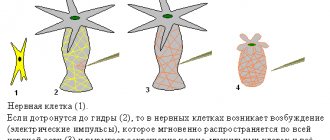
One interesting point to explore further is the team's observation of two nerve cells, or neurons, that seemed to be woven into a symmetrical dance. The images also showed the neurons' axons sending messages in complex spirals, unusual and mysterious whorls that looked like coiled snakes. “We have never seen anything like this,” the researchers write.
The brain is an incredibly complex computer, the mechanisms of which we have yet to understand.
This is interesting: How do brain cells map memories?
These extremely detailed maps of the brain are the culmination of years of research. This is truly a magical time in history when a variety of tools are available to scientists to create maps like these. Yet what these maps will ultimately reveal remains unknown—the study authors are cautious about whether they will lead to deep understanding of the brain. But in the future we will definitely have many amazing discoveries.
Diseases, disorders, brain lesions
Diseases, disorders, and brain lesions are varied. In further articles, we will focus on pathologies such as brain tumor, brain cyst (including arachnoid, retrocerebellar, liquor), trauma, concussion or bruise of the brain, brain cancer, hydrocephalus (hydropsis), vascular atherosclerosis, aneurysm, encephalopathy, demyelination, ischemia, ischemic or hemorrhagic stroke, heart attack, atrophy, spasm or vasoconstriction, glioblastoma, meningioma, dysfunction, dystonia, diffuse changes, hypoxia (oxygen starvation), encephalitis, inflammation, vascular diseases, atrophic changes. The clinical picture of such diseases depends on the type of pathology.
Brain treatment in Saratov, Russia
Sarklinik provides treatment for a number of diseases, diseases of the central and peripheral nervous system in Saratov, in Russia for children, adolescents, adults, boys, girls, guys, girls, men, women, brain treatment in Saratov . Hardware and non-hardware treatment methods make it possible to restore the functioning of the human nervous system.
Sign up for a consultation. There are contraindications. Specialist consultation is required.
Text: ® SARCLINIC | Sarclinic.com \ Sarlcinic.ru Photo: © pixologic / Photobank Photogenica / photogenica.ru The people depicted in the photo are models, do not suffer from the diseases described and/or all coincidences are excluded.
Related posts:
Apraxia, treatment of apraxia, treat apraxia in children, adults
Claude H Claude syndrome, alternating peduncular syndrome
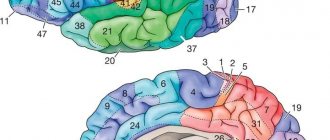
Zones of the cerebral cortex, functions, human cerebral cortex: structure, activity
Nocturnal enuresis code according to ICD 10 F 98.0
Nocturnal enuresis, urinary incontinence, children's enuresis, treatment
Comments ()
Research methods
Diagnosis of the functional state and activity of the trunk is carried out using clinical and instrumental laboratory methods. The first includes:
- neurological study of the activity of cranial nerves;
- study of voluntary movements;
- diagnostics of coordination of limbs and body;
- sensitivity study;
- Laboratory methods include:
- spinal puncture and examination of cerebrospinal fluid;
- X-ray of the skull;
- ventriculography;
- pneumoencephalography;
- Dopplerography;
- electroencephalography;
- magnetic resonance imaging;
- computed tomography.