Tunnel neuropathies account for 1/3 of diseases of the peripheral nervous system. More than 30 forms of tunnel neuropathies have been described in the literature [Levin O.S., 2005]. Various forms of compression-ischemic neuropathies have their own characteristics. We will first consider their general characteristics, then we will focus on the most common forms of hand tunnel syndromes (Table 1).
Causes
The anatomical narrowness of the canal is only a predisposing factor in the development of tunnel syndrome. In recent years, evidence has accumulated indicating that this anatomical feature is genetically determined. Another reason that can lead to the development of tunnel syndrome is the presence of congenital developmental anomalies in the form of additional fibrous cords, muscles and tendons, and rudimentary bone spurs. However, predisposing factors alone for the development of this disease are usually not enough. Some metabolic and endocrine diseases (diabetes mellitus, acromegaly, hypothyroidism), diseases accompanied by changes in joints, bone tissue and tendons (rheumatoid arthritis, rheumatism, gout), conditions accompanied by hormonal changes (pregnancy), space-occupying formations can contribute to the development of tunnel syndrome the nerve itself (schwannoma, neuroma) and outside the nerve (hemangioma, lipoma). The development of tunnel syndromes is facilitated by frequently repeated stereotypical movements and injuries. Therefore, the prevalence of carpal tunnel syndrome is significantly higher in people engaged in certain activities and in representatives of certain professions (for example, stenographers have carpal tunnel syndrome 3 times more often).
Symptoms and causes
Symptoms are related to the type of nerve damaged and may appear over a period of days, weeks, or years. Muscle weakness is the most common symptom of motor nerve damage. Other symptoms may include painful cramps and fasciculations (muscle twitching of the subcutaneous muscles), muscle atrophy, bone degeneration, and changes in the skin, hair, and nails. These general degenerative changes may also result from damage to the sensory fiber or autonomic fiber bundle.
Sensory nerve damage causes a wider range of symptoms because sensory nerves have a whole group of highly specialized functions. Large sensory fibers are surrounded by a myelin sheath and record vibration, tactile sensations and proprioception. Damage to large sensory fibers reduces the ability to sense vibration and touch, resulting in numbness, especially in the arms and legs. People may feel the sensation of wearing gloves or stockings. Many patients cannot distinguish the size of an object or its shape by touch. This damage to sensory fibers can contribute to loss of reflexes (as does motor nerve damage). Loss of proprioception (the sense of the body's position in space) makes it difficult to coordinate complex movements or balance with eyes closed. Neuropathic pain is difficult to treat and can have a serious impact on your emotional well-being and overall quality of life. Neuropathic pain often worsens at night, severely disrupting sleep, leading to further emotional distress.
Smaller sensory fibers without myelin sheaths transmit pain and temperature sensations. Damage to these fibers can impair the ability to feel pain or changes in temperature. People may not be able to feel the cut or the wound festering. Other patients may not feel the pain that is a warning sign during a life-threatening heart attack or other acute condition. Loss of pain sensation is a particularly serious problem in patients with diabetes, contributing to the high rate of lower extremity amputations in this population. Pain receptors in the skin may also become hypersensitive, so that patients experience severe pain (allodynia) from stimuli that are normally painless (for example, rubbing a cloth over the skin or lightly touching it). Symptoms of damage to bundles of autonomic nerve fibers are varied and depend on the organ innervated by them. Autonomic nerve dysfunction can be life-threatening and sometimes requires emergency medical attention, especially when breathing or heart rate is affected. Common symptoms of damage to the autonomic nerve fiber bundles include impaired sweating, which is necessary when overheated, impaired urinary function which can lead to urinary incontinence or bladder infections; and impaired control of the muscles responsible for contracting blood vessels, which can affect the maintenance of normal blood pressure. Loss of control of blood pressure can cause dizziness, nausea, or even fainting when a person suddenly falls when changing body position (a condition known as postural or orthostatic hypotension). Gastrointestinal symptoms often accompany autonomic neuropathy. The nerves that control contractions of the intestinal muscles malfunction, leading to diarrhea and constipation. Many patients also have problems with swallowing when the corresponding nerve fibers are damaged.
Peripheral neuropathy can be either inherited or acquired. Causes of acquired peripheral neuropathy include: nerve damage (trauma), tumors, intoxication, autoimmune reactions, nutritional disorders (vitamin deficiency), chronic alcoholism, and vascular and metabolic disorders. Acquired peripheral neuropathies are grouped into three broad categories: those caused by systemic disease, those caused by trauma from external factors, and those caused by infections or autoimmune disorders that damage nerve tissue. An example of acquired peripheral neuropathy is trigeminal neuralgia, in which damage to the trigeminal nerve causes episodic attacks of excruciating pain on one side of the face. In some cases, the cause is the consequences of a viral infection, as well as pressure on the nerve from tumor tissue or an enlarged blood vessel. In many cases, a specific cause cannot be identified. Doctors usually diagnose idiopathic neuropathy in such cases.
Traumatic injury is the most common cause of nerve damage. Injury or household trauma from car accidents, falls, or sports-related injuries can lead to nerve fraying, nerve compression, stretching, or complete separation from the spinal cord. Even minor injuries can also cause serious nerve damage. Broken or dislocated bones can put damaging pressure on nearby nerves, and nerve root compression can also occur with herniated discs.
Systemic diseases are conditions that affect the entire body and often cause peripheral neuropathy. These conditions may include: metabolic and endocrine disorders. Nerve tissue is very sensitive to changes in tissue metabolism and regeneration processes, which can change with systemic diseases. Diabetes mellitus, characterized by chronically high blood glucose levels, is a leading cause of peripheral neuropathy in some countries (USA). Approximately 60 to 70% of patients with diabetes have both moderate and severe forms of nervous system damage. Kidney disease can lead to excess toxic substances in the blood, which can seriously damage nerve tissue. Most patients requiring dialysis due to renal failure develop polyneuropathy. Some liver diseases also lead to neuropathies as a result of metabolic disorders.
Hormonal imbalances can alter normal metabolic processes and cause neuropathies. For example, a lack of thyroid hormones slows metabolism, leading to fluid retention and tissue swelling, which can put pressure on peripheral nerves. Excess growth hormone production can lead to acromegaly, a condition characterized by abnormal enlargement of many parts of the skeleton, including joints. The nerves supplying these altered joints are often damaged as well.
Vitamin deficiencies and chronic alcoholism can cause irreversible damage to nerve tissue. Vitamins. Vitamins E, B1, B6, B12, and niacin are very important for normal nerve function. Thiamine deficiency is particularly common in people with chronic alcoholism because these people have an impaired dietary intake of thiamine. Thiamine deficiency can cause quite painful neuropathy of the limbs. Some researchers believe that excessive alcohol consumption may, in itself, contribute to direct nerve damage, called alcoholic neuropathy. Vascular and blood diseases can reduce oxygen delivery to peripheral nerves and quickly lead to severe damage or death of nerve tissue (for example, acute brain hypoxia leads to stroke). Diabetes often leads to a narrowing of the blood vessel. Various forms of vasculitis often lead to thickening of the vessel wall and a decrease in the diameter of the vessels due to scar tissue. This category of nerve damage, in which isolated nerves in different areas are damaged, is called multifocal mononeuropathy.
Connective tissue diseases and chronic inflammation can cause direct or indirect nerve damage. When the tissue layers surrounding the nerves are in a prolonged inflammatory process, the inflammation can directly affect the nerve fibers. Chronic inflammation also leads to progressive destruction of connective tissue, exposing nerve fibers to greater risk of compression and infection. When joints become inflamed, they can swell and involve nerves, causing pain.
Cancer and benign tumors can grow and have a destructive effect on the nerves. Tumors can also form directly from nerve tissue cells. Quite often, polyneuropathy is associated with neurofibromatosis, a genetic disease in which multiple benign tumors form from nerve tissue. Neuroma formation may be one element of regional pain syndrome or sympathetic reflex dystrophy syndrome, which can be caused by traumatic causes or surgical trauma. Paraneoplastic syndrome, a group of rare degenerative disorders that are caused by a person's immune system's response to cancer, can also indirectly cause multiple nerve damage. Repeated stress exposure often leads to compression neuropathies. Cumulative damage can occur due to repeated excessive movements that require bending any group of joints for an extended period of time. As a result of such movements, inflammation and swelling of the tendons and muscles can occur, which can lead to narrowing of the channels through which some nerves pass. Such damage is not uncommon during pregnancy, likely because weight gain and fluid retention also narrow the nerve canals.
Toxic substances can also cause damage to peripheral nerves. People who are exposed to heavy metals (arsenic, lead, mercury, thallium), industrial toxins, or environmental toxins often develop neuropathies. Certain cancer drugs, anticonvulsants, antivirals, and antibiotics have side effects that may include peripheral nerve damage, which is sometimes a contraindication for long-term use.
Infections and autoimmune disorders can cause peripheral neuropathy. Viruses and bacteria that can affect nerve tissue include herpes zoster, Epstein-Barr virus, cytomegaly virus, and other types of herpes viruses. These viruses selectively damage sensory nerves, causing paroxysmal, sharp pain. Postherpetic neuralgia often occurs after an episode of shingles and can be very painful.
The human immunodeficiency virus (HIV) also causes significant damage in the central and peripheral nervous system. The virus can cause several different forms of neuropathy, each of which is clearly associated with a specific stage of immunodeficiency. Rapidly progressive, painful polyneuropathy involving the arms and legs is often the first clinical symptom of HIV infection.
Lyme disease, diphtheria, and leprosy are bacterial diseases characterized by extensive damage to peripheral nerves. Diphtheria and leprosy are now quite rare, but Lyme disease has become more common. Lyme disease can cause a wide range of neuropathic disorders, including the rapid development of a painful polyneuropathy, often within weeks of initial infection during a tick bite.
Viral and bacterial infections can also cause secondary nerve damage, causing autoimmune disorders in which the immune system attacks its own tissues. Autoimmune processes typically cause destruction of the myelin sheaths of nerves or axons (nerve fibers).
Some neuropathies are caused by inflammation resulting from the immune system's response rather than from direct damage from infectious agents. Inflammatory neuropathies can develop quickly or slowly, and chronic forms can have periods of both remission and relapse. Acute inflammatory demyelinating polyneuropathy, known as Guillain-Barré syndrome, can damage motor, sensory, and autonomic nerve fiber bundles. Most people recover after developing this syndrome, but sometimes severe forms are life-threatening, although severe cases can be life-threatening. Multifocal motor neuropathy is a form of inflammatory neuropathy that involves damage exclusively to motor neurons (can be either acute or chronic).
Hereditary forms of peripheral neuropathy are caused by congenital errors in the genetic code or mutations. Some genetic abnormalities lead to mild neuropathies with symptoms that begin in adolescence and then subside over time. More severe hereditary neuropathies often appear in infancy or childhood. The most common hereditary neuropathy is Charot-Marie-Touss disease. These neuropathies occur due to a disorder in the genes responsible for the formation of neurons or myelin sheaths. Signs of typical Charlotte-Marie-Tousse disease include extreme weakening of the leg and foot muscles, gait disturbance, disappearance of tendon reflexes, and numbness in the lower extremities.
Clinical manifestations
The full picture of tunnel syndrome includes sensory (pain, paresthesia, numbness), motor (decreased function, weakness, atrophy) and trophic disorders.
Various clinical course options are possible. Most often it starts with pain or other sensory disorders. Less commonly, it begins with movement disorders. Trophic changes are usually expressed insignificantly and only in advanced cases. The most characteristic feature of carpal tunnel syndrome is pain. Typically, pain appears during movement (load), then occurs at rest. Sometimes the pain wakes the patient up at night, which exhausts the patient and forces him to see a doctor. Pain in tunnel syndromes can include both a nociceptive component (pain caused by inflammatory changes occurring in the area of the nerve-canal conflict) and a neuropathic component (due to nerve damage). Tunnel syndromes are characterized by manifestations of neuropathic pain such as allodynia and hyperpathy, a sensation of electric current passing (electrical shooting), and burning pain. In later stages, pain may be due to muscle spasms. Therefore, when choosing pain therapy, it is necessary to be guided by the results of a thorough clinical analysis of the characteristics of the pain syndrome. Motor disorders arise as a result of damage to the motor branches of the nerve and manifest themselves in the form of decreased strength and rapid fatigue. In some cases, the progression of the disease leads to atrophy and the development of contractures (“clawed paw”, “monkey paw”).
With compression of arteries and veins, vascular disorders may develop, which is manifested by pallor, a decrease in local temperature, or the appearance of cyanosis and swelling in the affected area. With isolated nerve damage (in the absence of compression of arteries and veins), trophic changes are most often insignificantly expressed.
Diagnostics
As a rule, the diagnosis is established on the basis of the characteristic clinical manifestations described above. It is convenient for the clinician to use a number of clinical tests that allow differentiating different types of carpal tunnel syndromes. In some cases, it is necessary to conduct electroneuromyography (the speed of impulses along the nerve) to clarify the level of nerve damage. Nerve damage, space-occupying lesions or other pathological changes causing carpal tunnel syndrome can also be determined using ultrasound, thermal imaging, MRI [Horch RE et al., 1997].
Stop exposure to the pathogenic factor. Immobilization
The first thing to do is to stop physical impact on the affected area. Therefore, immobilization in the affected area is necessary. Recently, special devices have appeared in our country - orthoses, bandages, splints, which allow immobilization in the area of injury. At the same time, they are very convenient to use, they can be put on and taken off very easily, which allows the patient to maintain his social activity (Fig. 1). These funds are widely and successfully used abroad. Studies have appeared on the effectiveness of splinting, which have convincingly shown that it is quite comparable to the effectiveness of hormone injections and surgical operations. In our country, these devices are already used by traumatologists; They are clearly not yet sufficiently introduced into neurological practice.
Symptoms of radial neuritis
1. For dysfunctions of the upper arm:
- The arm does not bend at the elbow.
- The arm in the area of the wrist joint does not extend.
- The brush does not fix the specified position.
- The first finger is bent.
- The second and third fingers do not move well.
2. If the middle part is affected, the above symptoms remain, but at the same time:
- The forearm flexes.
- There is no numbness observed.
- Hand movements have a small amplitude.
3. If the lower arm is affected:
- The wrist joint does not extend.
- The hand permanently sags in one position, goes numb, and the fingers cannot straighten.
Change the usual locomotor stereotype and lifestyle
Tunnel syndromes are often the result not only of monotonous activity, but also of ergonomic disorders (improper posture, awkward position of the limb during work). Special exercises and recommendations for optimal organization of the workplace have been developed. To relieve pain and prevent relapse, orthoses and splints using the splinting principle are used. In rare cases, you have to change your profession. Training in special exercises and physical therapy are an important component of the treatment of tunnel neuropathies at the final stage of therapy.
Pain therapy
Physical influences (cold, heat).
In mild cases, ice compresses and sometimes “hot” compresses can help reduce pain. A doctor is usually consulted when these or other “home” methods “do not help.” • Anti-inflammatory therapy. Traditionally, for carpal tunnel syndromes, NSAIDs with a more pronounced analgesic and anti-inflammatory effect (diclofenac, ibuprofen) are used. It should be remembered that with long-term use of drugs in this group there is a risk of gastrointestinal and cardiovascular complications. In this regard, for moderate or severe pain, it is advisable to use a combination of low doses of the opioid analgesic tramadol (37.5 mg) and the safest analgesic/antipyretic paracetamol (325 mg). Thanks to this combination, a multiple increase in the general analgesic effect is achieved with a lower risk of side effects.
• Impact on the neuropathic component of pain. Often, with tunnel syndromes, the use of analgesics and NSAIDs is ineffective (it is in these cases that patients consult a doctor). This may be due to the fact that the dominant role in the formation of pain is played not by the nociceptive, but by the neuropathic mechanism. When pain is the result of neuropathic changes, it is necessary to prescribe drugs recommended for the treatment of neuropathic pain: anticonvulsants (pregabalin, gabapentin), antidepressants (venlafaxine, duloxetine), plates with 5% lidocaine. The choice of a particular drug should be made taking into account the clinical manifestations and individual characteristics of the patient (the possibility of developing side effects). It is important to inform the patient that drugs used for neuropathic pain, unlike “classical painkillers,” do not begin to act immediately (it is necessary to titrate the dose; the effect occurs several days or even weeks after starting the drug).
• Injections of anesthetic + hormones. A very effective and acceptable treatment method for most types of tunnel neuropathies is a blockade with the introduction of an anesthetic (Novocaine) and a hormone (hydrocortisone) into the area of infringement. Special guidelines describe techniques and doses of drugs for various tunnel syndromes [Zhulev N.M., 2005]. This procedure is usually resorted to if other measures are ineffective (cold compresses, the use of analgesics, NSAIDs), but in some cases, if the patient comes at a more advanced stage of the disease and experiences severe pain, it is advisable to immediately offer such a patient this manipulation.
• Other methods of pain relief. Currently, there are reports of the high effectiveness of injection of meloxicam with hydrocortisone into the tunnel area. An effective way to reduce pain and inflammation is electrophoresis, phonophoresis with dimexide and other anesthetics. They can be carried out in a clinic setting. Symptomatic treatment. For tunnel syndromes, decongestants, antioxidants, muscle relaxants, and drugs that improve the trophism and functioning of the nerve (ipidacrine, vitamins, etc.) are also used.
Surgical intervention. Surgical treatment is usually resorted to when other options for helping the patient have been exhausted. At the same time, for certain indications, it is advisable to immediately offer the patient surgical intervention. Surgery usually involves releasing the nerve from compression, “reconstructing the tunnel.” According to statistics, the effectiveness of surgical and conservative treatment does not differ significantly a year later (after the start of treatment or surgery). Therefore, after a successful surgical operation, it is important to remember about other measures that must be followed to achieve a full recovery (prevention of relapses): changing locomotor patterns, using devices that protect against stress (orthoses, splints, bandages), performing special exercises.
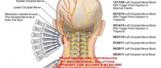
How is brachial neuritis diagnosed and treated?
The inflammatory process develops in the shoulder area and affects the nerve fibers of the brachial plexus. Treatment begins at the first signs of illness. Full recovery will take 2-4 weeks.
Causes of inflammation
The pain syndrome mainly affects the right side. Brachial plexus neuralgia develops due to a number of predisposing circumstances:
- due to hyporthermia (hypothermia);
- joint bruise or injury to the bones of the collarbone;
- ARVI, infection of soft tissues in the shoulder area by a virus;
- physical fatigue;
- inflammation of the ligaments.
The cause of brachial nerve neuritis is cold in rainy or frosty weather. Problems arise in the summer due to cold air from a working air conditioner, due to a draft. Staying in low temperature water for a long time will provoke a relapse. When diagnosing, it is important to know the root cause of the development of plexitis.
Pathology is caused by regular overload associated with work or sports. Age-related changes, hormonal imbalance, and diabetes mellitus affect the development of inflammation. If 1-2 sections of the nerve are affected, then sensitivity is impaired. If the entire nerve plexus is affected, arm paralysis is possible without proper treatment.
Manifestation of symptoms
The first bell is an involuntary muscle spasm, reminiscent of a trembling in the hand. Occurs temporarily, or the twitching becomes permanent. If you do not pay attention to the symptom, swelling of the muscle tissue will begin, which compresses the nerve endings. In this state of affairs, a dull, aching pain occurs. She worries constantly and is very exhausting. Acute resembles an attack. The pain intensifies with movement.
Gradually, the neuritis spreads to the entire arm. Other unpleasant sensations arise: numbness in certain areas of the shoulder, tingling, a feeling of crawling, difficulty moving the shoulder. The patient's temperature rises and general weakness appears. For treatment of pathology, contact a neurologist. The development of the inflammatory process is judged after examinations.
Treatment course
If you consult a doctor in a timely manner, treatment occurs quickly and there are no further relapses. After diagnosing the causes of inflammation, treatment of plexitis
from relieving pain attacks with the help of painkillers. The second step is to eliminate the inflammation with medications prescribed by the doctor. Conservative treatment is sufficient. Injections are prescribed. Injections suppress the effect of infection and remove harmful substances from the body.
Anesthetic ointments have an anti-inflammatory effect. Warming stimulates blood circulation and restores the functioning of muscle tissue. They are used after complete removal of swelling. The third stage is restoration. Rehabilitation measures include physiotherapy:
- laser therapy – the affected nerve is treated with light energy from a laser emitter;
- cryotherapy – the nerve plexus is cooled with a special medical device;
- electrophoresis is prescribed to introduce medicine through the skin using an electric current;
- magnetic therapy – a procedure to stimulate blood circulation;
- acupuncture (acupuncture);
- hirudotherapy – treat the sore spot with live medicinal leeches.
Physiotherapy helps relieve pain and reduce inflammation. After course procedures, muscle function returns to normal.
As prescribed by the doctor, they undergo a course of paraffin wraps, treatment with ozokerite, and take radon baths. Sea bathing and therapeutic exercises help. Preventive measures include swimming and physical exercise. For brachial neuralgia, therapeutic mud baths and massage are recommended, which will relax the spasmodic shoulder muscles and the swelling will go away. The massage procedure stimulates blood circulation in the affected area.
Therapeutic exercise restores the motor ability of the hand, because after exercise, blood flow in the joints improves and the muscular-ligamentous apparatus is strengthened. Special exercises work on muscle tissue. The training alternates between active and passive movements, gradually increasing the load.
Folk remedies for treatment
Traditional methods will help relieve pain and inflammation and speed up recovery. They are used as complementary therapy. Thermal procedure is used in consultation with the attending physician:
- Warm up the shoulder area with heated salt in a bag;
- Propolis tincture is used as a compress in combination with olive oil and crushed plantain;
- Rub the shoulder with horseradish juice, black radish, and fir oil;
- Warm baths with decoctions of sage, chamomile, oak bark, and walnut leaves relieve pain.
Traditional recipes:
- Geranium leaves are wrapped in a linen bag and tied to the shoulder for three hours.
- The inside of a boiled hot egg is applied to the sore area.
- An infusion of willow bark is useful. Pour 10 g of boiling water, the drink is infused for some time. Drink one tbsp 4 times a day. spoon.
- An ointment is prepared from boiled lilac buds. The gruel is mixed with pork fat.
- A drink made from dried mint leaves helps. They are poured with boiling water and allowed to settle. Drink half a glass of infusion in the morning and evening.
Brachial neuralgia does not depend on age.
It brings discomfort into the life of any person. It is important to consult a doctor in a timely manner and begin treatment in order to eliminate the symptoms that arise quickly. Do not self-medicate. Taking painkillers will give short-term results, but will not eliminate the root cause of the disease. Only a specialist will restore the motor ability of the hand. Author: K.M.N., Academician of the Russian Academy of Medical Sciences M.A. Bobyr
Carpal tunnel syndrome
Carpal tunnel syndrome (carpal tunnel syndrome) is the most common form of compression-ischemic neuropathy encountered in clinical practice. In the population, carpal tunnel syndrome occurs in 3% of women and 2% of men [Berzins Yu.E., 1989]. This syndrome is caused by compression of the median nerve as it passes through the carpal tunnel under the transverse carpal ligament. The exact cause of carpal tunnel syndrome is not known. The following factors most often contribute to compression of the median nerve in the wrist area: • Trauma (accompanied by local swelling, tendon stretching). • Ergonomic factors. Chronic microtraumatization (often found among construction workers), microtraumatization associated with frequent repeated movements (among typists, with constant long-term work with a computer). • Diseases and conditions accompanied by metabolic disorders, edema, tendon and bone deformities (rheumatoid arthritis, diabetes mellitus, hypothyroidism, acromegaly, amyloidosis, pregnancy). • Massive formations of the median nerve itself (neurofibroma, schwannoma) or outside it in the wrist area (hemangioma, lipoma).
Arm pain
Gout
Arthritis
Rheumatism
Ulcer
27923 March 26
IMPORTANT!
The information in this section cannot be used for self-diagnosis and self-treatment.
In case of pain or other exacerbation of the disease, diagnostic tests should be prescribed only by the attending physician. To make a diagnosis and properly prescribe treatment, you should contact your doctor. Pain in the hand: causes of occurrence, what diseases it occurs with, diagnosis and treatment methods.
Definition
Pain can be accompanied by various pathological processes of the upper extremities - injuries, degenerative and inflammatory lesions, neurological syndromes. Pain in the arm can be one of the symptoms of diseases of the cardiovascular, respiratory and digestive systems of the body.
Types of hand pain
The variety of pain syndromes is determined not only by the complexity of the structure of the upper limbs, but also by the variety of functional loads. By its nature, the pain can be pulling, shooting, aching. It may bother you when there is strain on the arm, or it may occur at night and disturb sleep. In addition, irradiation (return) of pain to the arm is possible due to myocardial infarction, angina pectoris, colic, cholecystitis, and stomach ulcers.
Possible causes of arm pain
Arm pain can be physiological in nature and be caused by muscle fatigue
after heavy or unusual stress.
It occurs due to the accumulation of anaerobic metabolic products (lactate) in muscle tissue and resolves within two to three days.
After significant physical activity exceeding the muscle endurance threshold, delayed pain may occur (1–2 days after physical activity). Experts believe that they are caused by damage to muscle cells, their membranes, and connections between microfibrils. Such pain is usually long-lasting.
Injuries
– bruises, fractures, tendon sprains, muscle tears – are characterized by a sharp onset of pain and its intensity. Bruises and fractures are accompanied by severe pain, swelling, and hemorrhage from small or large vessels.
A sprain occurs when there is a sudden movement in the joint, lifting significant weights, or a fall with emphasis on the arm and is characterized by varying degrees of damage to the connective tissue fibers. This injury is accompanied by severe pain, swelling and limited mobility in the joint. Often, sprains occur in the area of the wrist and elbow joints after heavy loads or monotonous movements repeated many times.
Muscle ruptures and tears occur when overused. In addition to severe pain, there is hemorrhage in the area of injury and the inability to tense the muscle.
One of the largest groups of diseases that causes severe pain in the arm is tunnel syndromes
caused by compression and inflammation of the nerve in a narrow space (tunnel) formed by the muscles, ligaments and bones of the arm.
Additional factors that increase the risk of such inflammation are endocrine diseases (diabetes mellitus, hypothyroidism), impaired joint mobility due to arthritis or rheumatism, tumor formations in the nerve area (neurofibroma, schwannoma) or beyond it (hemangioma, lipoma). As a rule, tunnel syndromes develop during prolonged similar movements (when working at a computer, playing tennis) or injuries. This is facilitated by incorrect posture, scoliosis, and osteochondrosis.
Inflammation of tendons and joint ligaments
also develops with significant physical stress and injury, especially in cases where the tendons are attached to powerful muscles. Stretching and microtrauma at the attachment site are accompanied by aching, aching pain, which can become unbearable during physical activity. Inflammation leads to swelling and limited mobility in the joint. A common pathology of the hand is epicondylitis, in which inflammation affects the junction of the muscle and ligament of the elbow joint. In this case, the pain is localized in the elbow area and accompanies movement in the elbow, hand and fingers.
Joint damage due to arthritis
causes not only pain, but also stiffness of movement. The causes of such damage may be osteoarthritis, gout, etc.
In any case, all signs of inflammation are present: pain, swelling, local increase in temperature, redness of the skin in the projection of the joint, impaired movement in the joint.
Rheumatoid
arthritis
is characterized by damage to first the small and then large joints of the hand, which is accompanied by stiffness of movement in the morning. In addition to increased pain, joint deformation and limited mobility, the disease is also characterized by general manifestations of the disease - fatigue, sweating of the palms and feet, weight loss.
Deposition of uric acid salts (urates) in the joints is the main manifestation of gout
– leads to severe pain. Its intensity is so great that even simply touching the sore joints causes discomfort. The basis of the disease is a metabolic disorder.
With inflammation of the ulnar periarticular synovial bursa - bursitis
– pain occurs in the elbow and is accompanied by swelling of the joint. In addition to severe pain in the arm, which limits movement, local redness of the skin and increased temperature over the joint area may be observed.
Arm pain may be due to anterior scalene syndrome
and be accompanied by muscle spasm and compression of the nerves and vessels of the shoulder girdle. The disease may be based on degenerative changes in the vertebrae (osteochondrosis) of the cervical spine or trauma.
The patient is bothered by pain in the neck and shoulders, which prevents him from raising his arms up and to the sides, taking a deep breath, or tilting his head.
Osteochondrosis of the cervical spine and its complications
(disc protrusion, intervertebral hernia) cause pain in the arm due to pinching of the nerve that innervates it. In this case, not only sharp pain may occur, but also numbness of the hand and impaired sensitivity.
There is another reason that should always be kept in mind when pain in the arm occurs - myocardial infarction
. The patient feels severe pain behind the sternum, which can radiate to the neck, back and left arm. Such pain is not the only sign of this dangerous condition. As a rule, shortness of breath, cold sweat, and difficulty breathing are present.
Diagnosis and examinations for arm pain
To diagnose a condition that may be causing arm pain, it is necessary to consider how acute the pain is and the events that precede its onset. With severe injuries, making a diagnosis is usually not difficult, but to distinguish a bruise from a fracture, radiography is necessary.
Clinical manifestations
Carpal tunnel syndrome is characterized by pain, numbness, paresthesia and weakness in the arm and hand.
Pain and numbness extend to the palmar surface of the thumb, index, middle and 1/2 ring finger, as well as to the dorsum of the index and middle finger. Initially, symptoms occur when performing any activities using a brush (working on a computer, drawing, driving), then numbness and pain appear at rest, sometimes occurring at night. The following tests are suggested to verify the diagnosis of carpal tunnel syndrome. Tinel test: tapping the wrist (above the median nerve) with a neurological hammer causes a tingling sensation in the fingers or pain radiating (electrical shooting) to the fingers (Fig. 2). Pain may also be felt in the tapping area. A positive Tinel sign is found in 26–73% of patients with carpal tunnel syndrome [Al Zamil M.H., 2008]. Durkan's test: compression of the wrist in the area of the median nerve causes numbness and/or pain in the 1st–3rd, half of the 4th fingers (as with Tinel's symptom). Phalen Test: Wrist flexion (or extension) 90 degrees produces numbness, tingling, or pain in less than 60 seconds (Figure 3). A healthy person may also develop similar sensations, but not earlier than after 1 minute. Opposition test: with severe thenar weakness (which occurs at a later stage), the patient cannot connect the thumb and little finger (Fig. 4); or the doctor (researcher) can easily separate the patient’s closed thumb and little finger.
Types of damage
Damage to the nerves of the hand is represented by the following groups.
- Complete rupture of a nerve is called neurotmesis. Surgical treatment is indicated. Recovery occurs over months or years depending on the severity of the damage.
- Partial rupture accompanied by various disorders - neuropraxia - occurs with closed injuries. A condition with preserved nerve sensitivity, when the lack of conduction is temporary.
- Neuropathy is a disorder resulting from a fracture, bruise or cut in the hand area.
- A pinched nerve occurs when the patient cannot bend the wrist. The little finger is completely immobilized, the ring finger is partially immobilized, the thumb moves with difficulty. The pain radiates to the little finger.
Differential diagnosis
Carpal tunnel syndrome should be differentiated from arthritis of the carpo-metacarpal joint of the thumb, cervical radiculopathy, and diabetic polyneuropathy. Patients with arthritis will show characteristic bone changes on x-rays. In cervical radiculopathy, reflex, sensory and motor changes will be associated with neck pain, while in carpal tunnel syndrome these changes are limited to distal manifestations. Diabetic polyneuropathy is usually a bilateral, symmetrical process involving other nerves (not just the median nerve). At the same time, a combination of polyneuropathy and carpal tunnel syndrome in diabetes mellitus cannot be ruled out.
Treatment
In mild cases of carpal tunnel syndrome, ice compresses and a decrease in load can help. If this does not help, the following measures must be taken: 1. Immobilization of the wrist. There are special devices (splints, orthoses) that immobilize the wrist and are convenient to use (Fig. 1). Immobilization should be carried out at least overnight, and preferably for 24 hours (at least in the acute period). 2. NSAIDs. Drugs from the NSAID group will be effective if the inflammatory process dominates in the pain mechanism. 3. If the use of NSAIDs turns out to be ineffective, it is advisable to inject novocaine with hydrocortisone into the wrist area. As a rule, this procedure is very effective. 4. In outpatient settings, electrophoresis can be performed with anesthetics and corticosteroids. 5. Surgical treatment. For mild or moderate carpal tunnel syndrome, conservative treatment is more effective. In cases where all conservative treatment options have been exhausted, surgical treatment is resorted to. Surgical treatment consists of partial or complete resection of the transverse ligament and releasing the median nerve from compression. Recently, endoscopic surgical methods have been successfully used in the treatment of carpal syndrome.
Pronator teres syndrome (Seyfarth syndrome)
Entrapment of the median nerve in the proximal part of the forearm between the pronator teres fascicles is called pronator syndrome.
This syndrome usually begins to appear after significant muscle activity over many hours involving the pronator and flexor digitorum muscles. Such types of activities are often found among musicians (pianists, violinists, flutists, and especially often among guitarists), dentists, and athletes [Zhulev N.M., 2005]. Long-term tissue compression is of great importance in the development of pronator teres syndrome. This can happen, for example, during deep sleep when the newlywed’s head is positioned for a long time on the partner’s forearm or shoulder. In this case, the median nerve in the pronator snuffbox is compressed, or the radial nerve in the spiral canal is compressed when the partner’s head is located on the outer surface of the shoulder (see radial nerve compression syndrome at the level of the middle third of the shoulder). In this regard, to designate this syndrome in foreign literature, the terms “honeymoon paralysis” (honeymoon paralysis, newlywed paralysis) and “lovers paralysis” (lovers paralysis) have been adopted. Pronator teres syndrome sometimes occurs in nursing mothers. In them, compression of the nerve in the area of the pronator teres occurs when the baby’s head lies on the forearm, he is breastfed, lulled to sleep, and the sleeping person is left in this position for a long time.
Clinical manifestations
With the development of pronator teres syndrome, the patient complains of pain and burning 4–5 cm below the elbow joint, along the anterior surface of the forearm and pain radiating to the 1st–4th fingers and palm.
Tinel's syndrome. In case of pronator teres syndrome, Tinel's sign will be positive when tapping with a neurological hammer in the area of the pronator snuff box (on the inside of the forearm). Pronator-flexor test. Pronating the forearm with a tightly clenched fist while creating resistance to this movement (counteraction) leads to increased pain. Increased pain can also be observed when writing (prototype of this test). When examining sensitivity, a sensitivity disorder is revealed, involving the palmar surface of the first three and a half fingers and the palm. The sensory branch of the median nerve, innervating the palmar surface of the hand, usually passes above the transverse carpal ligament. The occurrence of sensory disturbances on the palmar surface of the first finger, the dorsal and palmar surfaces of the second and fourth fingers, with preservation of sensitivity in the palm, allows one to confidently differentiate carpal tunnel syndrome from pronator teres syndrome. Thenar atrophy in pronator teres syndrome is usually not as severe as in progressive carpal tunnel syndromes.
Supracondylar process syndrome of the shoulder (Strother's band syndrome, Coulomb, Lord and Bedossier syndrome)
In the population, in 0.5–1% of cases, a variant of the development of the humerus is observed, in which a “spur” or supracondylar process (apophysis) is found on its distal anteromedial surface. Due to the accessory process, the median nerve is displaced and stretched (like a bowstring). This makes him vulnerable to defeat. This tunnel syndrome, described in 1963 by Coulomb, Lord and Bedossier, has almost complete similarities with the clinical manifestations of pronator teres syndrome: pain, paresthesia, and decreased flexion strength of the hand and fingers are detected in the zone of innervation of the median nerve. In contrast to pronator teres syndrome, when the median nerve is damaged under Strather's ligament, mechanical compression of the brachial artery with corresponding vascular disorders is possible, as well as severe weakness of the pronator teres (teres and minor). The following test is useful in diagnosing supracondylar process syndrome. When extending the forearm and pronation in combination with formed flexion of the fingers, painful sensations are provoked with localization characteristic of compression of the median nerve. If it is suspected that the compression is caused by a “spur” of the humerus, an x-ray examination is indicated. Treatment involves resection of the supracondylar process (“spur”) of the humerus and ligament.
Exercise therapy for radiation neuritis
Therapeutic exercise complex (PT) is considered one of the effective methods for treating radial neuritis. When treating radial nerve neuritis, various rehabilitation exercises are combined, taking into account the individual characteristics of the patient and the condition of the “sick” nerve of the upper extremities.
If the radial nerve is damaged, the emphasis is on exercises aimed at flexing the hand and fingers, abducting the first finger.
Then, sequentially, as the patient’s condition improves, the exercises “increase”, become more complex, and become more dynamic.
Exercise therapy helps improve blood supply, restore nerves, provide muscle tissue with nerve cells, and also helps accelerate the rehabilitation of lost functions.
Cubital tunnel syndrome
Cubital tunnel syndrome (Sulcus Ulnaris Syndrome) is compression of the ulnar nerve in the cubital canal (Mouchet's canal) in the area of the elbow joint between the internal epicondyle of the humerus and the ulna bone and is the second most common after carpal tunnel syndrome. Cubital tunnel syndrome develops for a number of reasons. Cubital tunnel syndrome can be caused by repetitive bending of the elbow joint. Therefore, cubital tunnel syndrome is classified as a disorder called accumulated trauma disorder (overuse syndrome). Those. the disorder may occur during normal, repetitive movements (most often associated with a specific occupational activity) in the absence of obvious traumatic injury. Direct trauma can also contribute to the development of cubital tunnel syndrome, such as leaning on the elbow while sitting. Patients with diabetes and alcoholism are at greater risk of developing cubital tunnel syndrome.
What are the causes of damage to the radial, ulnar and median nerves?
The radial, ulnar and median nerves pass through narrow canals formed by bones, ligaments, tendons and muscles. Nerve tissue is very delicate and vulnerable. Often we detect nerve suffering even with slight narrowing or deformation of the nerve canal. Narrowing or deformation of the canals of the radial, ulnar and median nerves occurs:
- In case of injury (bone fracture, bruise, hemorrhage, sprain or wound); Often, when injured, the nerve is pulled into scarred tissue or compressed by a bone fragment; nerve injury can also be represented by a nerve bruise, partial or complete interruption.
- When squeezed in an uncomfortable position (tucked or pinned hand while unconscious or intoxicated, under anesthesia);
- With thickening and deformation of the elbow or wrist joints and ligaments , with chronic injury and overload of the joints, often associated with the type of activity (athletes, musicians, drivers, cooks, dentists, working with vibration and metalworking tools). Very often, such changes are facilitated by diseases of the cervical spine. These types of neuropathy are called tunnel syndromes of the ulnar, radial and median nerves (cubital tunnel syndrome, carpal tunnel syndrome) .
Stimulation electromyography of the upper extremities, electroneuromyography (ENMG) of the upper extremities / EMG of the arms
The function of the nerves is to conduct electrical impulses from the brain to the muscles and organs, and to the brain from the sensory receptors of the body. Any damage to the nerve leads to disruption of the conduction of excitation along the nerve, which means disconnection of the affected part of the body from the brain.
Therefore, if the radial, ulnar or median nerve suffers, the following are possible:
- Decreased strength and weight loss of arm muscles
- Decreased sensitivity (numbness).
Clinical manifestations
The main symptoms of cubital tunnel syndrome are pain, numbness and/or tingling. Pain and paresthesia are felt in the lateral part of the shoulder and radiate to the little finger and half of the fourth finger. At first, discomfort and pain occur only when pressure is applied to the elbow or after prolonged bending. In a more severe stage, pain and numbness are felt constantly. Another sign of the disease is weakness in the arm. It manifests itself as a loss of “confidence” in the hand: suddenly objects begin to fall out of it during some habitual actions. For example, it becomes difficult for a person to pour water from a kettle. In advanced stages, the hand on the sore arm begins to lose weight, and pits appear between the bones due to muscle atrophy.
Diagnostics
In the early stages of the disease, the only manifestation (besides weakness of the forearm muscles) may be loss of sensation on the ulnar side of the little finger. If the clinical picture is blurred, the following tests can help verify the diagnosis of Cubital Tunnel Syndrome: Tinel test - the occurrence of pain in the lateral part of the shoulder, radiating to the ring finger and little finger when tapping with a hammer over the area where the nerve passes in the area of the medial epicondyle. Equivalent to Phalen's sign, sudden flexion of the elbow will cause paresthesia in the ring and little fingers. Frohman's test. Because of weakness of the abductor policis brevis and flexor policis brevis, excessive flexion of the interphalangeal joint of the thumb on the affected hand may be detected in response to a request to hold a paper between the thumb and index finger (Figure 5). Wartenberg test. Patients with more severe muscle weakness may complain that when putting their hand in a pocket, the little finger is pulled to the side (does not fit into the pocket) (Fig. 6).
Treatment
At the initial stages of the disease, conservative treatment is carried out.
Changing the load on the elbow and eliminating elbow flexion as much as possible can significantly reduce pressure on the nerve. It is recommended to fix the elbow joint in an extension position at night with the help of orthoses, hold the car steering wheel with your arms straightened at the elbows, straighten the elbow when using a computer mouse, etc. If the use of traditional drugs (NSAIDs, COX-2 inhibitors, splinting) for 1 week does not have a positive effect, an injection of an anesthetic with hydrocortisone is recommended. If the effectiveness of these measures is insufficient, then an operation is performed. There are several techniques for surgical release of the nerve, but all of them in one way or another involve moving the nerve anteriorly from the internal epicondyle. After surgery, treatment is prescribed aimed at quickly restoring nerve conduction. Guyon's tunnel syndrome Guyon's tunnel syndrome develops due to compression of the deep branch of the ulnar nerve in the canal formed by the pisiform bone, the hook of the hamate, the palmar metacarpal ligament and the palmaris brevis muscle. There are burning pains and sensitivity disorders in the 4th–5th fingers, difficulty in pinching movements, adduction and extension of the fingers.
Tunnel ulnar syndrome is very often the result of prolonged pressure from working tools, for example, vibrating tools, screwdrivers, pliers, and therefore occurs more often in representatives of certain professions (gardeners, leather cutters, tailors, violinists, people working with jackhammers). Sometimes the syndrome develops after using a cane or crutch. Pathological factors that can cause compression also include enlarged lymphatic ganglia, fractures, arthrosis, arthritis, ulnar artery aneurysm, tumors and anatomical formations around Guyon's canal. Differential diagnosis. The difference between Guyon's canal syndrome and ulnar canal syndrome is indicated by the fact that when a nerve is damaged in the hand area, pain occurs in the hypothenar and base of the hand, as well as intensification and irradiation in the distal direction during provoking tests. In this case, sensitivity disorders occupy only the palmar surface of the 4th–5th fingers. On the back of the hand, sensitivity is not impaired, since it is provided by the dorsal branch of the ulnar nerve, which arises from the main trunk at the level of the distal third of the forearm.
When making a differential diagnosis with radicular syndrome (C8), it should be taken into account that paresthesia and sensitivity disorders can also appear along the ulnar edge of the hand. Paresis and hypotrophy of the hypothenar muscles are possible. But with C8 radicular syndrome, the zone of sensory disorders is much larger than with Guyon’s canal, and there is no hypotrophy and paresis of the interosseous muscles. If the diagnosis is made early, limiting activity may help. Patients can be recommended to use fixators (orthoses, splints) at night or during the day to reduce trauma. If conservative measures fail, surgical treatment is performed aimed at reconstructing the canal in order to free the nerve from compression.
Physical education for radial neuritis
To restore function in case of radial nerve neuritis, exercises with resistance, weights, as well as exercises on a ladder consisting of vertical racks with transverse bars and other gymnastic devices are recommended.
— Gymnastics, which the patient does in a sitting position, is called passive. The exercises are aimed at developing flexion movements of the hand and fingers, the method of correctly positioning the forearm, and an attempt to place the first finger in opposition to the others.
— Gymnastics that restores and strengthens damaged muscles is called active healing. Here they practice exercises for the hand, first and second fingers, for the forearm, etc.
Exercises to treat radial neuritis
Specialized training for recovery from radial neuritis promotes the development of more active contraction of the radial and elbow flexors and extensors of the hand.
- Place the cushion under the wrist joint. Move the hand upward and to the starting position.
- Place a small pillow under the wrist joint. From this position, move your hand from side to side.
- Place a small pad under the wrist joint. Vigorously extend the hand upward, first without effort, and then with maximum effort through resistance.
- With the uninjured hand, bend and straighten first the first, then the second phalanges of the index, middle, ring and little fingers, while the thumb should be inside the palm.
- Squeeze your fingers into a fist and straighten.
- Pour water into a basin and place a small towel in it. Moving your bent fingers “on your bellies” from one edge to the other, squeeze and lightly knead the material in your hands.
Radial nerve compression syndrome
Three variants of compression damage to the radial nerve can be distinguished: 1. Compression in the armpit area.
Rarely seen. It occurs as a result of the use of a crutch (“crutch paralysis”), and paralysis of the extensors of the forearm, hand, main phalanges of the fingers, abductor pollicis muscle, and supinator develops. The flexion of the forearm is weakened, the reflex from the triceps muscle fades. Sensitivity is lost on the dorsal surface of the shoulder, forearm, and partly the hand and fingers. 2. Compression at the level of the middle third of the shoulder (spiral canal syndrome, “Saturday night paralysis”, “park bench”, “bench” syndrome). It occurs much more often. The radial nerve, emerging from the axillary region, bends around the humerus, where it is located in the bony spiral groove (groove), which becomes the musculoskeletal tunnel, since the two heads of the triceps muscle are attached to this groove. During the period of contraction of this muscle, the nerve is displaced along the humerus and as a result can be injured during forced repeated movements in the shoulder and elbow joints. But most often, compression occurs due to compression of the nerve on the outer-posterior surface of the shoulder. This usually occurs during deep sleep (deep sleep often occurs after drinking alcohol, which is why it is called “Saturday night syndrome”), in the absence of a soft bed (“park bench syndrome”). Pressure on the nerve may be due to the location of the partner's head on the outer surface of the shoulder. 3. Compressive neuropathy of the deep (posterior) branch of the radial nerve in the subulnar region (supinator syndrome, Froese syndrome, Thomson-Kopell syndrome, “tennis elbow” syndrome). Tennis elbow, tennis elbow, or epicondyllitis of the lateral epicondyle of the humerus is a chronic disease caused by a degenerative process in the area of muscle attachment to the lateral epicondyle of the humerus. Compression syndrome of the posterior (deep) branch of the radial nerve under the aponeurotic edge of the short extensor carpi radialis or in the tunnel between the superficial and deep bundles of the supinator muscle of the forearm can be caused by muscle overload with the development of myofasciopathies or pathological changes in perineural tissues. It manifests itself as pain in the extensor muscles of the forearm, their weakness and hypotrophy. Dorsal flexion and supination of the hand, active extension of the fingers against resistance provoke pain. Active extension of the third finger while pressing it and simultaneously straightening the arm at the elbow joint causes intense pain in the elbow and upper forearm. Treatment includes general etiotropic therapy and local effects. Take into account the possible connection of tunnel syndrome with rheumatism, brucellosis, arthrosis of metabolic origin, hormonal disorders and other conditions that contribute to compression of the nerve by surrounding tissues. Anesthetics and glucocorticoids are injected locally into the area of the pinched nerve. Complex treatment includes physical therapy, the prescription of vasoactive, decongestant and nootropic drugs, antihypoxants and antioxidants, muscle relaxants, ganglion blockers, etc. Surgical decompression with dissection of the tissues compressing the nerve is indicated if conservative treatment is unsuccessful. Thus, hand tunnel syndromes are a type of damage to the peripheral nervous system caused by both endogenous and exogenous influences. The outcome depends on the timeliness and adequacy of treatment, correct preventive recommendations, and the patient’s orientation in choosing or changing a profession that predisposes to the development of tunnel neuropathy.
The article uses drawings from the book by S. Waldman. Atlas of commom pain syndromes. – Saunders Elsevier. – 2008.
Treatment of radial neuritis
To cure radial neuritis, it is necessary to first eliminate the cause of its development. Specific measures here depend on its nature:
- if neuritis is provoked by an infectious disease, then antibiotic and antiviral agents are prescribed;
- if the disease is caused by blood supply disorders, then in most cases vasodilator drugs are prescribed;
- If the disease is traumatic, it is necessary, first of all, to completely immobilize the affected limb and eliminate its use as much as possible.
To stop inflammation, the doctor prescribes anti-inflammatory and decongestant drugs, analgesics, and B vitamins. Anticholinesterase drugs and biogenic stimulants are also successfully used.
Successful treatment of neuritis is facilitated by a course of physiotherapy, which can be carried out after 1 week of drug treatment. Physiotherapeutic agents that have proven effectiveness in the treatment of radial neuritis include:
- ultraphonophoresis;
- electrophoresis;
- exposure to pulsed currents;
- electrical stimulation;
- UHF therapy;
- acupuncture.
Massages of the area innervated by it and exercise therapy can also speed up the restoration of the functions of the affected nerve.