A little about history
Many scientists were involved in constructing a map of the surface of the brain: Bailey, Betz, Economo and others. Their maps differed significantly from each other in the shape of the fields, their size, and number. In modern neuroanatomy, Brodmann's brain fields have received the greatest recognition. There are 52 fields in total.
Pavlov, in turn, divided all fields into two large groups:
- centers of the first signal system;
- centers of the second signaling system.
Each center consists of a core, which plays a key role in performing the function of a specific center, and analyzers surrounding the core. It is noteworthy that centers in the cerebral cortex regulate the functioning of organs on the opposite side of the body. This is due to the fact that the nerve fiber pathways cross on their way from the center to the periphery.
Brodmann's brain fields are designated by Arabic numerals, some also have a designation from which the function of a particular field can be understood.
First Signal System: Location
The centers of the first signaling system are located in Brodmann's fields, which are present in both animals and humans. They are responsible for a simple reaction to an external stimulus, the formation of sensations and ideas. These centers are present in both the right and left hemispheres of the cerebral cortex. Brodmann fields of the first signaling system are present in humans from birth and normally do not undergo changes throughout life.
These fields include:
- 1 - 3 - are located in the parietal lobe of the cerebral cortex behind the central gyrus;
- 4, 6 - located in the frontal lobe anterior to the central gyrus, they contain Betz pyramidal cells;
- 8 - this field is located anterior to the 6th, closer to the frontal part of the frontal cortex;
- 46 - located on the outer surface of the frontal lobe;
- 41, 42, 52 - located on the so-called Heschle convolutions, on the basal part of the temporal lobe of the brain;
- 40 - located in the parietal lobe behind 1 - 3 fields, closer to the temporal part;
- 17 and 19 - located in the occipital part of the brain, most dorsal from the other fields;
- 11 is one of the most ancient structures, located in the hippocampus.
Divisions of the brain
- Rear. Connects the medulla oblongata, cerebellum, and pons.
- Average. The smallest part of the brain.
- Front. Refers to the largest part, occupying more than 2/3 of the volume, includes the telencephalon and diencephalon.
Each department performs certain functions, but at the same time they closely interact with each other.
Posterior
Located at the back of the skull. If we consider the medulla oblongata, which is part of this section, then it is a kind of connecting link between the spinal cord and the head. At the same time, it is responsible for regulating the important functions of coordinating the work of the heart and maintaining unconditioned reflexes. This section contains the entrance and exit of nerve endings; signals are transmitted from the spinal cord to the head.
Cerebellum
A small but very important part responsible for human coordination and adaptation of the body to new conditions. The cerebellum regulates muscle activity, helps maintain balance or stabilize posture, and allows for sequential actions.
Pons
The anatomical structure of the human brain includes a special transverse wave or pons. This part unites the cerebellum, medulla oblongata and cerebral cortex.
Middle section
The structure of the middle section presupposes the presence of a quadrigeminal region in the upper part, which performs a number of important functions regarding various perceptions of information. Tissues provide reflex transformations of auditory and visual reflexes.
Anterior section
It consists of main parts - the diencephalon and the telencephalon, which also additionally have certain functional elements. Intermediate includes:
- Thalamus. Occupies 80% of the diencephalon; almost all signals are processed through the thalamus and only then enter the cerebral cortex. Regulates sensitive abilities, visual, tactile, olfactory and others.
- Hypothalamus. Controls the functioning of internal organs, promotes normal temperature exchange, is responsible for the autonomic system, heart rate, libido, memory and a number of other important physiological and behavioral features.
- Epithalamus. It includes the pineal gland, which regulates the cycle of wakefulness and sleep, synthesizes the hormone melatonin, affects metabolic processes, and the concentration of hormonal compounds.
The hypothalamus connects to the pituitary gland, one of the important glands of the endocrine system. It is she who synthesizes hormones that help the thyroid gland function during labor, lactation, and also in a number of other metabolic processes.
Finite brain
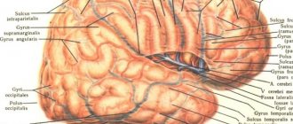
The anatomy of the human brain involves two hemispheres, a connecting sulcus and some other tissues that combine to form the terminal section. The surface of the hemispheres is conventionally divided into the following lobes:
- Frontal. Mainly affects logical abilities, speech, motor activity.
- Parietal. Responsible for the ability to touch, taste, smell, and to some extent verbal memory.
- Occipital. Perceives information received by the visual organs and the retina of the eye.
- Temporal. Helps perceive sounds and process signals. The temporal part is associated with the ability to remember, perceive speech and other important functions.
The structure of the brain is relatively complex, it includes many separate structures that perform various functions, thanks to which a person can live, feel, and realize his needs.
First signaling system: functions
The functions of Brodmann's fields in the first signaling system differ depending on the location of the center and the characteristics of its histological structure. In general, these kernels perform the following functions:
- implementation of the motor process;
- recognition of objects by touch;
- hearing;
- vision.
To carry out precise movement, simultaneous activation of several Broca's areas is necessary:
- Centers 4 and 6, whose pyramidal cells carry impulses to skeletal muscles and ensure their contraction.
- Field number 40, where the centers for performing complex movements that are stereotypical for a particular person are located. These centers are formed during the life of an individual, as a rule, during professional activity.
- Sometimes it is necessary to activate field 46, which is responsible for the synchronous rotation of the eyes along with the head.
Fields numbered 5 and 7 are involved in recognizing objects by touch, or stereognosis.
Fields 41, 42 and 52 are necessary for a person to perceive the sounds of the surrounding world. Moreover, fibers from two ears at once approach the center of hearing on one side. Therefore, damage to the cortex on one side does not lead to hearing impairment. The center located in field 41 is responsible for the primary analysis of information. In field 42 there are auditory memory centers. And with the help of field number 52, a person can navigate in space.
In fields 17 to 19 there is a visual analyzer. By analogy with the auditory centers, primary analysis of information occurs in field 17, visual memory is located in field 18, and evaluation centers and orientation are located in field 19.
In field 11 there are centers of smell, in field 43 there are centers of taste.
Notes
- ↑Brodmann Korbinian Vergleichende Lokalisationslehre der Grosshirnrinde: in ihren Principien dargestellt auf Grund des Zellenbaues. — Leipzig: Johann Ambrosius Barth Verlag, 1909.
- ↑Sapin M.R., Bilich G.L. Human anatomy. - M.:: “Higher School”, 1989. - P. 417. - 544 p. — 100,000 copies. — ISBN 5-06-001145-3
- ↑Gerhardt von Bonin {amp}amp; Percival Bailey The Neocortex of Macaca Mulatta. — Urbana, Illinois: The University of Illinois Press, 1925.
- ↑E. D. Chomsky Neuropsychology, 4th edition. — Peter, 2008.
| This article lacks links to sources of information. Information must be verifiable, otherwise it may be questioned and deleted. You can edit this article, adding links to authoritative sources. This mark is set June 12, 2012 . |
Second Signal System: Location
The presence of a second signaling system is characteristic only of humans. It is these centers that provide higher thinking, which includes generalization of information, dreams, and logic. In fact, for normal thinking and speech, activation of all Brodmann fields is necessary, but centers can be distinguished that have their own specific functions:
- 44 - located in the posterior part of the inferior frontal gyrus;
- 45 - located anterior to field 44, in the anterior portion of the frontal gyrus;
- 47 - located below the two previous fields, closer to the basal part of the frontal lobe;
- 22 - one of the most anterior parts of the temporal lobe;
- 39 - located in the posterior part of the superior temporal gyrus.
Paul Brodmann
- Fields 3, 1 and 2 - somatosensory area, primary zone. Located in the postcentral gyrus. Due to the commonality of functions, the term “fields 3, 1 and 2” is used (front to back)
- Field 4 is the motor area. Located within the precentral gyrus
- Area 5 is the secondary somatosensory area. Located within the superior parietal lobule
- Area 6 - premotor cortex and supplementary motor cortex (secondary motor area). Located in the anterior sections of the precentral and posterior sections of the superior and middle frontal gyri.
- Field 7 is the tertiary zone. Located in the upper parts of the parietal lobe between the postcentral gyrus and the occipital lobe
- Field 8 - located in the posterior parts of the superior and middle frontal gyri. Includes the center of voluntary eye movements
- Area 9 - dorsolateral prefrontal cortex
- Area 10 - anterior prefrontal cortex
- Area 11 - olfactory area
- Field 12 -
- Field 13 -
- Field 14 -
- Field 15 —
- Field 16 -
- Field 17 - nuclear zone of the visual analyzer - visual area, primary zone
- Field 18 - nuclear zone of the visual analyzer - center of perception of written speech, secondary zone
- Field 19 - nuclear zone of the visual analyzer, secondary zone
- Area 20 - inferior temporal gyrus (center of the vestibular analyzer)
- Area 21 - middle temporal gyrus (center of the vestibular analyzer)
- Field 22 - nuclear zone of the sound analyzer
- Field 23 -
- Field 24—error detector
- Field 25 —
- Field 26 -
- Field 27 -
- Field 28 - projection fields and associative zone of the olfactory system
- Field 29 -
- Field 30 —
- Field 31 -
- Area 32 - dorsal zone of the anterior cingulate cortex
- Field 33 -
- Field 34 -
- Field 35 —
- Field 36 -
- Field 37 - tertiary zone
- Field 38 -
- Area 39 - angular gyrus, part of Wernicke's area (center of the visual analyzer of written speech)
- Area 40 - marginal gyrus, part of Wernicke's area (motor analyzer of complex professional, labor and everyday skills)
- Field 41 - nuclear zone of the sound analyzer, primary zone
- Field 42 - nuclear zone of the sound analyzer, secondary zone
- Field 43 - taste area
- Field 44 - Broca Center
- Area 45 - triangular part of Brodmann area (musical motor center)
- Field 46 - motor analyzer of combined rotation of the head and eyes in different directions
- Field 47 —
- Field 48 -
- Field 49 —
- Field 50 —
- Field 51 -
- Field 52 -
Second signaling system: functions
As noted above, Brodmann’s cytoarchitectonic fields of the second signaling system are necessary for the implementation of higher nervous activity. And the main difference between a person and an animal is the ability to speak.
In field 45 is the Broca Center. It is necessary for normal speech motor skills. It is thanks to the presence of this center that a person is able to pronounce words. When it is damaged, a condition called “motor aphasia” develops.
In field 44 there is the center of written speech. Impulses from this area of the cortex travel to the skeletal muscles of the fingers and hand. When it is destroyed, a person loses the ability to write, which is called “agraphia.”
Field 47 is responsible for singing. It is during the normal functioning of this center that a person can pronounce words into a chant.
In field 22 there is the Wernicke center. This is where auditory speech analysis takes place. Thanks to the normal operation of the 22nd field, a person perceives words by ear.
Field 39 is the center of visual speech. The functioning of this field allows a person to distinguish between characters written on paper. When it is damaged, a person loses the ability to read, which is called sensory alexia.