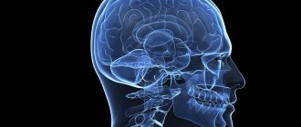
The spinal cord is the most important internal organ related to the structure of the central nervous system. The surface of the spinal cord has 3 membranes - arachnoid, hard and soft. The anatomy of the spinal cord is designed in such a way that the internal organ is the dominant system that supports the vital functions of the entire organism.
Structure of the spinal cord
Anatomical features
The spinal cord is located in the cavity of the spinal canal, which is formed by the processes of the vertebrae and their bodies. The beginning of the structure of the spinal cord is the foramen magnum of the brain. Next, the spinal cord is located in the canal, representing a 40-centimeter “cord” surrounded by three membranes.
The internal organ ends with a cluster of nerve fibers at the level of the first vertebrae in the lumbar region, called the cauda equina. This is where the narrowing begins, and then the internal organ is “stretched” into a terminal (terminal, terminal) filament, the diameter of which is 1 mm. The filum terminale extends to the coccygeal region, where it fuses with the periosteum.
The lower part of the trailing thread is tightly wrapped in ponytail fibers. When pain occurs in the coccyx area, doctors talk about a syndrome with the same name. The structure of the human spinal cord is such that the medulla itself is under constant protection - this is provided by the membranes and the spinal column itself.
The external structure is the shells and the space between them.
Where is gray matter located?
The gray matter of the brain is represented mainly by an accumulation of a large number of neurons with unmyelinated axons interwoven into glial tissues, their dendrites and blood capillaries that ensure their metabolism.
The largest accumulation of gray neurons forms the cerebral cortex, which covers the surface of the terminal section. The thickness of this structure is no more than 0.5 cm throughout, but it occupies more than 40% of the volume of the telencephalon, and its surface is many times greater than the plane of the cerebral hemispheres. This characteristic is determined by the presence of wrinkles and convolutions, which contain up to 2/3 of the area of the entire cortex.
Also, accumulations of gray matter in the brain form special nerve centers or nuclei, which have a characteristic shape and their functional purpose. The peculiarity of the structure of this structure is that the term “nucleus” means a paired or dispersed formation of neuron cells that do not have a myelin sheath.
There are a large number of nuclei of the nervous system, which, for the sake of a general concept and ease of perception, are usually identified according to the operation they perform, as well as their appearance. This distribution does not always correctly reflect reality, since the brain is a poorly understood structure of the central nervous system and sometimes scientists make mistakes.
The main cluster of nuclei is located within the brainstem, for example in the thalamus or hypothalamus. At the same time, the basal ganglia are located in the anterior section, which to some extent influence a person’s emotional behavior and are involved in maintaining muscle tone.
The gray matter of the cerebellum, like the terminal cortex, covers the hemispheres and the vermis at the periphery. Also, its individual ones form paired nuclei deep in the body of this rudiment.
Anatomically, the following types of nuclei are distinguished:
- Serrated. Located in the lower part of the white matter of the cerebellum, its pathways are responsible for the motor function of skeletal muscles, as well as for a person’s visual-spatial orientation in space.
- Spherical and cork-shaped. They process information received from the worm, and also receive afferent signals from parts of the brain responsible for somatosensory, auditory and visual data.
- Tent core. It is located in the tent of the cerebellar vermis and receives information about the position of the human body in space according to data received from the sense organs and the vestibular apparatus.
A characteristic feature of the structure of the spinal cord is that the gray substance in the form of nuclei is located inside the white component, but at the same time is an integral part of it. This arrangement can be seen in most detail when studying the spinal part of the central nervous system in a cross section, where a clear transition of gray matter into white matter from the center to the periphery will be clearly visible.
Blood supply
To ensure adequate nutrition of the brain, many large, medium and small blood vessels are connected to it. They originate from the aorta and vertebral arteries. The process involves the spinal arteries, anterior and posterior. The vertebral arteries supply the upper cervical segments.
Many additional vessels flow into the spinal arteries along the entire length of the spinal cord. These are radicular-spinal arteries, through which blood passes directly from the aorta. They are also divided into rear and front. The number of vessels may vary among different people, being an individual feature. Typically, a person has 6-8 radicular-spinal arteries. They have different diameters. The thickest ones feed the cervical and lumbar thickenings.
The inferior radicular-spinal artery (artery of Adamkiewicz) is the largest. Some people also have an additional artery (radicular-spinal) that arises from the sacral arteries. There are more radicular-spinal posterior arteries (15-20), but they are significantly narrower. They provide blood supply to the posterior third of the spinal cord along the entire cross section.
The vessels are connected to each other. These places are called anastomosis. They provide better nutrition to different parts of the spinal cord. The anastomosis protects it from possible blood clots. If a separate vessel is blocked by a blood clot, blood will still flow through the anastomosis to the desired area. This will save neurons from death.
In addition to the arteries, the spinal cord is generously supplied by veins, which are closely connected with the cranial plexuses. This is a whole system of vessels through which blood then flows from the spinal cord into the vena cava. To prevent blood from flowing back, there are many special valves in the vessels.
Interesting facts about the spinal cord
This organ has not yet been fully studied - it still hides many secrets from doctors, and their solution in the future may lead to the cure of currently incurable diseases of the nervous system. Here are some interesting facts about this amazing organ:
- If the spine grows over 20 years, then the spinal cord only grows for 5 years.
- Stress leads to a serious decrease in the number of neurons. If the normal number of neurons is 13-14 million , then as a result of stress their number drops by half - this is especially true for pregnant women.
- In the process of evolution of vertebrate organisms, the spinal cord appeared first, and only then the brain. The first performed all the simplest functions, including reflex ones.
- Some living beings are able to live after the loss of the brain , remaining only with the spinal cord.
- Damage to a specific area of the organ not only causes loss of sensation below the rupture site, but also the ability to sweat. This forces people with injuries to spend more time in the shadows, as the body has partially lost its thermoregulatory function, which is extremely important for life.
- Scientists have not yet come to a general conclusion, and cannot establish the mechanism of hair loss throughout the body in people with spinal cord injuries .
- If the thoracic organ has been affected, the person may lose the ability to cough.
- A biopsy and analysis of the white matter of an organ can detect hundreds and thousands of human diseases.
- The spinal cord very sensitively senses the rhythm of music, and therefore is automatically able to send signals that will make the body move to the rhythm.
- People with a healthy spine are much more active in their sexual life.
Meninges
- Hard shell. It is located immediately behind the periosteum of the spine, but does not adhere closely to it. The epidural space is located between the periosteum and the dura mater. The tissue of the hard shell is connective; it contains vessels, lymphatic and circulatory. The epidural space is filled with fatty tissue. Venous plexuses are also located here.
- The arachnoid membrane is a network of thin plates of connective tissue, similar in structure to a spider’s web. The plates are composed of collagen and elastic fibers. Between the arachnoid and soft membrane there is a subarochnoid space with cerebrospinal fluid, which ensures the exchange and nutrition of neurons.
- Soft shell. This is a vascular environment that has serrated ligaments for fixation and provides communication and nutrition between the cerebrospinal fluid and the brain.
| Program | Notes | Test papers |
Biology Tutor Notes
On this page you can find a summary of the topic “Spinal Cord”.
Of course, I cannot post all the materials that I use to prepare applicants for exams. However, you will be able to evaluate the level of the prepared material. I hope that when you turn to me for help, you will no longer buy a pig in a poke. You will know that your child or you is being taught by a specialist who knows his business - a biology tutor. You can read more detailed information about me here.
SPINAL CORD
The spinal cord is an oblong, somewhat flattened cylindrical cord, and therefore its transverse diameter throughout its entire length is usually larger than the anterior one. Located in the spinal canal from the level of the base of the skull to the I-II lumbar vertebrae, the spinal cord has the same curves as the spinal column, cervical and thoracic curves. The upper parts of the spinal cord pass into the brain, the lower parts end with the conus medullaris, the apex of which continues into a thin filum terminale. The length of the spinal cord in an adult is on average 43 cm, weight is about 34-38 g. Due to the metamerism of the structure of the human body, the spinal cord is divided into segments, or neuromeres. A segment is a section of the spinal cord with the right and left anterior (motor) roots emerging from it and the right and left dorsal (sensitive) roots penetrating into it.
Figure 1. Spinal cord.
| A, B - front view: | B - rear view: |
| 1 - bridge; | 1 - rhomboid fossa; |
| 2- medulla oblongata; | 2 - posterior median groove; |
| 3 - intersection of pyramids; | 3 - dorsal roots of the spinal nerves. |
| 4 - anterior median fissure; | |
| 5 - cervical thickening; | |
| 6-anterior roots of the spinal nerves; | |
| 7 - lumbosacral thickening; | |
| 8 - conus medullaris; | |
| 9 - ponytail; | |
| 10 - terminal thread. |
Along the entire length, 31 pairs of anterior and posterior roots depart from each side of the spinal cord, which, merging, form 31 pairs of right and left spinal nerves . Each segment of the spinal cord corresponds to a specific part of the body that receives innervation from this segment.
In the cervical and lumbar sections of the spinal cord, cervical and lumbosacral thickenings are found, the appearance of which is explained by the fact that these sections provide innervation to the upper and lower extremities, respectively.
Starting from the 4th month of fetal development, the spinal cord lags behind the growth of the spine. In this regard, there is a change in the direction of the roots. In an adult, the roots of the cranial segments still retain a horizontal course; in the thoracic and upper lumbar regions the roots follow obliquely - downwards and laterally; in the lower lumbar and sacrococcygeal regions, the roots, heading to the corresponding intervertebral lumbar and sacral foramina, are located almost vertically in the spinal canal. The combination of the anterior and posterior roots of the lower lumbar and sacrococcygeal nerves surrounds the filum terminale like a cauda equina.
Along the entire anterior surface of the spinal cord in the median fissure, and along the posterior surface is the posterior median sulcus. They serve as boundaries dividing the spinal cord into two symmetrical halves.
On the anterior surface, slightly lateral to the median sulcus, two anterior lateral grooves stretch - this is where the anterior roots leave the spinal cord on the right and left. On the posterior surface there are posterior lateral grooves - places where the dorsal roots enter the spinal cord on both sides.
The spinal cord contains gray and white matter. The central canal passes through the gray matter, the upper end of which communicates with the fourth ventricle.
The gray matter along the spinal cord forms two vertical columns located to the right and left of the central canal. In each column there are front and rear pillars . At the level of the lower cervical, all thoracic and two upper lumbar segments of the spinal cord, a lateral column , which is absent in other parts of the spinal cord.
On a cross section of the spinal cord, the gray matter has the shape of a butterfly or the letter “H”, it has a wider anterior horn and a narrow posterior horn . The anterior horns contain large nerve cells called motor neurons.
The gray matter of the dorsal horns of the spinal cord is heterogeneous. The bulk of the nerve cells of the posterior horn form their own nucleus, and at the base of the posterior horn, a well-defined layer of white matter, the thoracic nucleus , consisting of large nerve cells, is noticeable.
The cells of all nuclei of the dorsal horns of the gray matter are, as a rule, intercalary, intermediate neurons, the processes of which go in the white matter of the spinal cord to the brain.
The intermediate zone, located between the anterior and posterior horns, is represented by the lateral horn. The latter contains the centers of the sympathetic part of the autonomic nervous system.
The white matter of the spinal cord is located on the periphery of the gray matter. The grooves of the spinal cord divide it into sevenfold: anterior, middle and posterior cords. The anterior cord is located between the anterior median fissure and the anterior lateral sulcus, the posterior cord is between the posterior middle and posterior lateral sulci, the lateral cord is between the anterior and posterior lateral sulci.
The white matter of the spinal cord is represented by processes of nerve cells (sensory, intercalary and motor neurons), and the totality of processes of nerve cells in the cords of the spinal cord makes up three systems of bundles - tracts, or pathways of the spinal cord:
- short bundles of associative fibers connect segments of the spinal cord located at different levels;
- ascending (afferent, sensory) bundles are directed to the centers of the brain or to the cerebellum;
- descending (motor, efferent) bundles go from the brain to the cells of the anterior horns of the spinal cord. The ascending tracts are located in the white matter of the posterior cords. In the anterior and lateral funiculi there are ascending and descending fiber systems.
The anterior funiculi contain the following pathways:
1) anterior, motor, corticospinal (pyramidal) pathway. This path contains processes of pyramidal cells of the cortex of the anterior central gyrus, which end on the motor cells of the anterior horn of the opposite side, transmits impulses of motor reactions from the cerebral cortex to the anterior horns of the spinal cord;
2) the anterior spinothalamic tract in the middle part of the anterior cord provides the conduction of impulses of tactile sensitivity (touch and pressure);
3) on the border of the anterior cord with the lateral cord there is the vestibular cord, which originates from the vestibular nuclei of the VIII pair of cranial nerves located in the medulla oblongata and goes to the motor cells of the anterior horns. The presence of the tract allows you to maintain balance and coordinate movements.
The lateral funiculi contain the following pathways:
1) the posterior spinocerebellar tract occupies the posterior lateral sections of the lateral funiculi and is a conductor of reflex proprioceptive impulses directed to the cerebellum;
2) the anterior spinocerebellar tract is located in the anterolateral sections of the lateral funiculi, it follows into the cerebellar cortex;
3) lateral spinothalamic tract - the path for conducting impulses of pain and temperature sensitivity, located in the anterior sections of the lateral cord. Of the descending tracts in the lateral cords there are the lateral corticospinal (pyramidal) tract and the extrapyramidal - red nuclear spinal tract;
4) the lateral corticospinal tract is represented by fibers of the main motor pyramidal tract (the path of impulses that causes conscious movements), which lie medial to the posterior spinal cerebellar tract and occupy a significant part of the lateral cord, especially in the upper segments of the spinal cord;
5) the red nuclear-spinal tract is located ventral to the lateral corticospinal (pyramidal) tract. This pathway is a reflex motor efferent pathway.
The posterior cords contain pathways of conscious prioprioceptive sensitivity (conscious joint-muscular feeling), which are sent to the cerebral cortex and deliver information about the position of the body and its parts in space to the cortical analyzers. At the level of the cervical and upper thoracic segments, the posterior cords of the spinal cord are divided into two bundles by the posterior and intermediate grooves: a thin bundle (Gaull's bundle), lying more medially, and a wedge-shaped bundle (Burdach's bundle), adjacent to the posterior horn.
SPINAL CORD PATHWAYS
The spinal cord contains a number of neurons that give rise to long ascending pathways to various brain structures. The spinal cord also receives a large number of descending tracts formed by the axons of nerve cells localized in the cerebral cortex, midbrain and medulla oblongata. All these projections, along with the pathways connecting the cells of various spinal segments, form a system of pathways formed in the form of white matter, where each tract occupies a very specific position.
MAIN ASCENDING PATHWAYS OF THE SPINAL CORD
| Ascending (sensitive) pathways | Pathways | Columns of the spinal cord | Physiological significance |
| 1 | Thin beam (Gaull beam) | Rear | Tactile sensitivity, senses of body position, passive body movements, vibration |
| 2 | Wedge-shaped bundle (Burdach bundle) | >> | Same |
| 3 | Dorsolateral | Lateral | Pathways of pain and temperature sensitivity |
| 4 | Dorsal spinocerebellar Flexig | >> | Impulses from proprioceptors of muscles, tendons, ligaments; feeling of pressure and touch from the skin |
| 5 | Ventral spinocerebellar (Goversa) | >> | Same |
| 6 | Dorsal spinothalamic | >> | Pain and temperature sensitivity |
| 7 | Spinotectal | >> | Sensory pathways of visual-motor reflexes (?) and pain sensitivity (?) |
| 8 | Ventral spinothalamic | Front | Tactile sensitivity |
Some of them are fibers of primary afferent (sensitive) neurons running without interruption. These fibers, the thin (Gaull's bundle) and wedge-shaped (Burdach's bundle) bundles, are part of the dorsal funiculi of the white matter and end in the medulla oblongata near the neutron relay nuclei, called the nuclei of the dorsal funiculus, or the nuclei of Gaulle and Burdach. The fibers of the dorsal cord are conductors of skin-mechanical sensitivity.
The remaining ascending pathways begin from neurons located in the gray matter of the spinal cord. Because these neurons receive synaptic inputs from primary afferent neurons, they are commonly referred to as second-order neurons, or secondary afferent neurons. The bulk of fibers from secondary afferent neurons pass within the lateral funiculus of the white matter. The spinothalamic tract is located here. The axons of spinothalamic neurons cross and reach without interruption through the medulla oblongata and midbrain to the thalamic nuclei, where they form synapses with thalamic neurons. The spinothalamic tract carries impulses from skin receptors.
The lateral funiculi contain fibers of the spinocerebellar tracts, dorsal and ventral, carrying impulses from skin and muscle receptors to the cerebellar cortex.
The lateral cord also contains fibers of the spinocervical tract, the ends of which form synapses with relay neurons of the cervical spinal cord - neurons of the cervical nucleus. After switching in the cervical nucleus, this pathway goes to the cerebellum and brainstem nuclei.
The pain sensitivity pathway is localized in the ventral columns of white matter. In addition, the spinal cord’s own pathways pass through the posterior, lateral and anterior columns, ensuring the integration of functions and reflex activity of its centers.
| Descending (motor) tracts | Pathways | Columns of the spinal cord | Physiological significance |
| 1. | Lateral corticospinal (pyramidal) | Lateral. | Impulses to skeletal muscles. Voluntary movements. |
| 2. | Rubrospinal (Monakova) | « | Impulses supporting skeletal muscle tone |
| 3. | Dorsal vestibulospinal | « | Impulses that maintain body posture and balance |
| 4. | Olivospinal (Helweg) | « | Function unknown. Perhaps it is involved in the implementation of thalamospinal reflexes |
| 5. | Reticulospinal | Front | autonomic centers and sensitivity of muscle spindles, proprioceptors of skeletal muscles |
| 6. | Ventral vestibulospinal | « | Impulses that maintain body posture and balance |
| 7. | Tectospinal | « | Impulses that ensure the implementation of visual and auditory motor reflexes (reflexes of the quadrigeminal region) |
| 8. | Ventral corticospinal (pyramidal) | Front | Impulses to skeletal muscles, voluntary movements |
The descending tracts of the spinal cord are also divided into several independent tracts that occupy a specific position in the lateral and ventral cords of the white matter.
Evolutionarily more ancient descending pathways originate from neurons whose nuclei are located within the medulla oblongata and the pons. These are the reticulospinal and vestibular tracts. The reticulospinal tract is formed by the axons of neurons of the reticular formation of the hindbrain.
The evolutionarily younger descending pathway is the rubospinal tract, which reaches its greatest development only in mammals. Rubrospinal fibers are axons of neurons of the red nucleus located in the midbrain. The rubospinal tract crosses over and runs as part of the lateral funiculi of the white matter.
The most important descending pathway is the corticospinal or pyramidal tract, whose neurons are located in the motor zone of the cerebral hemispheres. The pyramidal tract is evolutionarily the youngest. It appears only in mammals and is most developed in primates and humans.
MEMBRANES OF THE SPINAL CORD
Externally, the spinal cord is surrounded by three membranes.
The outer - hard shell of the spinal cord is fixed by the ligamentous apparatus in the spinal canal and is separated from the periosteum of this canal by the epindual space filled with fatty tissue and venous plexus, the middle - thin, transparent, called the arachnoid membrane of the spinal cord; Internally, it is closely adjacent to the spinal cord and contains blood vessels that feed it - this is the pia mater of the spinal cord.
The dura mater of the spinal cord is an oblong sac with strong and thick walls, located in the spinal canal and containing the spinal cord with roots and membranes.
The inner surface of the dura mater of the spinal cord is separated from the arachnoid membrane by a narrow slit-like subdural space, which is penetrated by a large number of thin connective tissue crossbars. At the top, the subdural space of the spinal cord communicates freely with a similar space in the cranial cavity. Below, this space ends blindly at the level of the II sacral vertebra.
The arachnoid membrane of the spinal cord is tightly adjacent to the spinal cord. There are two layers in it - inner and outer, between which blood vessels are located. The inner layer of the membrane is very firmly fused with the tissue of the spinal cord and, in the form of processes, is embedded into it along with blood vessels.
Blood supply to the spinal cord is carried out by the vertebral artery - a branch of the subclavian artery, as well as from the posterior intercostal, lumbar and lateral sacral arteries of the spinal cord: the former spinal artery, unpaired, lying in the anterior longitudinal fissure of the spinal cord, and the paired posterior spinal artery adjacent to the posterolateral surface spinal cord. Numerous branches arise from these arteries and the substance of the brain.
Bibliography:
1. Human anatomy R.P. Samusev Yu.M. Celine M.: Medicine 1995
2.Human physiology /ed. G. and Kositsky M.: Medicine 1985
Materials from the site https://km.ru
Sincerely, Doctor of Biological Sciences, Leading Researcher at the Research Institute of Obstetrics and Gynecology named after. D.O. Otta tutor in chemistry and biology Sokolov Dmitry Igorevich
Conclusion
Thus, the loss of certain functions, for example, leg movements, makes it possible to determine which part was damaged. Injuries to this organ are among the most serious, and the damage is often irreparable. The main thing is to monitor the health of your spine and not overload it unless absolutely necessary.
The organ is located in the spinal canal, and its length is no more than 45 cm, which is less than the length of the spine itself. This is due to the fact that the brain grows only until the age of five, and the spine, as a rule, until the end of puberty.
Peculiarities
The internal structure of the spinal cord has thickenings in the lumbosacral and cervical regions. This structure is formed because in the corresponding parts of the spine there are a large number of exiting nerves that are directed to the lower or upper extremities.
- The cervical thickening is located at the level of the third and fourth cervical vertebrae and lasts until the second thoracic vertebra.
- The lumbosacral thickening is located from the level of the 9-10 thoracic vertebra and lasts until the 1st lumbar vertebra.
What is gray matter made of?
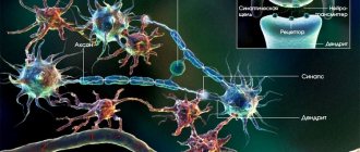
As mentioned earlier, brain tissue has a complex structure. The main components of the human nervous system, as well as other mammals, are gray and white matter, with the first component being a dense accumulation of neuron bodies, their dendrites and glial cells, which are the basis or backbone of this substance.
Basically, the gray matter of brain tissue is formed by clusters of cell bodies of various neurons and their dendrites. The functional feature of this NS unit is that these cells are capable of being excited by a special impulse, processing, transmitting and storing the information thus obtained.
Like any other living cell in the body, it has its own core, membrane and processes that unite a group of similar structures into a single whole. The study of this NS unit is complicated not only by its small size, but also by its location, since the largest concentrations of them are most often located in hard-to-reach places, intervention in which is fraught with disastrous consequences.
The functional significance of glial cells is very diverse: they serve as a barrier to other structures of the body, but in some cases they perform a protective function. A special feature of glia is the ability to repair and divide, which other nerve cells cannot boast of. A layer of them forms a special tissue called neuroglia and is located in all parts of the NS.
Since neurons are deprived of protection from the negative effects of the environment and are helpless against mechanical damage, in some cases glia are capable of phagocytizing or assimilating an incoming foreign antigen that poses a danger to gray cells.
What diseases are possible?
Since this organ regulates the transmission of impulses to all systems and organs, the main sign of disruption of its activity is loss of sensitivity. Due to the fact that this organ is part of the central nervous system, diseases are associated with neurological characteristics. Typically, various spinal cord lesions cause the following symptoms:
- disturbances in the movement of limbs;
- pain syndrome of the cervical and lumbar regions;
- disorders of skin sensitivity;
- paralysis;
- urinary incontinence;
- loss of muscle sensitivity;
- increased temperature in affected areas;
- muscle pain.
You may be interested in: Spinal cord stroke: types, main signs, treatment methods
These symptoms can develop in different sequences, depending on the area in which the lesion is located. Depending on the causes of diseases, there are 3 groups:
- All kinds of developmental defects, including postpartum ones. The most common are congenital anomalies.
- Diseases that involve circulatory problems or various tumors. It happens that such pathological processes also cause hereditary diseases.
- All kinds of injuries (bruises, fractures) that disrupt the functioning of the spinal cord. These could be injuries resulting from car accidents, falls from a height, domestic injuries, or as a result of a bullet or knife wound.
Any spinal cord injury or disease that causes such consequences is very dangerous because it often deprives many people of the ability to walk and live a full life. You should consult a doctor as soon as possible to begin treatment on time if the above symptoms or the following disorders are observed after an injury or illness:
- loss of consciousness;
- blurred vision;
- frequent seizures;
- difficulty breathing.
Otherwise, the disease can progress and cause the following complications:
- chronic inflammatory processes;
- disruption of the gastrointestinal tract;
- disturbance in the functioning of the heart;
- circulatory disorders.
Therefore, you should seek help from a doctor in time to receive the correct treatment. After all, thanks to this you can save your sensitivity and protect yourself from pathological processes in the body that can lead to a wheelchair.
How gray matter influences some human abilities
The gray tissue of the brain, regulating the processing of signals from the outside and generating effector impulses, is not only responsible for the functioning of the entire human nervous system, but also affects his abilities: mental, cognitive, physical, etc.
Various experiments by scientists have shown that a person’s abilities depend on the volume of the gray substance, while changes in the amount of white substance did not show any noticeable changes.
Experiments by British scientists have shown that the thinner the cerebral cortex, therefore, the smaller the volume of gray substance, the worse a person copes with solving logical problems, the less various abilities he has, and also with a low volume of substance, subjects often had problems with reaction speed , speech dysfunctions, memory problems and poor intellectual abilities.
At the same time, studies have shown that learning foreign languages, memorizing poetry, scientific or artistic works, and playing music affect the enlargement of the cerebral cortex. The longer and more intense the learning process, the greater the volume of gray substance becomes, therefore, the more abilities, including mental ones, a person displays.
A decrease in the amount of gray matter is affected by:
- a person’s lifestyle is a sedentary, inert, inactive, from a physical and mental point of view, way of life;
- abuse of bad habits - alcohol, drug addiction and smoking reduce the volume of gray substance.
For example: those suffering from alcoholism experience a significant decrease in the amount of brain tissue, which is reflected in behavior and mental functions: incoherent speech, problems with memory and perception, inhibition of thought processes.
White and gray matter of the spinal cord
The cross-sectional diagram of the structure of the spinal cord is similar to the wings of a butterfly; this part of the internal organ is called gray matter. On the outside, gray matter is surrounded by white matter, but the cellular structure and functions of these substances differ significantly.
Gray matter consists of interneurons and motor neurons:
- interneurons provide close connections between the neurons themselves;
- motor neurons contribute to the transmission of motor reflexes.
The white matter contains axons - these are nerve processes that create fibers of descending and ascending pathways.
Roots
When considering the functions of the spinal cord and its structure, one cannot fail to mention the so-called spinal cord roots.
In short, the roots of the spinal cord are bundles of nerve fibers that enter any segment of the organ and form the spinal nerves.
The roots form the sensitive part of the spinal nerve. The root consists of motor nerve fibers, which are processes of the anterior horns of the gray matter.
This is interesting! How we are made: human structure - internal organs in detailed description and location diagram
Internal structure
Gray and white matter together form all the spinal cord pathways. They represent its internal composition. Gray matter is located in the center, and white matter is located throughout the periphery. Gray matter is formed as a result of the accumulation of short processes of neuronal cells and consists of 3 projections that form gray pillars. They are located along the entire length of this organ and in cross-section form:
- anterior horn, containing large motor neurons;
- the dorsal horn, formed with the help of small neurons that contribute to the formation of sensory columns;
- side horn
The gray matter of this organ of the nervous system also suggests the presence of kidney cells. They, located along the entire length of the gray matter, form tuft cells that conduct connections between all segments of the dorsal bridge.
The main part of the white matter is made up of long processes of neurons that have a myelin sheath, which gives the white tint to neurons. The white matter on both sides of the spinal cord is connected by the white commissure. The neurons of the white matter of the spinal cord are collected in special bundles; they are separated into 3 cords of the spinal cord using three grooves.
You may be interested in: Anterior roots of the spinal cord: structure, cross-sectional structure and main functions
In the cervical and thoracic sections of this organ there is a posterior cord, which is divided into thin and wedge-shaped. They continue in the initial part of the brain. In the sacral and coccygeal regions, these cords merge into one and are almost indistinguishable.
Of course, the white and gray matter together do not have a homogeneous structure, but they form an interconnection with each other, thanks to which nerve impulses are transmitted from the central nervous system to all peripheral nerves. Because of such a close connection with the brain, many doctors do not separate these two components of the human nervous system, as they consider them one whole. Therefore, it is very important to take care of preserving their functions, which are vital for every person.
How the nervous system works, what is white matter, gray matter
The human nervous system has a complex structure. Conventionally, experts distinguish between the peripheral and central nervous systems of humans.
The human central nervous system includes all parts of the brain (terminal, middle, medulla, intermediate, cerebellum), as well as the spinal cord. These components control the functioning of all body systems, connect them with each other and ensure their coordinated operation in response to external influences.
Functional features of the central nervous system:
- The human brain is located in the cranium and plays a controlling role: it participates in the processing of information received from the environment and regulates the vital functions of all systems of the human body, and is a kind of steering wheel.
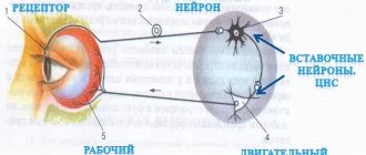
- The main function of the spinal region of the central nervous system is to transmit information from nerve centers located in other parts of the body to the brain. Also, with its support, motor reactions to external stimuli are performed (using reflexes).
The peripheral nervous system includes all branches of the spinal cord and brain that are located outside the central nervous system or, in other words, on the periphery. It includes cranial and spinal nerves, as well as autonomic nerve fibers that connect the structures of the central nervous system with other parts of the human body. With its help, unconscious (at the level of reflexes) control of the vital functions of certain organs occurs, be it heartbeat or automatic muscle contraction in response to external stimuli (for example, blinking).
This part of the nervous system is especially vulnerable to various toxins or mechanical damage, since it does not have protection in the form of bone tissue or a special barrier separating the blood and its components.
Peripheral NS includes:
- Vegetative or autonomous NS. It is controlled by the human subconscious and controls the performance of vital functions of the body. The main task of this part of the NS is to regulate the internal environment of the body, through the circulatory, endocrine system, as well as various endocrine and exocrine glands. Anatomically, it is divided into sympathetic, parasympathetic and metasympathetic NS. In this case, centers or vegetative nuclei, consisting of a gray cerebral component, are located in the spinal and head sections of the central nervous system, and the latter is represented by clusters of neurons located in the walls of the bladder, gastric tract and other organs.
- Somatic NS. Responsible for human motor function - with its help, afferent (incoming) signals are transmitted to the neurons of the central nervous system, from where, after processing, information is received through efferent (descending motor) fibers to the limbs and organs of the human body to reproduce the corresponding movement. Its neurons have a special structure that allows them to transmit data over long distances. Thus, most often the body of a neuron is located in close proximity to parts of the central nervous system or enters it, but at the same time its axon extends further, eventually reaching the surface of the skin or muscles. Through this part of the NS, various protective reflexes are carried out, which are carried out at the subconscious level. This feature is achieved by the presence of reflex arcs, which allow the action to be performed without the participation of the main center, since in this case the nerve fibers connect the dorsal part of the central nervous system with the area of the body directly. In this case, the final point of information perception is the cerebral cortex, where memories of all actions performed remain. Thus, the somatic nervous system is involved in learning, protection and the ability to process information received from the environment.
- Some experts classify the human sensory nervous system as the peripheral nervous system. It includes several groups of neurons located on the periphery of the central nervous system, which are responsible for the perception of information from the environment through the organs of hearing, vision, touch, taste and smell. Responsible for the physical perception of concepts such as temperature, pressure, sound.
As mentioned earlier, the structures of the human nervous system are represented by a white and gray substance, each of which has its own structure and contains different types of nerve cells that differ in appearance and functionality.
Thus, the white matter mainly performs a conductive function and transmits nerve impulses from one part of the brain matter to another. This feature is due to the structure of the neurons of this structure, the bulk of which consists of long processes or axons covered with myelin, which has a high conductivity of electrical impulses (about 100 m/s).
Neuron axons can be divided into 2 main groups:
- Long (intracortical), connect distant areas, located in the depths of the medulla.
- Short processes that connect the gray cells of the cortex and nearby structures of the white matter have a second name - subcortical.
Also, depending on the location and functionality of the nerve cell fibers of the white matter, it is customary to distinguish the following groups:
- Associative. They differ in size: they can be either long or short and perform various tasks, but at the same time they are concentrated in one of the hemispheres. Long axons are responsible for connecting distant convolutions, while short axons connect nearby structures.
- Commissural. They connect the 2 hemispheres and ensure their coordinated work, located in opposite parts. Such axons can be considered during the anatomical study of this organ, since they consist of the anterior commissure, the corpus callosum, and the fornix commissure. Projection axons connect the cortex with other centers of the central nervous system, including the spinal cord. There are several types of such fibers: some connect the thalamus with the cortex, the second - the cortex with the nuclei of the bridge, and the third conduct impulses, thanks to which the command and control of certain limbs is carried out.
There are 2 types of similar fibers, which differ in the direction of the transmitted information:
- Afferent. Through them, information comes from the underlying structures of the brain, organ systems and tissues to the cortex and subcortical structures that process the incoming information.
- Efferent. They carry out a response impulse from the centers of higher mental activity to controlled structures.
The opposite of the white medulla is the gray component, which, like its predecessor, consists of a cluster of neurons - with their help, all functions of higher nervous activity of a person are performed.
Its main part is located on the surface of the white medullary component located in the head and makes up the cortex, which has a conventionally gray color. It also lies deep in the brain and along the entire length of the spinal cord in the form of nuclei. The gray matter includes several groups of nerve cells, their dendrites and axons, as well as glial tissues that perform an auxiliary function.
Branched processes of neurons or dendrites, through synapses, receive and transmit information from the axons of neighboring cells to their own. The quality of the impulse depends on the density of their branching - the more developed the branches of the main fiber and the more extensive the network of synapses, the more data will flow to the cell nucleus from neighboring ones.
Since neurons and, accordingly, the nuclei of gray matter cells are located close to each other, they do not require long axons, while the main flow of information is transmitted through the dendritic synaptic connection of nearby cells. For the same reason, their axons do not require a myelin sheath.
Individual accumulations of gray matter are called nuclei, each of which controls the performance of a specific vital function of the body, and they can be divided into 2 large groups: those related to the central nervous system and those responsible for the peripheral nervous system.
The anatomical structure of gray matter neurons in all parts of the central nervous system has a similar structure and approximately the same composition. Therefore, the pattern of arrangement of neurons in the terminal section is no different from the totality of these elements in other structures.
Functions of the organ
The structure and functions of the spinal cord are the most important system that supports the normal functioning of the human body.
The functionality of the spinal cord is divided into 2 parts:
- reflex - these are the simplest motor reflexes of the body, for example, when a hand is burned, a person begins to pull his hands away from the source of injury, or when he hits his knee with a hammer, a reflexive extension of the knee occurs;
- The conduction function is the transmission of nerve impulses from the brain region to the inner part of the spinal cord, as well as the transmission of nerve impulses from the brain to the internal organs of the human body.
With the help of conductive communication, almost every mental action is carried out - get up, walk, lie down, sit down, draw, pass, cut, etc. A person does not even think about most of the actions, performing them at a reflex level in everyday life.
What is the gray matter of the brain responsible for?
Work on studying the brain as a control organ began in the 18th century and continues to this day. Perhaps this process would have gone much faster if there had not been a ban on the anatomical study of brain tissue and dissection of the body of a deceased person for a long time. The situation is also complicated by the fact that the brain is a rather inaccessible organ, which is reliably protected from the outside by the bones of the skull and a large number of membranes, damage to which can negatively affect the experimental subject.
So, the human brain includes several functional clusters of gray matter neurons, be it its cortex or nuclei, responsible for performing individual movements or controlling the activity of some vital systems of the body.
The cerebral cortex is a relatively young structure that began to form in the process of human evolutionary development. Its presence and degree of development is a distinctive feature of the human brain, since in most mammals the gray matter of the cortex is limited in size and not as functional.
The main function of the gray matter of the cerebral cortex is to perform higher psychiatric tasks that the individual sets for himself in the process of learning new skills, while experience can be gained from other sources or the environment. Also, an expression of the work of the cerebral cortex is the sound reproduction of speech and its internal manifestation, which is also popularly designated by the concept “to oneself.”
Gray matter also forms nuclei and small plates, which are also present in other parts of the brain.
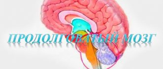
The medulla oblongata, as a functional continuation of the spinal section, combines the characteristic structural features of both sections of the central nervous system. Just like the dorsal one, it includes a large number of conducting fibers, the main task of which is to communicate the terminal section with the spinal part. In this case, the gray matter of the medulla oblongata no longer has a characteristic continuous structure, as in the cerebral cortex, but lies in the form of nuclei.
This department, like the entire central nervous system, regulates the physiological processes on which human life depends. These include the following operations: breathing, heartbeat, elimination, digestion, as well as protective reflex movements (such as blinking or sneezing) and muscle tone. Nerve pathways and centers pass through it, responsible for the coordination and spatial position of the body in the environment through the nuclei of the vestibular apparatus.
A characteristic feature of the location and structure of the gray matter in the middle section of the brain is that it combines the structural features of the medulla oblongata and terminal section, with paired clusters of gray matter forming nuclei, and individually scattered neurons forming the central periaqueductal structure and the so-called substantia nigra.
The anatomical structure of the nuclei and this section does not differ from the structure of this structure in the medulla oblongata. The main task of these centers is to perceive information from the environment through the organs of hearing, vision, smell, and also participate in the performance of some conditioned reflexes, for example, turning the head towards a loud sound or bright light.
Other structures of the middle section require special attention: the central gray matter and the substantia nigra. They have a number of features due to their structure and purpose.
A layer of substantia nigra conditionally separates the cerebral peduncle from the tegmentum and regulates the motor function of the limbs. It has been noted that when this component of the nervous system is damaged, the patient develops Parkinson's disease, tremors of the limbs, and a decrease in motor skills is also noted.
The central periaqueductal gray matter is a sparse, scattered collection of unmyelinated neurons surrounding the aqueduct. Serves as a conductor and store of information from underlying structures (reticular formation, nuclei of the vestibular apparatus, hypothalamus, etc.), and also participates in the formation of painful sensations of aggressive behavior and controls human sexual behavior.
Segments
Spinal cord segment
-
this is a separate part of an organ
that is responsible for certain parts of the body, as well as for the functioning of all organs. There are 31 segments in total. To make it easier to understand the functions of each of the segments, which together make up the departments, you need to create a simple table.
Sections of the spinal cord and their functions:
| Departments | What are they responsible for? |
| Cervical | Movement of the diaphragm and elbow joints |
| Breasts | Skin sensitivity, muscle function, lungs, heart, digestive system |
| Lumbar | Work of the prostate, kidneys, bladder, adrenal glands, uterus, ureters |
| Sacral | Centers for defecation, urination and erection |
What is white matter responsible for?
As mentioned earlier, the white matter of the brain performs several tasks: first of all, it is a link between the gray matter of the cortex and other functional clusters of neurons located in deep structures.
Other functions of the white matter of the brain are also known - it acts as a link between the cerebral hemispheres through the corpus callosum, and also ensures the interaction of remote areas of the cortex with other parts of the nervous system, including the spinal cord, using specific fibers.
Its main feature and distinctive feature is that the white matter is formed by a cluster of long nerve processes or fibers covered with a myelin sheath, which ensures rapid transmission of electrical impulses and relevant information to functional centers.
The white matter of the telencephalon forms the cerebral hemispheres, which are the most developed and massive structure of the central nervous system. This feature is due to the presence of a large number of projection fields in the cortex, which require a developed network of connecting fibers for their normal functioning. Otherwise, the connection and parallel execution of higher mental functions of the brain are disrupted: for example, speech becomes slow and inarticulate.
In the middle part of the brain, the white matter is located mainly over its entire surface, as well as ventral to the gray matter of the quadrigeminal colliculi. It also consists of the upper legs, which connect the midbrain with the cerebellum and transmit efferent information from this motor center to other parts of the central nervous system.
The white matter of the oblong section includes all types of fibers: both long and short. The long ones perform a transient function and connect the descending pyramidal tracts with the spinal nerve cords, and also carry out the coordinated work of the medulla oblongata with the thalamic structures, while the short ones form a connection between the nuclei of this department and send information to the overlying structures of the central nervous system.
Organization of pathways
The pathways of the entire body are usually divided into:
- associative;
- afferent;
- efferent.
The task of associative pathways is to connect neurons between all segments. These connections are considered short.
Afferents provide sensitivity. These are ascending pathways that receive information from all receptors and send it to the brain. Efferent pathways transmit signals from the brain to neurons throughout the body. They belong to the descending paths.
Where is the white matter located?
The white matter of the brain begins to form by 6 months of intrauterine development of a person, while its formation does not stop throughout the subsequent years of life. This feature allows the body to train and accumulate experience.
White matter itself is the opposite of gray matter and is a dense network of neuron branches that transmit information from the cerebral cortex to the underlying nerve centers of the spinal cord and brain. At the same time, the functioning of the connection is influenced by the quantity and quality of the formed nerve pathways: the denser and stronger the connection between the structures, the more developed and talented the individual turns out to be.
The largest accumulation of white matter is located in the cranium and is represented by large lobes. This is understandable: all the control centers of the body are located in the brain, and also in its structures the formation and fulfillment of higher mental tasks occurs, the presence of which distinguishes humans from the rest of the animal world. Moreover, in addition to the main one, the white matter also performs a protective function: in appearance and physical characteristics, it is a gelatinous, fat-like mass that plays the role of a shock absorber for the underlying structures.
Also, the white matter forms the peripheral meninges for the gray matter of the spinal cord - like the head of the central nervous system, it contains all types of fibers (commissural, associative and projection), with a characteristic myelin color, which are collected in special bundles that provide communication between the spinal cord and other parts peripheral and central NS.