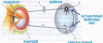
Anatomy of the human midbrain - information:
The midbrain, mesencephalon , develops in the process of phylogenesis under the predominant influence of the visual receptor, therefore its most important formations are related to the innervation of the eye. Hearing centers were also formed here, which, together with the vision centers, later grew in the form of four mounds of the roof of the midbrain.
With the appearance in higher animals and humans of the cortical end of the auditory and visual analyzers in the forebrain cortex, the auditory and visual centers of the midbrain themselves fell into a subordinate position and became intermediate, subcortical. With the development of the forebrain in higher mammals and humans, pathways began to pass through the midbrain, connecting the telencephalon cortex with the spinal cord (cerebral peduncles). As a result, the human midbrain contains:
- subcortical centers of vision and nuclei of nerves innervating the muscles of the eye;
- subcortical auditory centers;
- all ascending and descending pathways connecting the cerebral cortex with the spinal cord and passing through the midbrain;
- bundles of white matter connecting the midbrain with other parts of the central nervous system.
Accordingly, the midbrain, which is the smallest and most simply structured part of the brain in humans, has two main parts: the roof, where the subcortical centers of hearing and vision are located, and the cerebral peduncles, where the pathways predominantly pass.
- Dorsal part, roof of the midbrain, tectum mesencephali. It is hidden under the posterior end of the corpus callosum and is divided by two criss-crossing grooves - longitudinal and transverse - into four colliculi, arranged in pairs. The upper two hillocks, colliculi superiores, are subcortical centers of vision, the two lower ones, colliculi inferiores, are subcortical centers of hearing. The pineal body lies in a flat groove between the superior tubercles. Each hillock passes into the so-called handle of the hillock, brachium colliculi, heading laterally, anteriorly and upward, to the diencephalon. The handle of the superior colliculus, brachium colliculi superioris, goes under the pillow, pulvinar, of the thalamus to the lateral geniculate body, corpus geniculatum laterale. The handle of the lower colliculus, brachium colliculi inferioris, passing along the upper edge of the trigonum lemnisci to the sulcus lateralis mesencephali, disappears under the medial geniculate body, corpus geniculatum mediale. The named geniculate bodies already belong to the diencephalon.
- The ventral part, the cerebral peduncles, pedunculi cerebri, contains all the pathways to the forebrain. The cerebral peduncles look like two thick semi-cylindrical white cords that diverge from the edge of the pons at an angle and plunge into the thickness of the cerebral hemispheres.
- The midbrain cavity, which is a remnant of the primary cavity of the midbrain bladder, has the appearance of a narrow canal and is called the cerebral aqueduct, aqueductus cerebri. It is a narrow, ependymal-lined canal 1.5-2.0 cm long, connecting the IV ventricle with the III. Dorsally, the aqueduct is limited by the roof of the midbrain, ventrally by the tegmentum of the cerebral peduncles. Internal structure of the midbrain.
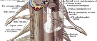
In a cross section of the midbrain, three main parts are distinguished:
- roof plate, lamina tecti;
- tire, tegmentum, representing the upper section of the pedunculi cerebri;
- ventral section of the pedunculi cerebri, or the base of the cerebral peduncle, basis pedunculi cerebralis.
According to the development of the midbrain under the influence of the visual receptor, it contains various nuclei related to the innervation of the eye. In lower vertebrates, the superior colliculus serves as the main termination of the optic nerve and is the main visual center. In mammals and in humans, with the transfer of visual centers to the forebrain, the remaining connection of the optic nerve with the superior colliculus is important only for reflexes. The fibers of the auditory loop (lemniscus lateralis) end in the nucleus of the inferior colliculus, as well as in the medial geniculate body. The roof of the midbrain has a bilateral connection with the spinal cord - tractus spinotectalis and tractus tectobulbaris et tectospinalis. The latter, after crossing in the tegmentum, go to the muscle nuclei in the medulla oblongata and spinal cord. This is the so-called visual-sound reflex pathway, which was mentioned when describing the spinal cord.
Thus, the plate of the roof of the midbrain can be considered as a reflex center for various types of movements that arise mainly under the influence of visual and auditory stimuli.
The cerebral aqueduct is surrounded by central gray matter, which in its function is related to the autonomic system. In it, under the ventral wall of the aqueduct, in the tegmentum of the cerebral peduncle, the nuclei of two motor cranial nerves are located - n. oculomotorius (III pair) at the level of the superior colliculus and n. trochlearis (IV pair) at the level of the inferior colliculus.
The nucleus of the oculomotor nerve consists of several sections, corresponding to the innervation of several muscles of the eyeball. Medial and posterior to it there is also a small, also paired, vegetative accessory nucleus , nucleus accessories, and an unpaired median nucleus .
The accessory nucleus and the unpaired median nucleus innervate the involuntary muscles of the eye, m. ciliaris and m. sphincter pupillae. This part of the oculomotor nerve belongs to the parasympathetic system. Above (rostral) the nucleus of the oculomotor nerve in the tegmentum of the cerebral peduncle is the nucleus of the medial longitudinal fasciculus. Lateral to the cerebral aqueduct is the nucleus of the midbrain tract of the trigeminal nerve , nucleus mesencephalicus n. trigemini.
The cerebral peduncles are divided into the ventral part, or the base of the cerebral peduncle, basis pedunculi cerebralis, and the tegmentum, tegmentum. The boundary between them is the black substance, substantia nigra, which owes its color to the black pigment contained in its constituent nerve cells - melanin. The tegmentum mesencephali is a part of the midbrain located between its roof and the black substance (substantia nigra) of the cerebral peduncles. The tractus tegmentalis centralis - the central tegmental tract - is a projection descending nerve pathway located in the central part of the midbrain tegmentum. It contains fibers coming from the thalamus, globus pallidus, red nucleus and reticular formation of the midbrain to the reticular formation and olive of the medulla oblongata; belongs to the extrapyramidal system.
Substantia nigra extends along the entire length of the cerebral peduncle from the pons to the diencephalon; according to its function it belongs to the extrapyramidal system. Located ventral to the substantia nigra, the base of the cerebral peduncle contains longitudinal nerve fibers descending from the cerebral cortex to all underlying parts of the central nervous system (tractus corticopontinus, corticonuclearis, corticospinalis, etc.). The tegmentum, located dorsal to the substantia nigra, contains predominantly ascending fibers, including the medial and lateral lemniscus. As part of these loops, all sensory pathways ascend to the cerebrum, with the exception of the visual and olfactory ones.
Among the nuclei of gray matter, the most significant is the red nucleus, nucleus ruber. This elongated, sausage-shaped formation extends in the tegmentum of the cerebral peduncle from the hypothalamus of the diencephalon to the inferior colliculus, where it begins an important descending tract, the tractus rubrospinal, connecting the red nucleus with the anterior horns of the spinal cord. This bundle, after leaving the red nucleus, intersects with a similar bundle of the opposite side in the ventral part of the median suture - the ventral decussation of the tegmentum.
Nucleus ruber is a very important coordination center of the extrapyramidal system, connected with its other parts. Fibers pass to it from the cerebellum as part of the upper peduncles of the latter after their decussation under the roof of the midbrain, ventrally from the aqueductus cerebri, as well as from the pallidum - the lowest and most ancient of the subcortical nodes of the brain that are part of the extrapyramidal system. Thanks to these connections, the cerebellum and the extrapyramidal system, through the red nucleus and the tractus rubrospinal extending from it, influence the entire skeletal muscle in the sense of regulating unconscious automatic movements. The reticular formation, formatio reticularis, and fasciculus longitudindlis medialis also continue into the tegmentum of the midbrain. The latter originates in various places. One of its parts begins from the vestibular nuclei, passes on both sides along the sides of the midline, directly under the gray matter of the bottom of the aqueduct and the IV ventricle, and consists of ascending and descending fibers going to the nuclei of the III, IV, VI and XI cranial nerves . The medial longitudinal fasciculus is an important associative pathway that connects the various nuclei of the nerves of the eye muscles with each other, which determines the combined movements of the eyes when they are deviated in one direction or another. Its function is also associated with eye and head movements that occur when the balance apparatus is stimulated.
What role does gray matter play?
Millions of years of evolution, natural selection and the origin of species have given the human being a unique structure - a relatively thick cerebral cortex. It is known that the structure of gray matter is properly developed only in representatives of the human species. Unlike lower and even higher mammals, the gray substance has endowed a person with the opportunity to have a unique property of matter, the object of study of all neurosciences and philosophy - consciousness and self-awareness, the resulting abstract thinking, developed memory, inner speech and many other specific attributes of higher nervous activity a reasonable person.
It must be remembered that gray matter is a collection of nerve cells, namely neurons. When talking about the function of gray matter, we are talking about the function of all clusters of neurons with short processes. So, the functions of gray matter are diverse:
Physiological tasks: generation, transmission, reception and processing of electrical signals. Neurophysiological: perception, speech, thinking, memory, vision, emotions, attention. Psychological: personality formation, worldview, motivation, will.
For a long time, scientists have wondered what the gray matter of the brain is responsible for.
Back in the 18th century, Franz Gall drew attention to the dark brain substance. For the first time, the scientist managed to localize some mental functions on the cortex
Subsequent studies were carried out by removing a section of the cortex and observing which brain function was lost. A serious impetus for further research was the study of the work of the cortex by Academician Pavlov, who studied basic reflexes and the principles of strengthening the conditioned reflex. In parallel, his French colleagues found a speech center in the cortex - the lower part of the frontal gyrus. Modern science, although it knows many properties of the cerebral cortex, claims that the percentage of knowledge on it is no more than one thousandth.
One blind spot in the empirical knowledge of the brain and its formation is the question of what heterotopia of the gray matter of the brain is. In particular, this question is often raised in the field of clinical medicine, where treatment is only symptomatic, that is, the symptoms are removed alone. As is known, heterotopia is a defective accumulation of neurons that have stopped in a certain place and have not reached their histological place. So, if the cause of the pathology is found, there will be an etiological treatment. A variant of the manifestation of heterotopia is childhood epilepsy.
Important Akinesia, hypokinesia, oligokinesia, bradykinesia: causes, symptoms, treatment
Notes
- Mosby's Medical, Nursing & Allied Health Dictionary, Fourth Edition, Mosby-Year Book 1994, p. 981
- Breedlove, Watson, & Rosenzweig. Biological Psychology, 6th Edition, 2010, pp. 45-46
- ↑ Martin. Neuroanatomy Text and Atlas, Second edition. 1996, pp. 522-525.
- Collins Dictionary of Biology, 3rd ed. WG Hale, VA Saunders, JP Margham 2005
- Haines, Duane E.
Neuroanatomy: an atlas of structures, sections, and systems. — 8th. — Philadelphia : Wolters Kluwer/ Lippincott Williams & Wilkins Health. - P. 42. - ISBN 978-1-60547-653-7. - Kolb, E. M., E. L. Rezende, L. Holness, A. Radtke, S. K. Lee, A. Obenaus, and Garland T., Jr. 2013. Mice selectively bred for high voluntary wheel running have larger midbrains: support for the mosaic model of brain evolution. Journal of Experimental Biology 216:515–523.
- Martin. Neuroanatomy Text and Atlas, Second Edition, 1996, pp. 35-36.
Functions
Mission – organization of mental activity.
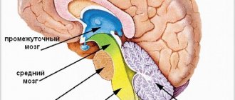
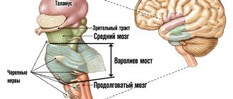
Intermediate
Participates in coordinating the work of organs, regulating body movement, maintaining temperature, metabolism, and emotional background.
Thalamus. The main task is to sort information. It works like a relay - it processes and sends data coming from receptors and pathways to the brain. The thalamus affects the level of consciousness, attention, sleep, wakefulness. Supports speech functioning.
Epithalamus. Interaction with other structures occurs through melatonin, a hormone produced by the pineal gland in the dark (therefore, it is not recommended to sleep in the light). A derivative of serotonin - the “happiness hormone”. Melatonin is a participant in the regulation of circadian rhythms, being a natural sleep aid, it affects memory and cognitive processes. It affects the localization of skin pigments (not to be confused with melanin), puberty, and suppresses the growth of a number of cells, including cancer cells. Through connections with the basal ganglia, the epithalamus participates in the optimization of motor activity, and through connections with the limbic system, in the regulation of emotions.
Subthalamus. Controls the body's muscle responses.
Hypothalamus. Forms a functional complex with the pituitary gland and directs its work. The complex controls the endocrine system. The hormones it produces help cope with distress and maintain homeostasis.
Thirst and hunger centers are located in the hypothalamus. The department coordinates emotions, human behavior, sleep, wakefulness, and thermoregulation. Endorphins similar in action to opiates are found here, which help to endure pain.
Hemispheres
They act together with subcortical structures and the brain stem. Main destination:
- Organization of interaction of an organism with the environment through its behavior.
- Consolidation of the body.
Corpus callosum
The corpus callosum received attention after operations to dissect it in the treatment of epilepsy. Operations relieved seizures while changing a person's personality
It was found that the hemispheres are adapted to work independently. However, to coordinate activities, information exchange between them is necessary. The corpus callosum is the main transmitter of information.
Striatum
- Reduces muscle tone.
- Contributes to the coordination of internal organ function and behavior.
- Participates in the formation of conditioned reflexes.
The olfactory brain contains centers that control the sense of smell.
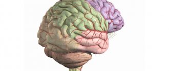
Cerebral cortex
Head of mental processes. Controls sensory and motor functions. Consists of 4 layers.
The ancient layer is responsible for elementary responses (for example, aggression) characteristic of humans and animals.
The old layer is involved in the formation of attachment and laying the foundations of altruism. Thanks to the layer we are happy or angry.
The intermediate layer is a formation of a transitional type, since the modification of old formations into new ones is carried out gradually. Ensures the activity of the new and old cortex.
The neocortex concentrates information from subcortical structures and the brainstem. Thanks to it, living beings think, talk, remember, and create.
5 cerebral lobes
The occipital lobe is the central section of the visual analyzer. Provides visual pattern recognition.
Parietal lobe:
- controls movements;
- orients in time and space;
- provides perception of information from skin receptors.
Thanks to the temporal lobe, living things perceive a variety of sounds.
The frontal lobe regulates voluntary processes, movements, motor speech, abstract thinking, writing, self-criticism, and coordinates the work of other areas of the cortex.
The insula is responsible for the formation of consciousness, the formation of an emotional response and the maintenance of homeostasis.
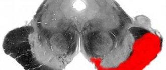
What does the bear have to do with it?
The bear face is an excellent association for remembering the basic structures of the midbrain. If you turn our drawing over, you will see this:
You can also use the picture from Wikipedia:
Here we see:
- Eyes (red cores);
- Ears (brain legs);
- The line separating the head and ears (substantia nigra);
- Nose (central gray matter);
- Nostrils (Sylvius aqueduct).
The triangles inside and next to the gray matter are a schematic representation of the nuclei of the cranial nerves; we will definitely analyze them in more detail in separate lessons. The green plates that are located laterally are the midbrain pathways; now I will publish this article and immediately begin the second part devoted exclusively to them.
Let's mark all the anatomical formations we have analyzed in a separate figure: