The auditory cortex selectively hears what we listen to
Rice. 1.
Areas of the superior temporal gyrus responsible for the perception of spoken language.
primary auditory
cortex receives information from the thalamus, where it arrives (through several intermediate stages) from the organ of hearing - the cochlea.
This information is structured from the very beginning - divided into frequencies. auditory cortex
takes the first steps toward understanding what is heard by filtering auditory information and combining it with data from other senses.
Wernicke 's area
, which occupies the most posterior part of the superior temporal gyrus, recognizes words and plays a key role in speech understanding. Image from D. Purves, G. J. Augustine, D. Fitzpatrick, et al. editors. Neuroscience. 2nd edition. Sunderland (MA): Sinauer Associates; 2001
People are able to listen and understand each other even in a company where everyone is talking at the same time. How the brain selects the necessary sounds from a complex acoustic background is unknown. American neuroscientists, working with patients who had electrodes implanted into the superior temporal gyrus during treatment for epilepsy, discovered that the activity of neurons in the secondary auditory cortex reflects the speech of the person to whom the subject is listening. Based on the activity of these neurons, a specially trained computer program can determine which of the two speakers the subject is listening to and reconstruct the words heard.
Isolating the speech of one specific person from a multi-voice choir is a technically extremely difficult task, as developers of automatic speech recognition systems are well aware of. Our brain, however, easily copes with it, but how it does it is not really known. It can be assumed that at some stage in the processing of auditory information, the speech of the person we are listening to is cleared of “extraneous impurities,” but when and where this happens is again unclear.
Nima Mesgarani and Edward Chang from the University of California, San Francisco, studied the functioning of neurons in the secondary auditory cortex (Fig. 1) in three patients with epilepsy who, in preparation for surgery, had microelectrodes implanted in the superior temporal lobe. gyrus (Fig. 2).
Rice. 2.
Location of electrodes on the subjects' brains.
Shades of red
show how different the signal from the electrode is when perceiving speech and in silence.
Image from the discussed article in Nature
Previously it was shown that neurons in the secondary auditory cortex “encode” (reflect) the spoken language perceived by a person. Computer programs have been developed that, after special training, are able, based on data on the activity of these neurons, to reconstruct the timbre of the speaker’s voice and even recognize spoken words (Formisano et al., 2008. “Who” is saying “what”? Brain-based decoding of human voice and speech Pasley et al., 2012. Reconstructing Speech from Human Auditory Cortex). But these experiments were carried out on subjects who were allowed to listen to the speech of only one speaker. Mesgarani and Chang decided to find out what information the auditory cortex neurons would reflect if there were two speakers, but the subject was asked to listen to only one of them.
The experiments used recordings of two voices - male and female. They uttered meaningless seven-word phrases such as “ready tiger go to red two now” or “ready ringo go to green five now.” The first, third, fourth and seventh words were always the same. The second word - tiger or ringo - served as a conditioned signal for the subject. One of these words was displayed on the screen in front of him, and he had to listen to which of the two speakers would pronounce this word. In fifth place was a word denoting one of three colors (red, blue or green), in sixth place was one of three numerals (two, five or seven). The subject had to answer what number and what color was named by the one of the two speakers who uttered the key word. The phrases were combined in such a way that two voices simultaneously called different numbers and colors.
The authors used a previously developed program to reconstruct the sound signal from data on the activity of neurons in the auditory cortex. The program was previously “trained”, and during the training the subjects were allowed to listen to voices one at a time, and not both at the same time. When the program learned to reconstruct spectrograms of single phrases well, the main phase of the experiment began. Now the subjects listened to two voices simultaneously, and the spectrograms reconstructed by the program from data on neuronal activity were compared with real spectrograms of phrases spoken by two speakers.
It turned out that in those cases when the subject successfully completed the task (that is, correctly named the color and number pronounced in the voice that said the key word), the spectrogram reconstructed from his neurons reflected the speech of only one of the two speakers - the one who was needed listen (Fig. 3). If the subject was mistaken, the reconstructed spectrogram was not similar to the speech of the “correct” speaker, but reflected either an unintelligible mixture or correlated with the spectrogram of the second, “distracting” speaker. As a rule, in the first case the subject could not correctly reproduce the words of either of the two speakers, and in the second he indicated the number and color, called the “distractor” voice.
Rice. 3.
Examples of oscillograms and spectrograms of spoken phrases (
a
–
d
) and reconstructions of spectrograms made by a computer program based on data on the operation of neurons in the auditory cortex (
e
–
h
).
a
,
b
- phrases spoken by two voices -
SP1
(male) and
SP2
(female) - separately.
c
,
d
- phrases spoken by two voices simultaneously (in figure
d, blue
and
red colors
show the areas in which the voice of the first or second speaker is louder, respectively).
e
,
f -
spectrograms reconstructed by a computer program based on the work of neurons in the auditory cortex when listening to two phrases one by one.
g
,
h
- the same, when listening to both phrases simultaneously (
g
- the subject listens to the first voice,
h
- to the second).
Image from the discussed article in Nature
At the final stage, the authors used a computer program - a regularized linear classifier (see Linear classifier), trained to distinguish between two voices and spoken words based on the activity of neurons in the auditory cortex when listening to single phrases. When this program was asked to process data on the work of the same neurons when listening to two voices at the same time, it successfully identified both the voice (male or female) and the words (color and number) spoken by the speaker to whom the subject was listening. In those experiments in which the subject completed the task, based on the work of his neurons, the program successfully identified the voice in 93%, the color in 77.2%, and the number in 80.2% of cases. In experiments where the subject made a mistake, the program either produced a random result or recognized the “distracting” voice and the words spoken by it.
Thus, the study showed that in the secondary auditory cortex, speech information is reflected in a “filtered” form: the work of neurons encodes the speech of the person to whom the subject is listening. Although we still do not know the mechanisms of this filtering, it is already possible to determine from the activity of neurons in the auditory cortex which of the two speakers a person is listening to and identify the words heard.
Source:
Nima Mesgarani, Edward F. Chang.
Selective cortical representation of attended speaker in multi-talker speech perception // Nature
. 2012. V. 485. P. 233–236.
See also:
Hearing is responsible for the integration of hearing and touch, “Elements”, 10.24.2005.
Alexander Markov
LECTURES ON NEUROPSYCHOLOGY: Lecture No. 10 5/5 (2)
Lecture No. 10 Neuropsychological syndromes of damage to the cortical parts of the cerebral hemispheres.
1 Projection zones of the cerebral cortex 2. Syndrome in neuropsychology 3. Cortical neuropsychological syndromes a. Syndromes of damage to the posterior parts of the PD cortex b. Syndromes of damage to the anterior parts of the cortex B P
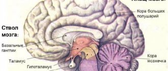
- Brain zones There are three projection zones in the cerebral cortex. Primary projection zone - occupies the central part of the core of the brain analyzer. This is a collection of the most differentiated neurons in which the highest analysis and synthesis of information occurs, and clear and complex sensations arise there. Impulses approach these neurons along a specific impulse transmission pathway in the cerebral cortex (spinothalamic tract).
The secondary projection zone is located around the primary one, is part of the core of the brain section of the analyzer and receives impulses from the primary projection zone. Provides complex perception. When this area is damaged, a complex dysfunction occurs.
The tertiary projection zone, the association zone, consists of multimodal neurons scattered throughout the cerebral cortex. They receive impulses from the associative nuclei of the thalamus and converge impulses of different modalities. Provides connections between various analyzers and plays a role in the formation of conditioned reflexes.
Anatomical information In each hemisphere, the following surfaces are distinguished: 1. convex superolateral surface (facies superolateralis), adjacent to the inner surface of the bones of the cranial vault 2. lower surface (lat. facies inferior), the anterior and middle sections of which are located on the inner surface of the base of the skull, in areas of the anterior and middle cranial fossae, and the posterior ones - on the tentorium of the cerebellum 3. the medial surface (lat. facies medialis), directed towards the longitudinal fissure of the brain.
These three surfaces of each hemisphere, passing one into another, form three edges. The superior edge (lat. margo superior) separates the superolateral and medial surfaces. The inferolateral edge (lat. margo inferolateralis) separates the superolateral surface from the bottom. The inferomedial edge (lat. margo inferomedialis) is located between the lower and medial surfaces. In each hemisphere, the most prominent places are distinguished: in front - the frontal pole (lat. polus frontalis), in the back - occipital (lat. polus occipitalis), and on the side - temporal (lat. polus temporalis).
The hemisphere is divided into five lobes. Four of them are adjacent to the corresponding bones of the cranial vault: 1. frontal lobe (lat. lobus frontalis) 2. parietal lobe (lat. lobus parietalis) 3. occipital lobe (lat. lobus occipitalis) 4. temporal lobe (lat. lobus temporalis) 5. insular lobe (lat. lobus insularis) (island) (lat. insula) - located deep in the lateral fossa of the cerebrum (lat. fossa lateralis cerebri), separating the frontal lobe from the temporal lobe.
- Syndrome in neuropsychology In neuropsychology, the term “syndrome” has two meanings. The first concept is “neuropsychological syndrome” - a natural combination of HMF disorders that arise as a result of local brain damage and are based on a pathological change in one (or several) factors. The second meaning is a grossly expressed violation of any one function. In these cases, the expression “agnosia syndrome”, “semantic aphasia syndrome”, etc. is used. The study of neuropsychological syndromes is the main task of clinical neuropsychology (or syndromology) - the main direction of modern neuropsychology. Violations of the HMF can occur in different forms: 1. in the form of a gross dysfunction (or loss of function), 2. in the form of a pathological weakening (or strengthening) of the function 3. in the form of a decrease in the level of function performance.
By dysfunction, as a rule, we mean the collapse of its psychological structure. The classification of neuropsychological syndromes proposed by A. R. Luria is based on the topical principle, that is, on the principle of identifying the area of brain damage - the morphological basis of the neuropsychological factor.
In accordance with this principle, neuropsychological syndromes are divided: 1. into syndromes of damage to the cortical parts of the cerebral hemispheres and the “proximal subcortex”; 2. syndromes of damage to deep subcortical structures of the brain.
Cortical neuropsychological syndromes, in turn, are divided into lesion syndromes: ♦ lateral (convexital); ♦ basal; ♦ medial cortex of the cerebral hemispheres.
Subcortical neuropsychological syndromes are divided into syndromes involving: ♦ median nonspecific structures; ♦ median commissures (corpus callosum, etc.); ♦ structures located deep in the hemispheres (basal ganglia, etc.).
A special category consists of neuropsychological syndromes (cortical and subcortical) that arise from massive (tumor, traumatic, vascular) brain lesions involving both cortical and subcortical structures. In addition to the topical principle of classifying syndromes, the nosological principle is sometimes used. In these cases, syndromes are divided into “tumor”, “vascular”, “traumatic”, etc. Such a classification has predominantly clinical significance and emphasizes the features of neuropsychological syndromes associated with the nature of the disease. Sometimes syndromes are characterized depending on the patient’s age: “children’s” neuropsychological syndromes and “senile” neuropsychological syndromes.
HMF disorders included in the neuropsychological syndrome never occur in isolation from neurological disorders and other clinical symptoms of the disease. Therefore, neuropsychological (syndromic) analysis of mental processes disorders should always be combined with an analysis of the general clinical picture of the disease.
3 Cortical neuropsychological syndromes of the PD cortex Syndromes of damage to the cortical parts of the cerebral hemispheres (primarily the left hemisphere) have been studied in most detail in modern neuropsychology. Among cortical neuropsychological syndromes, the greatest attention has been paid to damage to the lateral (convexital) cortex.
Cortical neuropsychological syndromes occur when the secondary and tertiary fields of the cerebral cortex are damaged. Damage to the primary fields leads only to neurological symptoms - elementary disorders of sensory and motor functions. Cortical neuropsychological syndromes can generally be divided into two large categories: ♦ syndromes associated with damage to the posterior parts of the brain; ♦ syndromes associated with damage to the anterior parts of the cerebral hemispheres (left and right).
- Neuropsychological syndromes of damage to the posterior parts of the cerebral cortex.
The posterior sections of the cerebral cortex, located posterior to the Rolandic fissure, include the cortical nuclear zones of three main analyzer systems: 1. visual, 2. auditory, 3. cutaneous-kinesthetic.
They are divided into primary (17, 41, 3rd), secondary (18,19,42,22,1,2,5, 7th) and tertiary (37,39,40,21st) fields (total 52 fields pp. 55-60). Neuropsychological syndromes of damage to the posterior parts of the cerebral cortex have common features - they are based mainly on gnostic, mnestic and intellectual disorders associated with a violation of various modality-specific factors.
Modern neuropsychology describes the following neuropsychological syndromes that occur when the posterior convexital parts of the cerebral cortex are damaged.
- Syndromes of damage to the occipital and occipito-parietal cortex. These syndromes are based on disturbances in modality-specific visual and visuospatial factors associated with damage to the secondary cortical fields of the visual analyzer and adjacent parts of the parietal cortex.
Inability to combine complexes of visual stimuli into specific groups. Disturbances take on various forms and primarily manifest themselves in disorders of visual gnosis in the form of visual agnosia (objective, simultaneous, color, facial, alphabetic, optical-spatial), i.e., in disorders of visual perceptual activity.
Visual agnosia in its form depends on the side of the brain lesion - color, facial and optical-spatial agnosia are more often manifested when the right hemisphere of the brain is damaged. Letter and object agnosia - left (in right-handers).
According to another point of view, object agnosia in its expanded form is observed only with bilateral pathological lesions.
A special group of symptoms of damage to these parts of the brain consists of disturbances in visual memory and visual representations, which, in particular, manifest themselves in drawing defects (more often also in right-sided lesions). Lesions of the occipito-parietal parts of the cerebral cortex are often accompanied by symptoms of impaired visual (modality-specific) attention in the form of ignoring one part of the visual space.
An independent complex of neuropsychological symptoms in these lesions is associated with disturbances in optical-spatial analysis and synthesis. These disturbances manifest themselves in difficulties in orienting in external visual space (in one’s room, on the street, etc.), as well as in difficulties in perceiving spatial characteristics of objects and their images (maps, diagrams, watches, drawings, etc.). Visuospatial impairments can also manifest themselves in the motor sphere. In these cases, the spatial organization of motor acts suffers, as a result of which the praxis of the posture is disrupted and spatial (constructive) motor apraxia occurs.
There may be a combination of optical-spatial and motor-spatial disorders, which is called apraktoagnosia. Finally, an independent group of symptoms with damage to the occipito-parietal cortex (on the border with the temporal secondary fields) consists of disturbances of speech functions in the form of optical-mnestic aphasia. A feature of this form of speech disorders is disturbances in visual-figurative representations, as a result of which it becomes difficult to remember words denoting specific objects.
Thus, neuropsychological syndromes of damage to the occipital and occipital-parietal parts of the cerebral cortex include gnostic, mnestic, intellectual, motor and speech disorders caused by disturbances in modality-specific visual and visuospatial factors.
- Syndromes of damage to the TPO zone - the temporo-parietal-occipital sections of the cerebral cortex. These syndromes are based on violations of more complex - integrative ("associative") factors associated with the work of the tertiary fields of the cortex.
At the same time, when the SRW zone is damaged, the spatial analysis and synthesis itself is often disrupted. Damage to the SRW zone manifests itself in the following symptoms.
- difficulties in orientation in external visual space (especially in right-left spatial coordinates).
- motor-spatial disorders in the form of constructive apraxia, difficulties in writing letters (symptom of mirror copying).
The specificity of these syndromes consists in violations of complex intellectual functions associated with processes at the “quasi-spatial” level. These include, first of all, speech disorders of a special kind, known in neuropsychology under the name “so-called semantic aphasia.” In this case, impossible constructions include various logical-grammatical structures that arose relatively late in the history of language and express different types of relationships (spaces, sequences, etc.) using case endings or the arrangement of words in a sentence.
Complex semantic disorders with damage to the TPO area also include violations of symbolic “quasi-spatial” categories in the form of primary acalculia. They are expressed in the disintegration of understanding of the bit structure of numbers and, as a consequence, in the disruption of mental counting actions.
Intellectual disorders in this category of patients also manifest themselves at the speech level in the form of a lack of understanding of the corresponding logical and grammatical structures, which prevents the successful implementation of a number of verbal and logical intellectual operations. In their expanded form, neuropsychological syndromes of damage to the TPO zone occur with lesions in the left hemisphere of the brain (in right-handed people). With right-sided lesions in the TPO syndrome, there are no phenomena of semantic aphasia, but other problems include violations of counting and visual-figurative thinking. However, the problem of lateral differences in neuropsychological syndromes that arise when the TPO zone is damaged has not yet been sufficiently studied, as has the entire problem of the functions of the right hemisphere of the brain in general.
- Syndromes of damage to the parietal cortex of the brain. The parietal postcentral zones of the cerebral cortex occupy a large area, including a number of fields (secondary and tertiary). “Parietal” syndromes are associated with damage to the secondary cortical fields of the skin-kinesthetic analyzer, as well as tertiary parietal fields.
These syndromes are based on violations of modality-specific skin-kinesthetic factors. “Parietal” syndromes include various gnostic, mnestic, motor and speech disorders associated with the disintegration of tactile (or “tactile”) simultaneous syntheses. In neuropsychology, two main types of syndromes of damage to the parietal region of the brain are known: inferior parietal and superior parietal (independently).
- Syndromes of damage to the convexital cortex of the temporal region of the brain. The main factors causing the appearance of temporal cortex lesion syndromes are modality-specific - associated with the processing of sound information (speech and non-speech sounds).
Describing syndromes of damage to the temporal parts of the cortex of the left hemisphere, they distinguish: • a syndrome associated with damage to the T1 zone (“nuclear zone” of the auditory analyzer cortex), which is based on a phonemic hearing disorder, • a syndrome associated with damage to the T2 zone (areas located on the border temporal and parieto-occipital cortex), the basis of which is impairment of auditory-verbal memory. Damage to the temporal cortex of the right hemisphere leads to impairments in non-speech and musical hearing, as well as memory for non-speech sounds and musical memory. In these cases, the patient is unable to determine the meaning of various everyday sounds and noises (auditory agnosia) or does not recognize and cannot reproduce familiar melodies (amusia).
- Syndromes of damage to the mediobasal cortex of the temporal region of the brain. Damage to this cortex zone leads to disruption of modality-nonspecific factors.
The mediobasal regions of the temporal cortex are part of the limbic system of the brain, which is characterized by very complex functions. These include regulation of the level of wakefulness of the brain, emotional states, participation in the processes of memory, consciousness, etc. Three groups of symptoms included in these syndromes have been most studied.
The first group of symptoms is modality-nonspecific memory impairment (auditory-speech and other types). Defects in “general memory” manifest themselves in these patients as difficulties in directly retaining traces, i.e., in primary impairments of short-term memory.
The second group of symptoms is associated with disturbances in the emotional sphere. Damage to the temporal regions of the brain leads to distinct emotional disorders, which are classified in the psychiatric literature as affective paroxysms (in the form of attacks of fear, melancholy, horror), accompanied by violent vegetative reactions.
Such paroxysms usually precede general convulsive epileptic seizures. Long-term shifts in affective tone are also common. The nature of emotional disorders to a certain extent depends on the side of the lesion.
The third group of symptoms consists of symptoms of impaired consciousness. In some cases, these are drowsy states, confusion, and sometimes hallucinations; in others - difficulties in orientation in place, time, confabulation. With general epileptic seizures, a complete blackout of consciousness occurs, followed by amnesia for what happened.