Lumbar reflex syndromes
Lumbago - acute pain in the lower back (lumbago). Develops after physical activity. Manifests itself with sharp pain in the lumbar region. The antalgic posture and tension of the lumbar muscles are objectively determined. Neurological symptoms of loss of function of the roots or nerves of the lumbosacral region, as a rule, are not detected.
Lumbodynia is chronic lower back pain. It manifests itself as dull aching pain in the lower back. Palpation determines the pain of the spinous processes and interspinous ligaments and facet joints (at a distance of 2-2.5 cm from the midline) in the lumbar region. Movement in the lumbar region is limited. Sensory disorders are not defined.
Symptoms of vertebrogenic headache
Symptoms of vertebrogenic headache depend on its cause.
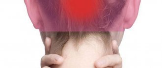
If your head hurts due to a lack of oxygen in the brain, i.e. compression of blood vessels, the following complaints may appear:
- burning, pulsation in the occipital, parietal, temporal regions;
- pain “radiates” to the eye sockets;
- nausea leading to vomiting;
- noise in ears;
- darkening of the eyes.
IMPORTANT! These symptoms are combined into vertebrobasilar insufficiency syndrome, or cerebral blood supply disorder.
In some cases, a person notes insomnia, tearing and redness of the facial skin.
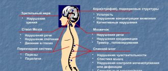
When the structures of the nervous tissue are compressed, the patient complains of:
- acute intense unilateral pain;
- less often - impaired swallowing and difficulty speaking.
IMPORTANT! If the pain is caused by a lack of blood supply, it will intensify with loud sounds and bright light, with compression of the nerve roots - during physical activity.
Symptoms appear more clearly if osteochondrosis is accompanied by hypertension or increased intracranial pressure.
Make an appointment
To register, choose any method:
- call the clinic +7 (495) 103-99-55,
- order a call back,
- leave a request for an appointment using a convenient form on the website:
Headache is a seemingly symptom that occurs periodically in all people.
IMPORTANT! However, vertebrogenic headache is a more serious symptom that indicates the development of osteochondrosis of the cervical spine.
In this case, the patient experiences many other unpleasant symptoms, ranging from a burning sensation in various parts of the head to tinnitus and darkening of the eyes. Vertebrogenic headache cannot be ignored - if treatment is not started in time, complications in the form of intensive development of osteochondrosis will not keep you waiting. Book a consultation with an experienced neurologist at our center to eliminate vertebrogenic headaches!
SIGN UP
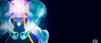
Piriformis syndrome
The piriformis muscle begins at the anterior edge of the upper sacrum and attaches to the inner surface of the greater trochanter of the femur. Its main function is hip abduction. The sciatic nerve passes between the piriformis muscle and the sacrospinous ligament. Therefore, when the piriformis muscle is tense, compression of the nerve is possible, which occurs in some cases with lumbar osteochondrosis.
The clinical picture of piriformis muscle syndrome is characterized by sharp pain in the subgluteal region radiating along the posterior surface of the lower limb. Adduction of the hip causes pain (Bonnet test), the Achilles reflex is reduced. The pain syndrome is accompanied by regional autonomic and vasomotor disorders, the severity of which depends on the position of the body - pain and autonomic disorders decrease in the supine position and intensify when walking.
Coccydynia – pain in the sacral area. A polyetiological clinical syndrome that may be caused by discopathy of the first coccygeal disc, causing reflex tension of the pelvic floor muscles, or ligament pathology. No sensory disorders are detected. A rectal examination reveals areas of tenderness in the muscles involved (usually the levator ani muscle).
Lumbar compression syndromes
Upper lumbar compression syndromes are a relatively rare location.
Compression of the LII root (LI-LII disc) is manifested by pain and loss of sensitivity along the inner and anterior surfaces of the thigh, and decreased knee reflexes.
Compression of the LIV root (LII-LIV disc) is manifested by pain along the anterior inner surface of the thigh, decreased strength and subsequent atrophy of the quadriceps femoris muscle, loss of the knee reflex.
Compression of the LV root (LIV-LV disc) is a common location. It manifests itself as pain in the lower back with irradiation along the outer surface of the thigh, the anterior surface of the leg, the inner surface of the foot and big toe. Hypotonia and wasting of the tibialis muscle and decreased strength of the dorsal flexors of the thumb are noted.
Compression of the SI root (LV-SI disc) is the most common location. It manifests itself as pain in the buttock, radiating along the outer edge of the thigh, lower leg and foot. The strength of the triceps surae muscle decreases, sensitivity in the areas of pain irradiation is impaired, and the Achilles reflex fades.
Compression syndromes of the cervical localization
At the cervical level, not only roots and vessels, but also the spinal cord can be subjected to compression. Compression of blood vessels and/or the spinal cord is manifested by a clinical syndrome of complete or, more often, partial transverse lesion of the spinal cord with mixed paresis of the arms and lower spastic paraparesis.
Root compression can be clinically divided into:
- root C3 – pain in the corresponding half of the neck;
- root C4 – pain in the area of the shoulder girdle, collarbone. Atrophy of the trapezius, splenius and longissimus muscles of the head and neck. Possible cardialgia;
- root C5 – pain in the neck, shoulder girdle, lateral surface of the shoulder, weakness and atrophy of the deltoid muscle;
- root C6 – pain in the neck, scapula, shoulder girdle, radiating along the radial edge of the arm to the thumb, weakness and hypotrophy of the biceps brachii muscle, decreased reflex from the tendon of this muscle;
- root C7 - pain in the neck and scapula, spreading along the outer surface of the forearm to the II and III fingers, weakness and atrophy of the triceps brachii muscle, decreased reflex from its tendon;
- root C8 – pain from the neck spreads along the inner edge of the forearm to the fifth finger of the hand, decreased carporadial reflex.
Cervical reflex syndromes
Clinically manifested by lumbago or chronic pain in the neck area with irradiation to the back of the head and shoulder girdle. On palpation, pain is detected in the area of the facet joints on the affected side. Sensitivity disorders, as a rule, do not occur.
It should be noted that the cause of pain in the neck, shoulder girdle, and scapula can be a combination of several factors, for example, reflex pain syndrome due to spinal osteochondrosis in combination with microtrauma of the tissues of the joints, tendons and other structures of the musculoskeletal system. Thus, with glenohumeral periarthrosis, many researchers note in such patients damage to the C5-C6 discs, as well as injury to the shoulder joint, or myocardial infarction, or other diseases that play the role of triggers.
Clinically, with glenohumeral periarthrosis, pain in the periarticular tissues of the shoulder joint and limitation of movements in it are noted. Only pendulum-like movements of the shoulder in the sagittal plane are possible (frozen shoulder syndrome). The adductor muscles of the shoulder and periarticular tissues are painful on palpation, especially in the area of the coracoid process and the subacromial zone. Sensory disorders are not determined, tendon reflexes are preserved, sometimes somewhat animated.
Reflex cervical syndromes include the anterior scalene muscle syndrome. The anterior scalene muscle connects the transverse processes of the middle and lower cervical vertebrae with the first rib. When this muscle is involved in the process, pain occurs along the anterior outer surface of the neck, radiating along the ulnar edge of the forearm and hand. When palpating the anterior scalene muscle (at the level of the middle of the sternocleidomastoid muscle, somewhat laterally), its tension is determined, and in the presence of muscle trigger points, pain distribution zones are reproduced in it - shoulder, chest, scapula, hand.
Vertebrogenic neurological complications in the thoracic spine with osteochondrosis are rare, since the bone frame of the chest limits displacement and compression. Pain in the thoracic region more often occurs in inflammatory (including specific) and inflammatory-degenerative diseases (ankylosing spondylitis, spondylitis, etc.).
In medical practice, the first place in terms of treatment is taken by lesions of the lumbar and lumbosacral spine.
What causes vertebrogenic headache?
Vertebrogenic headache is of a neuralgic nature.
It appears due to false impulses arising in the nerve fibers of the cervical spine, or due to disruption of the cerebral blood supply due to pinching of the vertebral arteries by intervertebral discs. Osteochondrosis occurs due to wear of the intervertebral discs, which is facilitated by excessive stress on the spine. They shift and become smaller in size (flatten), causing the nerve fibers along the spine to experience excessive pressure and also become more susceptible.
Displaced discs compress blood vessels, especially the vertebral arteries - the two largest vessels running along the spinal column, which are responsible for feeding the brain.
So, vertebrogenic headache occurs due to two main reasons:
- insufficient blood supply to the brain;
- pinched nerve roots.
The development of such conditions is facilitated by sedentary work, poor diet, lack of physical activity, exposure to tobacco smoke and chemical toxic substances, spinal injuries and poor environmental conditions.
Diagnosis of vertebrogenic headache
If you suspect a vertebrogenic headache, you can immediately contact a neurologist. You can also make an appointment with a therapist, who will write a referral to a specialist.
In addition to collecting anamnesis and examination, the doctor will need data from other studies.
- X-ray of the cervical spine.
- MRI of the cervical spine.
- Angiography of cerebral vessels.
- Dopplerography.
After a detailed diagnosis, a final diagnosis is made and treatment is selected.