general characteristics
The brain is the main organ of the nervous system. Doctors are still studying it. The adult human brain contains 90-95 billion neurons. The cell bodies of neurons form the gray matter of the brain, and the nerve fibers (neuron processes) form the white matter.
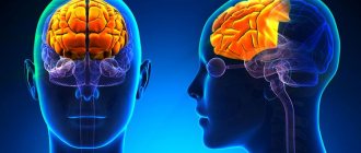
Rice. 1. Sections of the brain.
This organ has the following types of membranes:
- soft or vascular;
- arachnoid (cerebrospinal fluid - cerebrospinal fluid - circulates between the arachnoid and soft membrane, which serves as a kind of shock absorber and protects against shock, and also supports trophic and metabolic processes between the blood and the brain);
- hard.
The brains of men and women differ in their mass. In representatives of the stronger sex, its weight is 100-150 g more. However, mental development does not depend in any way on this indicator.
The functions of generator and impulse transmission are performed by neurons. Inside the brain there are ventricles (cavities) in which cerebrospinal fluid is formed. Paired cranial nerves extend from the brain to different parts of the human body. There are a total of 12 such pairs in the body.
Structure
The main organ of the nervous system consists of three parts:
TOP 4 articles
who are reading along with this
Spinal cord: structure and functions
Speech
Spinal cord: structure and functions
Memory
- two forebrain hemispheres;
- trunk;
- cerebellum.
It also has five departments:
- terminal or anterior, constituting 80% of the mass;
- intermediate;
- average;
- posterior, which includes the pons and cerebellum;
- oblong.
The white matter of the cerebral hemispheres of the forebrain is presented in the form of nerve fibers, which can be of three types:
- association – connect cortical areas in one hemisphere;
- commissural - connects the two hemispheres;
- projection – connect the cortex with the underlying formations.
Gray matter consists of neuron bodies; in the cerebral hemispheres, this substance forms the cortex and subcortical nuclei.
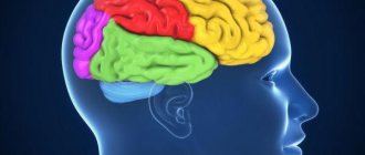
Rice. 2. Lobes of the cerebral cortex.
The following table will help you understand in more detail the structure and functions of various parts of the brain:
A comment
The work of the authors under the guidance of Dr. A.A. Zueva is devoted to the surgery of intracerebral tumors of functionally significant areas of the brain (both cortical areas and pathways using the example of the pyramidal tract). The authors have accumulated sufficient clinical material (65 patients), of which 14 operations with intraoperative awakening were performed. Neurophysiology was used (a combination of bipolar stimulation and transcranial evoked potentials). All patients underwent MRI control in the early postoperative period with an assessment of the extent of tumor resection; both early and 4-month follow-up were followed in all patients.
The dependence of current strength and pulse penetration depth is analyzed in detail, and modern Russian and foreign works on this topic are presented.
A small remark that does not reduce the value of the study is a small list of references, and the second thing to mention in the work, in addition to the pyramidal tract, is the need to map the associative conductors of the white matter.
The work is of scientific and practical interest; it should, of course, be supported and the authors wish further success in their work.
S.A. Goryainov, A.A. Potapov (Moscow)
Table “Structure and functions of the brain”
| Department | Structural features | Functions |
| End or front | It consists of two hemispheres, which are connected to each other by the corpus callosum. The surface of the hemispheres has many grooves and convolutions. | The right hemisphere is responsible for the left side of the body, and the left hemisphere is responsible for the right side. The centers for the regulation of voluntary movements, the centers of conditioned reflexes and mental functions are located here. The temporal lobe of the cerebral cortex regulates hearing, taste and smell, the occipital lobe regulates vision, the parietal lobe regulates touch; frontal – speech pronunciation. |
| Intermediate | Consists of the hypothalamus and thalamus. | The thalamus is a mediator in the transmission of stimuli to the hemispheres and helps to adequately adapt to changes in the environment. The hypothalamus regulates the functioning of metabolic processes and endocrine glands. Manages the work of the cardiovascular and digestive systems. Regulates sleep and wakefulness, manages food and drink needs. |
| Rear | It consists of the cerebellum and the pons, which is presented in the form of a white thick cushion located above the oblongata. The cerebellum is located behind the pons and has two hemispheres, the inferior and superior surfaces and the vermis. | This department provides the function of an intermediary in the transmission of impulses to higher parts of the brain. The cerebellum is responsible for coordination and precision of movements. |
| Average | Located from the anterior edge of the bridge to the optic tracts. | Responsible for hidden vision, as well as the implementation of orientation reflexes (turning the body in the direction of sound). |
| Oblong | It is a continuation of the spinal cord. | Controls balance, contains the centers of breathing, swallowing, sneezing and coughing, vomiting, cardiovascular and digestive. |
Biology lesson in 8th grade “Brain”
Topic: Brain
Goals:
Educational
- Study the structure and functions of the brain.
- Reveal the role of the medulla oblongata, midbrain, diencephalon and cerebellum in the implementation of conditioned reflexes and find out their meaning.
Developmental:
- continue to develop the skills to observe and describe an experiment.
Educational:
- cultivate accuracy and clarity when answering, the ability to observe the world around you.
Equipment:
- table “Brain structure”,
- disassembled models of the brain.
- Interactive complex
- VCR, TV.
During the classes
- Organizing time
Teacher: Hello friends!
The secrets of consciousness, the mysteries of nature excite people, they always attracted them. Reason and will motivated people to explore space, to sculpt cities.
Behavior, consciousness, will, reflexes Everything is studied in science by doctors Neurons, axons, reflex arcs, keys of the Central nervous system.
Teacher: what are we going to talk about today?
Study: about the central nervous system
Teacher: Correct, we continue to study the central nervous system and the topic of our lesson is “Brain”
Teacher: (brain model)
—What do you know about the brain?
Study: The brain belongs to the central nervous system. It consists of nerve cells - neurons, which form gray and white matter. All our actions occur with the participation of the brain.
Teacher:
— What would you like to know about the brain?
Studying:
The structure of the brain... What parts it is divided into and what functions it performs, what causes headaches
Teacher:
Then what goal will we set for today's lesson?
(Students themselves determine the purpose of the lesson during the conversation.)
Studying:
Find out: structural features of the brain, functions of the main parts.
Teacher: slide number 3
Lesson objectives:
- Study the structure and functions of the brain.
- Reveal the role of the medulla oblongata, midbrain, diencephalon and cerebellum in the implementation of conditioned reflexes and find out their meaning.
Teacher: open your notebooks and write down the date and topic of today's lesson
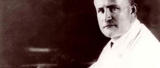
Teacher: the epigraph of our lesson will be the words of academician, doctor, scientist Vladimir Mikhailovich Bekhterev…..
slide number 3
.
Click
.
On the board there is an epigraph for the lesson: “The brain (head) must be protected, loaded in moderation and not injured.”
slide number 4
The mass of the brain in an adult varies from 1000 to 2000 g. Typically, the mass of the brain in women is slightly less than in men; this difference is due to the different mass of their bodies.
Many people think that the larger the brain, the smarter a person is. Brain weight I.S. Turgeneva 2012, V.I. Lenin 1340 Mendeleev 1571, Bekhterev 1720, Pavlov 1653 However, the mass of other gifted people was much smaller, for example, A. France - 1017. The idiot had the heaviest brain - 2850g.
Teacher: At the end of the lesson we will answer the question
slide number 5
Problem: is it possible to say that the larger the brain (head), the smarter the person?
Teacher: Man has long wanted to penetrate the mystery of the brain, to understand its role and significance in human life. Already in ancient times, the “father of medicine” Hippocrates connected consciousness and the brain.
Teacher: The human brain is a complex organ capable of perceiving and processing a huge amount of information. It is covered with three shells. Inside the brain are the cavities (ventricles) of the brain. 12 pairs of cranial nerves depart from it. Table
1 ( Appendix)
Of great importance for the brain is an abundant supply of blood, which brings oxygen and nutrients. Deterioration of blood supply leads to disturbances in brain function, memory suffers and performance decreases. With excess or poor nutrition, or with smoking, changes occur in the walls of blood vessels. They lose elasticity, become brittle due to salt deposition, they narrow and, as a result, pressure increases. Sudden attacks associated with increased blood pressure can lead to complications, such as stroke. A stroke is sometimes called a brain stroke. During a stroke, the blood supply to the brain is sharply disrupted, a person develops a severe headache, vomiting, confusion, loss of speech and sensitivity, and may experience paralysis.
The brain consists of 3 parts: Forebrain, midbrain, hindbrain, which are divided into 5 sections: cerebral hemispheres, diencephalon, medulla oblongata, cerebellum and pons.
There is a dummy brain on the demonstration table, let’s look at its structure: Slide No. 7
. Find all the parts of the brain on the model.
1.Cerebral hemispheres:
Formed by gray and white matter, grooves and convolutions are visible on the surface. The convolutions are folds of the cerebral cortex, and the depressions between them are furrows.
Working with disk.
Large grooves divide the hemispheres into lobes, there are 4 of them: frontal, parietal, occipital and temporal.
Parts of the cerebral cortex perform different functions, so they are divided into zones. Working with the disk
(“Biology 8th grade, Human”, Education 1 C)
- The cortex of the frontal lobes contains centers that regulate motor activity.
— In the cortex of the parietal lobes, located behind the frontal lobes, there are zones of bodily sensations, including touch and joint-muscular feeling.
— Adjacent to the parietal lobe is the temporal lobe, in which the primary auditory cortex, as well as the centers of speech and other higher functions, are located.
- The posterior parts of the brain are occupied by the occipital lobe, located above the cerebellum; its cortex contains areas of visual sensation.
2. Diencephalon
located under the corpus callosum and the fornix fuses on the sides with the cerebral hemispheres. Conducts impulses to the cerebral cortex from skin receptors and sensory organs.
It consists of the visual hillocks (thalamus) and the subthalamic region (hypothalamus).
In its parts of the diencephalon there are centers of thirst, hunger, and maintaining the constancy of the internal environment of the body. With the participation of the diencephalon, the functions of the endocrine glands and the autonomic nervous system
Teacher:
physical education and
“Freeze” commands.
Teacher:
What happened to the body after the command? the “Freeze” command caused you to stop moving and fixed a certain body position. With this command, movements in many joints of the body were simultaneously blocked. Under the influence of impulses coming from the diencephalon, the muscles simultaneously contract, fixing a new body position. We have observed the late reflex of the diencephalon.
Below the diencephalon is located:
3. Midbrain
– Located under the diencephalon, the lower border of its surface is the anterior edge of the pons. The midbrain is involved in the reflex regulation of various types of movements that arise under the influence of visual and auditory impulses.
For example, it provides a change in the size of the pupil, the curvature of the lens depending on the brightness of the light, or a rotation of the head and eyes towards the light source.
Experience No. 2
Open the textbook on page 61 (read paragraph 1 in a low voice).
(the ball suddenly bursts, the students look up: a new stimulus has caused an orienting reflex.)
Teacher: what did you hear, how did you react to the sound?
The irritant in this situation is a loud sound. This is an orienting reflex.
An indicative reflex is a reflex that occurs in response to any unexpected change in the situation or the appearance of new stimuli.
Teacher: Continuation of the spinal cord
is
4. Medulla oblongata:
The gray matter of the medulla oblongata is located in separate clusters - nuclei. Through the nuclei of the medulla oblongata, many reflex processes are carried out, for example, such as coughing, sneezing, lacrimation, etc. The nerve centers responsible for the acts of swallowing and the functioning of the digestive glands are also located here.
The medulla oblongata also contains vital centers involved in the regulation of respiration, the activity of the heart and blood vessels.
Damage to these centers leads to human death.
Experience No. 3
Teacher: The unconditioned reflex of the medulla oblongata cannot occur without irritation of the root of the tongue. Please make several swallowing movements at a fast pace. (Students, on command, make several swallowing movements at a fast pace in a row and make sure that in the absence of an irritant, in this case, saliva).
Is swallowing possible without saliva? No
Teacher: Why? What is the irritant? (saliva), Why doesn’t the swallowing movement occur? No saliva
Teacher: Correct. But if there is an irritant, any object, a swallowing movement occurs involuntarily, so small children should not be given small objects to play with. Babies often put things in their mouths and may involuntarily swallow small objects.
Teacher: And one more of the main parts of the brain:
5. Cerebellum.
It is located posterior to the pons and from the upper part of the medulla oblongata, filling most of the posterior cranial fossa.
Takes part in coordinating movements, making them precise and purposeful. When the cerebellum is damaged, a person’s movements are impaired, it is difficult for him to maintain balance, and his gait resembles the gait of a person who has lost his orientation.
Experience No. 4
We can observe the coordination of muscle work carried out by the cerebellum when performing the finger-nose cerebellar test.
Close your eyes, reach forward with your index finger extended and touch the tip of your nose with your fingertip.
Teacher: Did everyone succeed, touch the tip of their nose? More than 30 muscles were involved in this movement. The cerebellum receives impulses from many receptors and processes them. Thanks to the activity of the cerebellum, the body’s response occurs taking into account all external factors. But if the cerebellum is affected, then its work is disrupted.
Teacher: Give an example of a dysfunction of the cerebellum
(example of a drunk person: the effect of alcoholic drinks on the brain dulls the work of the cerebellum and a drunk person cannot coordinate his movements)
That's why a drunken person, trying to take one step, is forced by inertia to take several steps in the same direction?
Teacher:
Between the midbrain and medulla oblongata there is a bridge, which also has certain functions.
Bridge
- this is the place where the nerve fibers are located, along which nerve impulses go up to the cerebral cortex or back, down to the spinal cord, to the cerebellum, to the medulla oblongata. There are also centers associated with facial expressions and chewing functions.
Teacher: In today's lesson we are looking at the structure of the brain.
The epigraph of our lesson is the words of Bekhterev: “The brain must be protected, loaded in moderation and not injured,” who devoted his entire life to studying the brain. There is an institute that bears his name, the Bekhterev Brain Institute. Slide 8
Slide 9,10,11.
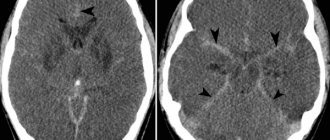
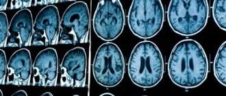
Studying the brain is impossible without special equipment - tomographs; on this slide you see several such devices that are used to take pictures of the brain
Slide 12.
Guys, let’s think and answer the question from our lesson: “
Is it possible to say that the larger the brain (head), the smarter the person?”
Write down your homework
Homework
: pp. Answer the questions in paragraph.
Slide number 13
“The human brain is the most complex object in the universe, the more we understand how it works, the better we can take care of it.”