Structure and functions of the brain in the table
| Part | Structure | Function |
| Oblong (stem section) | Extension of the spinal cord located on the trunk. It has a white substance on the outside and a gray substance on the inside. Gray matter is contained in the form of nuclei. | Conductive, nutritional, protective, control of respiratory rate, control of heart rate, control of vital reflexes responsible for sneezing, swallowing, hunger. |
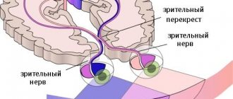
| Average | Connects the forebrain and hindbrain. Contains parts called quadrigeminal tuberosities. | Primary or subcortical centers of hearing and vision. Thanks to this, a person in the field of vision touches new objects or sound sources that appear. There are also centers responsible for muscle tone. |
| Intermediate | It consists of: thalamus, epithalamus, hypothalamus. The thalamus contains the centers of almost all sensory senses. The hypothalamus is the part of the intermedia that connects to and controls the pituitary gland. | Vision, tactile and taste sensations, sensations of body temperature and the environment, memory, sleep. |
| Cerebellum (hindbrain) | The subcortical part of the brain that has grooves. It consists of two hemispheres, which are held together by a worm. | Regulates coordination of movement, the ability to maintain the body in free space. |
| Cerebral hemispheres ( telencephalon) | It is formed from two parts (right and left), divided into grooves and convolutions, due to which the surface increases. They consist of a large amount of gray matter, which is located on the outside, and white on the inside. | Vision (occipital lobe), skin-articular sensitivity and muscle tone (parietal lobe). Memory, thinking, consciousness, speech (frontal lobe) and hearing (temporal lobe). |
Brain structure
The three largest parts of the brain are represented as: the cerebral hemispheres, the cerebellum and the brain stem. The list of the 5 main parts of the brain looks a little different:
- telencephalon (occupies 80% of the total mass);
- diencephalon;
- hindbrain (consists of the cerebellum and pons);
- midbrain;
- medulla.
The structure of the brain, pictures of which are presented below, can be considered in several aspects. So there are 5 main parts of the brain:
- final (80% of the total mass);
- intermediate;
- posterior (cerebellum and pons);
- average;
- oblong.
The brain is also divided into 3 parts:
- cerebral hemispheres;
- brain stem;
- cerebellum.
Structure of the brain: drawing with the names of the departments.
Structure of the brain: names of departments
Other GM departments and their functions
The structure of the human brain is divided into several sections.
The formation of brain activity occurs during intrauterine development thanks to the rhomboid, midbrain, and forebrain.
The parts of our brain are responsible for various tasks, characterized by the telencephalon and medulla oblongata, intermediate and middle brain, as well as the hindbrain, pons and cerebellum.
Their functions are shown in the table:
| Medulla | Another name for this zone is the bulbus, located in the back of the skull, between the cerebellar region, the pons and the dorsal segment. The bulbus is a continuation of the spinal cord. The white matter of the brain in this area is represented by neurons, and the gray matter by nuclei:
If the functioning of this department is disrupted, heart problems will arise and the transmission of impulses to the brain centers will be disrupted. |
| Diencephalon | This brain region “filters” the impulses of neurons. It will accept all incoming data and decide where and how it will go next. It consists of a lower zone and a posterior zone, consisting of the epithalamus and thalamus. This department is responsible for the functioning of the endocrine system. The hypothalamus is part of the inferior region. This dense neuronal bundle regulates body temperature and the cycle of wakefulness and sleep. It synthesizes hormonal compounds that “tell” a person when to drink or eat. This is the pleasure zone, responsible for interest in the opposite sex. The medullary zone is connected to the pituitary gland, which regulates all glands. Impulses come from the hypothalamic zone to the pituitary gland, and the order to synthesize or stop the release of hormones is “executed.” The thalamus processes impulses from receptors responsible for vision, taste, hearing, and tactile sensitivity. The signals are distributed to the corresponding brain areas. The epithalamus synthesizes the melatonin hormone, which is responsible for the cyclic processes of wakefulness, the emotional sphere, and puberty. |
| Midbrain | The brain section is small in size and consists of two halves: on the roof in the subcortex there are centers of hearing and vision, and conductive pathways are located on the legs. This brain segment includes substantia nigra with red nuclei. There is a temporoparietal node and nuclei of neurons that control the ocular myofibers and temporal zones, which process sound effects that are transformed into recognizable sounds. Reflex activity and reaction to the stimulus are controlled. This organ is responsible for spatial orientation. |
| Finite brain | This is the youngest part of the brain, the main part of the brain, responsible for higher nervous activity, and has numerous grooves with convolutions. The corpus callosum separates the right and left hemisphere zones. Each hemisphere is equipped with a nucleus, mantle, and olfactory brain. |
| Pons | This anatomical formation is part of the hindbrain, which contains the cerebellar region. The functions of the bridge are similar to its name; it consists of nerve fibers. Through it there are impulses passing from the body to the GM and vice versa. It makes up the brain stem, located between the midbrain and medulla oblongata. It contains the nuclei of nerves that control chewing, facial expressions, and some ocular myofibers. It receives signals from receptors responsible for the sensory organs, skin, and inner ear. Thanks to this department, a person feels taste, maintains balance, and hears sounds. |
| Cerebellum | Consists of 2 hemispheric areas and an unpaired formation connecting them. The cerebellar surface is covered by the cortex, which forms 2 nuclei in the thickness of the hemispheric zones. In the deep layers, the lobules consist of a white substance that connects the cerebellar segment with three pairs of legs with the spinal trunk and GM. Responsible for coordinating and regulating the movements of myofibers and muscle memory. Thanks to him, a person maintains a certain body position. |
The anatomical and physiological parameters of the GM have been studied by scientists for decades; they are different for each person, because there are not even two people who think the same way. Experts will eventually reveal these and other secrets of the brain.
From the editor: What is the name of the disease in which you forget everything?
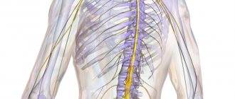
Parts of the human brain
The operation of all systems depends on the correct functioning of the vital organ.
Different parts of the human brain control many large and small functions, but they themselves need nutrition and a stable supply of oxygen. This work is carried out by the vessels that supply and drain blood. It is supplied to areas of the brain by 2 vertebral and 2 internal carotid arteries. Blood flows through the jugular veins. There are also two of them.
To improve blood circulation in the vessels of the head, it is necessary to spend more time in the fresh air, stimulate it with accessible physical exercises and, if necessary, take drugs such as Gingko Biloba. Cerebral circulation disorders result in headaches, dizziness, problems with perception and memory, absent-mindedness and problems with performance.
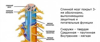
A vital organ is covered with several membranes:
- Solid. This is the outer layer that performs mechanical protective functions. It is mainly composed of collagen and elastin, the fibers of which are elastic and elastic. This shell is loosely attached to the cranial bones, merging with them along the edges of the bones, openings in the skull and at the exit points of the nerves.
- Arachnoid or arachnoid. This is the thinnest transparent membrane, which does not fit tightly to the soft one and forms the so-called subarachnoid space filled with cerebrospinal fluid. Where large grooves and depressions are located in the brain, the so-called cisterns containing cerebrospinal fluid are located. The fluid circulates through the ventricles of the brain and through the subarachnoid space.
- Soft. It forms the inner layer in the ventricles, forming the choroid plexuses. They produce cerebrospinal fluid. The shell consists of loose connective tissue, literally penetrated by a network of blood vessels. They perform the most important function of tissue nutrition.
All departments act as a single harmonious system, so a “failure” of one of them leads to disruption in others, causing internal problems and external symptoms. Parts of the organ and their activities
The main functions of the human brain are related to its anatomy and developmental characteristics. It consists of the following parts:
- Oblong. This peculiar continuation of the spinal cord has a similar structure. It controls coordination of movements, blood circulation, breathing, including the processes of sneezing and coughing, and is also involved in regulating metabolism. The oblongata, together with the middle, intermediate and pons forms the brain stem. This formation is involved in the control of articulate, coherent speech, breathing and heartbeat.
- The pons transmits information from the spinal cord to different parts of the brain.
- Cerebellum. It is located behind the bridge, covers the diamond-shaped fossa and occupies almost the entire rear one. Above it are the large hemispheres, separated from it by a transverse slit. The structure of the cerebellum has white and gray matter, as well as two hemispheres, which gives reason to call it the small brain. It is also involved in controlling movement coordination.
- Average. Occupies the area from the pons to the optic tract and papillary bodies, is responsible for hidden vision, and includes the center of the orientation reflex, thanks to which a person turns in the direction of the sound that appears.
- Large hemispheres. They are separated from each other by a longitudinal groove, in the depths of which there is the fornix and the corpus callosum. The right hemisphere controls the left half of the body, the left hemisphere controls the right. Each hemisphere consists of separate lobes: frontal, temporal, parietal and occipital, cortex and subcortex. The cortex forms numerous convolutions and grooves, consists of gray matter, and is divided into ancient, old and new. The cerebral hemispheres, or forebrain, are responsible for numerous functions, including intelligence and thinking.
Despite the fact that the structure of the brain of Homo sapiens is well known, its functions continue to be examined, periodically presenting scientists with real surprises.
The gray matter that makes up the human brain is a collection of neurons. There are about 25 billion of them. The entire brain is covered with 3 membranes:
- hard;
- soft;
- cobweb (cerebrospinal fluid, which circulates through the channels of this membrane, protects the brain from damage).
The weight of the brain of a man and a woman is slightly different: for women, its weight is on average 1245 g, and for representatives of the stronger sex - 1375 g. It is worth noting that its weight in no way affects the level of mental development of a person. First of all, it depends on the number of connections in the brain.
A person’s life activity depends entirely on how the various parts of the brain function. In this process, a special place is occupied by brain cells that generate and transmit impulses.
| part of the brain | features structure | functions performed |
| medulla | regulates metabolism, analyzes nerve impulses, where the centers of thirst and hunger are concentrated, receives information from the senses | coordination of movements |
| bridge | the centers of vision and hearing are concentrated, regulates the size of the pupil and the curvature of the lens, maintains the stability of the body when walking. | responsible for reflexes: coughing, working, sneezing, etc. inverts hearts and other internal organs |
| cerebellum | connects the front axle to the rear | consists of gray and white matter |
| average brain | consists of the diencephalon and cerebral hemispheres | the center is associated with the movement of the eyeballs and facial expressions. |
| forebrain | cylindrical cord similar to the spinal cord | the middle part and hemispheres having a cortex. |
The thickness of this surface is about 3 mm and covers both hemispheres. The cortex itself has 6 layers, which differ in width, size, density and shape of neurons:
- External granular;
- Molecular;
- External pyramidal;
- Internal grainy;
- Internal pyramidal;
- Fusiform.
The entire cerebral cortex consists of bundles of nerve fibers and neurons. There are more than 10 billion of them.
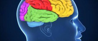
Each lobe of the cerebral cortex is responsible for some specific functions:
- occipital lobe - for vision;
- frontal - for movements, speech and complex thinking;
- temporal - smell and hearing;
- parietal - taste and touch.
In gray matter, all neurons communicate with each other. The white matter of the brain consists of nerve fibers. Some of them unite both cerebral hemispheres together. There are 3 types of fibers in the white matter:
- projection (perform a conductive function, thanks to them the cerebral cortex has a connection with other formations);
- association (play a connecting role between different cortical areas of one hemisphere);
- commissural (connects both hemispheres to each other).
The left lobe of the brain is responsible for logic and analytical thinking, the right lobe is responsible for creative thinking.
- vasomotor center - regulates the tone of the vessels (their degree of compression), i.e. blood pressure depends on its work;
- lungs (respiratory center) - regulates the frequency of inhalation and exhalation
- food - all digestive reflexes: salivation, swallowing, secretion of gastric juice, pancreas function;
- protection - coughing, sneezing, lacrimation and gag reflex;
The hindbrain - cerebellum (pons) - is essentially one structure - the pons connects the two parts of the cerebellum. The cerebellum determines the position of the body in space and is responsible for coordination.
The midbrain is essentially an extension of the pons; it is the human visual and sensory center.
This section contains the pituitary gland and hypothalamus - two glands that influence all other human glands.
Telencephalon - includes the hemispheres
| Share | Functions |
| Frontal lobe | perception of visual images; personal qualities of a person; |
| Parietal lobe | responsible for human sensations; determines the position of the body in space; coordination |
| Occipital lobe | visual centers |
| Temporal lobe | hearing centers and smell centers |
The cerebral cortex is considered a prerequisite for the presence of higher nervous activity in a person - conscious speech, creativity, intelligence, etc.
The cortex consists of gray matter, i.e. from neuron bodies. It forms furrows, and between them there are convolutions. Fissures divide the brain into lobes.
The more convolutions, the smarter a person? Not at all. The brains of outstanding scientists have as many convolutions as the brains of ordinary people.
Convolutions increase the surface area of the brain and reduce the length of nerve pathways.
- Postembryonic period of development
- Liver in the human body
- Structure of the spinal cord
- Human excretory system
- Female reproductive system
- Human circulatory system
Brain training
Every year our body ages, and this cannot be avoided - these are age-related features. The midbrain can develop simultaneously with the other hemispheres.
The following exercises are used for training and prevention:
- Aerial drawings. Use your fingers to make imaginary shapes in the air. You can draw geometric shapes, numbers, write names or more complex elements. During the process it is necessary to change hands.
- It is considered a good exercise to write texts with both hands at the same time. Take two sheets of paper, two pens. Choose a simple text. This will use all parts of the human brain.
- Computing. They teach you to count in your head at school. But over time, without proper training, this becomes more difficult to do. To keep your mind sharp, practice a few examples of addition, subtraction, division, or multiplication each day.
- List of words. This exercise develops memory. We write down any 10 words on paper and read the list several times. We remove the leaf. Then we remember all the written words. This will not only help train the brain, but also after reading the text, the necessary information will be stored in memory on its own.
- It is useful to do gymnastics for visual endings. There are many exercises for the eyes. Just 5 minutes every day can preserve your vision for many years.
- You can develop your sense of smell using essential oils. The main thing is not to smell many aromas at the same time, this can negatively affect the sensations.
Midbrain
It carries out righting and righting reflexes, thanks to which a person can walk and stand. The midbrain also influences the regulation of muscle tone and allows the body to turn towards the source of a sharp sound.
This section extends from the anterior edge of the pons to the papillary bodies and optic tracts. It contains a cluster of nuclei, which are called quadrigeminal tubercles. The midbrain is responsible for hidden vision. It also contains the center of the orienting reflex, which ensures the body turns in the direction of a sharp noise.
Pathologies and symptoms of damage
The main functions of the subcortical nuclei are to maintain posture and regulate motor activity; damage to this part of the brain affects the activity of the extrapyramidal system. Damage to the nuclei is accompanied by insufficient or excessive movements.
Dopamine deficiency, which correlates with the death of neurons in the substantia nigra, leads to the development of Parkinson's disease. One of the most common neurological pathologies (1 case per 200 people over 60 years of age) is manifested by symptoms:
- Rigidity (hardness) of skeletal muscles.
- Hypokinesia (insufficient motor activity, limitation of the volume and speed of voluntary movements).
- Tremor (frequent, rhythmic shaking) of the limbs and other parts of the body.
- Postural instability (inability to keep the body in balance, which leads to difficulty walking and frequent falls).
Dopamine deficiency is associated with the leading influence of the subcortical nuclei on the cortical parts of the brain. Damage to such parts of the brain as the putamen and caudate nucleus provokes the development of hypotonic-hyperkinetic syndrome, which is manifested by a decrease in the tone of skeletal muscles and hyperkinesis - pathological, uncontrolled movements that occur spontaneously at the wrong command of the brain. Types of dyskinesias (motor disorders) that occur:
- Hyperkinesis of choreic type. Jerky, erratic, varied movements, performed involuntarily, similar to normal movements, but differing from them in amplitude, intensity and adequacy of the situation.
- Athetosis. Tonic type spasms affecting the muscles of the face, limbs, and torso.
- Torsion spasm. Spastic muscle contraction of the tonic type, mainly in the torso area, leads to slow, erratic, involuntary movements, often rotational, corkscrew-like around the axis of the body.
- Hemiballism. Large, sweeping movements of great strength.
- Hemispasm in the face area. Repeated involuntary contraction of a muscle group in one half of the face.
- Tiki. Uncontrolled, monotonous, serial movements, for example, the formation and relaxation of skin folds on the forehead, raising and lowering the eyebrows, blinking.
- Tremor. Small, frequent trembling of the limbs, head and other parts of the body.
- Myoclonus. Muscle twitching at a fast pace.
- Torticollis of spastic type. Muscle spasm in the neck area, during which the head involuntarily tilts towards the spasmed muscle.
Unlike obsessive movements that appear as a result of traumatic brain injuries, physical and nervous fatigue, and psychologically traumatic situations, hyperkinesis cannot be delayed arbitrarily. Damage to the globus pallidus leads to disorders - hypomimia (absence or weakening of the activity of the facial muscles, lack of expression on the face, which resembles a frozen mask), physical inactivity (limited motor activity, decreased force of muscle contraction), monotonous speech devoid of expressive intonation.
Damage to the putamen is associated with the development of obsessive-compulsive disorder and ADHD (a syndrome reflecting attention deficit and increased motor activity). When the shell is damaged, trophic disorders develop (impaired cellular nutrition of tissues), which are more often manifested by damage to the skin - the appearance of ulcers. Violation of the functions of the shell negatively affects respiratory activity and the process of salivation (increases).
The basal ganglia are areas of gray matter accumulation that form functional brain structures responsible for motor activity and skeletal muscle tone. Damage to the basal ganglia is accompanied by motor and other disorders.
Unbelievable but true
Scientists have collected information that speaks about the phenomenal abilities of the human brain, namely:
1. The brain is considered gray. However, only dying cells turn gray. Living cells glow pink!
2. The organ works without rest from birth to death.
3. The organ contains 80 to 100 billion neurons. The left hemisphere contains more neurons.
4. According to information published in 2014, a woman’s head contains more “gray matter.”
5. Men have more white cerebrospinal fluid.
6. People with a humanitarian background have a higher percentage of “gray matter.”
7. Systematic physical exercise helps increase brain mass.
8. 60% of the brain is white matter, its color is determined by myelin, which increases the speed of electrical impulses.
9. Fat is very good for the brain.
10. The organ consumes up to 20% oxygen and needs the same amount of glucose.
From the editor: Anatomical features of the brain stem
11. The organ produces energy that can power a 25W light bulb!
12. It was found that organ size does not affect mental abilities.
13. The more convolutions, the more neurons, the better the memory.
14. You can increase the number of brain convolutions with the help of meditation.
15. When the process of yawning occurs, the organ cools down.
16. If a person neglects sleep, the temperature of the brain rises.
17. A person can process 70,000 thoughts per day.
18. Information in the organ moves through neurons at a speed of 1.5 to 440 km/h.
19. The organ is capable of instantly scanning and processing images in 13 milliseconds, while the blinking of the eye occurs in several hundred milliseconds.
20. According to statistics, approximately 20% of the population is left-handed. A right-handed person is most adapted to the conditions of civilization. People with left-handed ability have a harder time living.
21. Only 1% of the population can use both hands equally; they are called ambidexters.
hindbrain
This section consists of the pons located in front and the cerebellum located behind it. The structure of the cerebral pons: its dorsal surface is covered by the cerebellum, and its ventral surface has a fibrous structure. These fibers are directed transversely. On each side of the bridge they pass into the cerebellar middle peduncle.
The bridge itself looks like a white thick roller. It is located above the medulla oblongata. The nerve roots emerge from the bulbar-pontine groove. Hindbrain: structure and functions - on the frontal section of the bridge, it is noticeable that it consists of a large ventral (anterior) and a small dorsal (posterior) part. The border between them is the trapezoidal body. Its thick transverse fibers belong to the auditory tract. The hindbrain provides the conductive function.
The cerebellum, often called the cerebrum, is located posterior to the pons. It covers the rhomboid fossa and occupies almost the entire posterior fossa of the skull. Its mass is 120-150 g. The cerebral hemispheres hang above the cerebellum, separated from it by a transverse fissure of the brain. The inferior surface of the cerebellum is adjacent to the medulla oblongata.
It distinguishes 2 hemispheres, as well as the upper and lower surfaces and the worm. The boundary between them is called a deep horizontal gap. The surface of the cerebellum is cut by many slits, between which there are thin ridges (gyri) of the medulla. The groups of gyri located between the deep grooves are lobules, which, in turn, make up the lobes of the cerebellum (anterior, flocnonodular, posterior).
There are 2 types of substance in the cerebellum. Gray is on the periphery. It forms the cortex, which contains the molecular, pyriform neurons and granular layer. The white matter of the brain is always located under the cortex. Likewise, in the cerebellum it forms the brain body. It penetrates into all convolutions in the form of white stripes covered with gray matter.
It contains two elements of the human brain: the pons and the cerebellum. The pons consists of a dorsal surface, which is covered by the cerebellum, and a ventral fibrous surface. The fibers are arranged transversely in such a way that they pass directly from the pons into the middle cerebellar peduncle. The main function of the hindbrain is conduction.
The cerebellum, which is also sometimes called the small brain, occupies almost the entire posterior fossa of the skull. Its mass is 120-150 g. The cerebellum is divided from the cerebral hemispheres, which hang above it, by a transverse fissure. Conventionally, it can be divided into a worm, two hemispheres, a lower and an upper surface.
The cerebellum contains two substances: white and gray. The gray matter is the cortex, which in turn consists of the granular layer, the molecular layer and piriform neurons. The white matter is the medulla of the cerebellum. The coordination of human movements is entirely dependent on the functioning of the cerebellum.
Intermediate
This section has a common edge with the midbrain and telencephalon, is located along the fibers of the optic thalamus to the real surface, and from the ventral tegmentum in front of the optic chiasm.
Based on its functions, the intermediate section is divided into types: thalamus and hypothalamus.
Thalamus
The thalamus is responsible for processing information transmitted from receptors to the cortex. Includes approximately 120 cores, which are divided into specific and non-specific. Signals passing through the thalamus: muscle, skin, visual, auditory. Impulses sent by the cerebellum and brainstem nuclei also pass through.
Hypothalamus
This department is responsible for the centers of smell, regulation of energy and metabolism, the constancy of hemeostasis (the internal environment of the body), for the center of vegetative work through the nervous system. The functional participation of other parts of the brain allows a person not only to move, but also to perform a cycle of actions - jumping, running, swimming.
Since the diencephalon contains many vegetative nuclei, pineal gland, pituitary gland, and visual thalamus, it is also responsible for the following aspects:
- Performing work related to metabolic processes (water-salt and fat balance, protein and carbohydrate metabolism) and heat regulation, since it is one of the centers of the nervous autonomic system.
- The body's sensitivity to various stimuli, as well as the processing and comparison of this information.
- Emotions, behavior, facial expressions, gestures associated with changes in the functioning of internal organs.
- Hormonal background, production and regulation of hormones produced by the pituitary gland and epiphysis.
The diencephalon performs the following main functions:
- control of endocrine glands;
- thermal control;
- regulation of falling asleep, awakening and wakefulness;
- water balance;
- responsible for the center of satiety and hunger;
- responsible for feelings of pleasure and pain.
Finite brain
The structure of the brain cannot be briefly described, since without studying its structure it is impossible to understand its functions. The telencephalon extends from the occipital to the frontal bone. It distinguishes 2 large hemispheres: left and right. It differs from other parts of the brain by the presence of a large number of convolutions and grooves. The structure and development of the brain are closely interrelated. Experts distinguish 3 types of cerebral cortex:
- ancient, which includes the olfactory tubercle; perforated anterior substance; semilunar, subcallosal and lateral subcallosal gyri;
- old, which includes the hippocambus and dentate gyrus (fascia);
- new, represented by the rest of the cortex.
The structure of the cerebral hemispheres: they are separated by a longitudinal groove, in the depths of which the fornix and the corpus callosum are located. They connect the hemispheres of the brain. The corpus callosum is a new cortex made up of nerve fibers. There is a vault underneath it.
The structure of the cerebral hemispheres is presented as a multi-level system. So they distinguish between lobes (parietal, frontal, occipital, temporal), cortex and subcortex. The cerebral hemispheres perform many functions. The right hemisphere controls the left half of the body, and the left hemisphere controls the right. They complement each other.
The functions of the brain are difficult to understand without a thorough study of its structure and structure. The telencephalon consists of 2 cerebral hemispheres: right and left. The structure of the cerebral hemispheres differs from other parts in a large number of grooves and convolutions. Each hemisphere consists of:
- mantles;
- olfactory brain;
- kernels.
Experts conditionally divide the cerebral cortex into 3 types:
- ancient (consists of: olfactory tubercle, anterior substance, subcallosal, semilunar and lateral subcallosal gyrus);
- old (includes fascia (dentate gyrus) and hippocambus);
- new (it includes all other parts of the cortex).
Thus, the structure of the cerebral hemispheres is a multi-level system, where both hemispheres are separated by a groove in which the corpus callosum and fornix are located. Thanks to them, both hemispheres are connected to each other. The nerve fibers that make up the neocortex are called the corpus callosum.
In this multi-level system of the cerebral hemispheres, one can distinguish the frontal, parietal and occipital lobes, as well as the subcortex and cortex. Both hemispheres complement each other: thus, the left half of the body is controlled by the right, and the left is responsible for the right half.
Brain: structure and functions, general description
The pituitary gland is responsible for sex hormones, maturation and development.
The epithalamus controls biological rhythms, secretes hormones for sleep and wakefulness, reacts to light when eyes are closed and secretes hormones for awakening, and is responsible for metabolism.
Functions of departments
There are six main divisions.
- The medulla oblongata is responsible for connecting the brain with the spinal cord.
- The pons controls the contractions of all muscles during complex movements.
- The midbrain is responsible for hearing, vision and muscle tone.
- The diencephalon is responsible for interaction with the outside world.
- The cerebellum is responsible for coordination of movements, as well as orientation in space.
- The cerebral hemispheres are responsible for thought processes.
Medulla
This section is located in the skull, it is the beginning of the brain stem. In its rear part there is a groove and two cords, which are the connecting link with the spinal cord. This is where the white and gray substances are located, the first outside, the second inside.
The medulla oblongata is responsible for two main functions: reflex and conduction. Thanks to this, human cardiovascular activity, breathing, various types of reflexes are controlled here, and the connection between the brain and spinal cord is carried out.
The formation of this department is completed by the age of 7.
Pons
Functionally, the pons is responsible for contractions of the muscles of the entire torso and limbs that occur during complex movements. Here are centers similar to the spinal cord, but more developed.
This section changes by preschool age, when it shifts and occupies the position in which it will remain forever.
Cerebellum
This department is located above the previous two. It is divided into two hemispheres, which are connected by a structure called the “worm”. The parts of the brain and the cerebellum are united by nerve fibers, which, accordingly, form “legs” connecting it with the spinal cord and medulla oblongata.
Structure and functions
The cerebellum is formed from white and gray matter. The first is located under the bark, and the second is located outside, forming the cortex of the department.
The cerebellum is responsible for such important parameters as coordination of movements and maintaining body balance. This department is also responsible for muscle contraction.
People whose cerebellum is affected suffer from problems with spatial orientation, speech disorders and smooth movement. The growth of the department ends by the age of 15.
Midbrain
The lower nuclei are responsible for the functioning of the human auditory system.
They receive impulses produced in the outside world, implementing the human guard reflex, that is, the body can instantly engage in an action that requires a quick reaction.
Functions
This department plays an important role in fine motor skills and the acts of chewing and swallowing, ensuring their correct sequence. Like the parts of the brain described above, the midbrain is directly related to muscle function.
From the editor: Types and characteristics of organic dementia
Thus, it controls work during prolonged stress, for example, when some part of the body must remain in one position for a long time, then it maintains muscle tone so that it can suddenly move to another position.
The development of the midbrain directly depends on the formation of other parts.
Diencephalon
The hypothalamus is considered the most important element of the diencephalon, since it contains many autonomic centers.
It is responsible for metabolism, feelings of fear and rage, body temperature, nerve connections, etc.
The hypothalamus also produces cells that influence the functioning of the pituitary gland, which regulates some of the body's autonomic functions. The thermal stage of diencephalon development ends in adolescence.
Finite brain
The parts of the human brain directly depend on the functioning of the hemispheres, or telencephalon. The two hemispheres, which make up up to 80% of the mass of the entire brain, are connected through the corpus callosum and other commissures. The cortex covering the elements of the department consists of several layers of gray matter.
It is thanks to it that the realization of higher mental activity is possible. The work performed by both hemispheres is unequal. The left, dominant, is responsible for thought processes, counting, writing, the right is for the perception of signals from the outside world.
This department develops most actively until puberty; later the pace decreases.
Their totality has gone through long centuries of evolution, changing, improving and adapting to changes, which, in fact, ensured the survival of the human species.
The parts of the brain collectively and each individually are indispensable centers for controlling the autonomic functions of the body.
general description
The human brain consists of 25 billion neurons. These cells are the gray matter. The brain is covered with membranes:
- hard;
- soft;
- arachnoid (the so-called cerebrospinal fluid, which is cerebrospinal fluid, circulates through its channels). Liquor is a shock absorber that protects the brain from shock.
Despite the fact that the brains of women and men are equally developed, they have different masses. So, among representatives of the stronger sex, its weight is on average 1375 g, and among women - 1245 g. The weight of the brain is about 2% of the weight of a person of normal build. It has been established that the level of a person’s mental development is in no way related to his weight. It depends on the number of connections created by the brain.
Brain cells are neurons that generate and transmit impulses and glia that perform additional functions. Inside the brain there are cavities called ventricles. Paired cranial nerves (12 pairs) depart from it to different parts of the body. The functions of the parts of the brain are very different. The vital functions of the body completely depend on them.