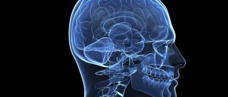
What pathologies can develop?
As a rule, symptoms depend on which segment of the organ is affected by disease or injury, as well as on what type of pathology develops. Signs of brain dysfunction include:
- impaired innervation of the legs and arms or other areas of the body;
- severe pain syndrome in the spinal region;
- unauthorized bowel movement;
- psychosomatic disorders;
- impaired mobility of the torso;
- severe muscle or joint pain;
- muscle atrophy.
The following diseases may be accompanied by similar symptoms:
- Tumor. This includes both malignant and benign neoplasms, which can be located extradurally, intradurally, or intramedullarily. Extradural tumor is characterized by rapid progression and is localized in hard tissues. An intradural neoplasm develops under hard tissues. Intramedullary neoplasms are characterized by their development in a liquid substance.
- Intervertebral hernia. The initial stage of hernia development is protrusion. When the fibrous ring of the disc is destroyed, the contents exit into the spinal canal. If the spinal cord was involved in the lesion, the development of myelopathy (not compression or chronic) is diagnosed.
- Chronic myelopathy. Often (with untimely treatment) osteochondrosis becomes the cause of the development of spondylosis, which is the final degenerative change in the structure of tissues. In this case, the appearance of osteophytes is observed, which subsequently serve to compress the brain canal.
- Heart attack. Caused by impaired circulation of the organ, the occurrence of necrotic processes and is characterized by the formation of blood clots and dissection of the aorta. It is recommended to immediately contact a specialist if pain occurs in this department. This is the only way to prevent irreversible consequences.
Anatomical features
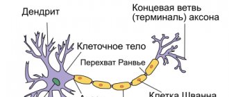
A rather thick tourniquet, white in color, located in the spinal canal - this is the human spinal cord. Its diameter is about 1-1.5 cm, and its length almost reaches half a meter (up to 45 cm). This organ weighs about 38 g.
- Reflexes
- Interesting Facts
- Reflex Bow
- Segmental structure
- What are the segments?
- Role and functions in the body
- Gray matter of the spinal cord
The narrow spinal canal is not only the location of an important organ, but also its protection. The core of the organ consists of a gray substance
It is covered by a white substance, which is also covered with protective and nourishing shells for the core. This is the general plan of the structure of the spinal cord.
Topography
The structure and functions of the spinal cord are quite complex. Neurosurgeon students study it in detail. Experts very carefully consider the development of the spinal cord. Ordinary people are interested in the question of what its topography is and familiarity with the leading role of this organ.
So, it is quite simple to describe the essence and goals that this body serves. The cervical spinal cord at the level of the back of the head in the area of the foramen passes into the cerebellum. The spinal cord ends at the level of the first 2 lumbar vertebrae. The conus spinal cord is located where a pair of vertebrae are located in the lumbar region. Next is the well-known “terminal thread”.
But this fragment is considered atrophied. It is called the “end” region. Nerve endings called “roots” are distributed along the entire circumference of the thread. The filum terminale is equipped with a substance containing a small proportion of nervous system tissue. But the outer part is not even equipped with a similar fabric.
The topography of the organ includes a pair of thickenings where the innervating processes emerge (cervical thickening of the spinal cord and lumbar). The outer and rear surfaces of the bundle are separated by slits called “middle”. The one in front is deeper, the back one is smoothed.
External structure
The general structure of the spinal cord suggests its division into a number of surfaces: posterior, anterior and two lateral. The spinal cord has faint grooves on the lateral surface. They are located longitudinally, and nerves extend from the grooves. They are also called “roots”. In the lumbar area, together with the terminal filament, they form a tail, which is commonly called a horse's tail. The grooves divide half of this cord into the following structures:
- front;
- lateral;
- posterior (cords).
The grooves of the spinal cord extend along the canal. The roots are divided into anterior ones - they are formed by efferent neurons, and posterior ones, created by afferent neurons. Their bodies converge into a knot. The roots unite and form a nerve. So, on all sides of the tourniquet there are over 30 nerve endings, forming exactly the same number of pairs. This is the external structure of the spinal cord.
White matter
All cords are made entirely of white matter of the spinal cord. They consist of longitudinal nerve fibers. These threads converge, forming peculiar conductors. Based on their functional purpose, fibers are divided into 3 types:
- motor;
- associative;
- sensitive.
The first are represented by short bundles and combine all parts into a single system. The second ones are called ascending. They give signals to the centers. Still others are descending. They provide signals from central structures to areas of the horns.
Important Trimethine (trimethadione): instructions for use, reviews, analogues
Gray matter
It structurally resembles grouped longitudinal plates consisting of homogeneous neurons. It contains not only neuronal bodies, but also neuropil, glial cells and capillaries. Along the entire spine it forms 2 columnar types, left and right. They are connected by gray adhesions.
The anterior horns contain the largest neurons. They form the motor nuclei of the spinal cord and inhibitory neurons. The structure of the gray matter of the background horns is not the same. It contains a huge number of intercalary type neurons.
The lateral horns of the spinal cord fill the centers of the ANS, the dilation of the pupil, the bases of innervation of the digestive system and other important organs of the human body. In the nucleus of the gray matter of the spinal cord there is a canal that neurosurgeons call “central.” It is filled with liquor. In adults, in some places it is filled with cerebrospinal fluid, and in others it is overgrown.
Shells
Anatomy of the spinal cord describes the membranes of the spinal cord:
- vascular soft;
- hard;
- avascular or arachnoid.
The characteristics of shell 1 are as follows: soft, penetrated by vessels and nerves. It is enveloped by the avascular part. There is some space here called “subarachnoid”. The cerebrospinal fluid generated in one of the systems flows into this niche. The last shell is made up of connective tissue; it is strong and flexible. The membranes of the spinal cord and brain are identical and form a single structure.
Reflexes
The functional diversity of neurons in spinal structures creates conditions for reflex activity, both with the participation of its own elements and central ones - the cortex and subcortical centers.
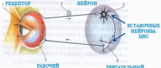
Each such reflex arc necessarily contains the following links:
- afferent – perceives impulses;
- intercalary – connects sensory and motor neurons;
- efferent - transmits the impulse to the immediate performer.
Among the motor reflexes, attention should be paid to myotatic ones, which are aimed at stretching/contracting muscles, as well as skin ones - plantar and abdominal. With their help, every person can perform voluntary and involuntary movements.
In addition to motor reflexes, there are other reflexes. Thus, autonomic ones are usually divided into sympathetic and parasympathetic. Their common task is to control and ensure the constancy of the internal environment of the body to maintain human life.
Interesting Facts
This organ has not yet been fully studied - it still hides many secrets from doctors, and their solution in the future may lead to the cure of currently incurable diseases of the nervous system. Here are some interesting facts about this organ:
- If the spine grows over 20 years, then the spinal cord only grows for 5 years.
- Stress leads to a serious decrease in the number of neurons. If the normal number of neurons is 13-14 million, then as a result of stress their number drops by half - this is especially true for pregnant women.
- In the process of evolution of vertebrate organisms, the spinal cord appeared first, and only then the brain. The first performed all the simplest functions, including reflex ones.
- Some living beings are able to live after losing their brain, remaining only with their spinal cord.
- Damage to a specific area of the organ not only causes loss of sensation below the rupture site, but also the ability to sweat. This forces people with injuries to spend more time in the shadows, as the body has partially lost its thermoregulatory function, which is extremely important for life.
- Scientists have not yet come to a general conclusion, and cannot establish the mechanism of hair loss throughout the body in people with spinal cord injuries.
- If the thoracic organ has been affected, the person may lose the ability to cough.
- A biopsy and analysis of the white matter of an organ can detect hundreds and thousands of human diseases.
- The spinal cord very sensitively senses the rhythm of music, and therefore is automatically able to send signals that will make the body move to the rhythm.
- People with a healthy spine are much more active in their sexual life.
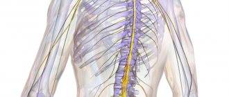
The organ is located in the spinal canal, and its length is no more than 45 cm, which is less than the length of the spine itself. This is due to the fact that the brain grows only until the age of five, and the spine, as a rule, until the end of puberty.
This organ controls absolutely all motor processes in the body, including contraction of the heart muscles, breathing and movement of the limbs.
We study the anatomy of the spinal cord:
Location of the spinal cord and its functions:
Thus, the loss of certain functions, for example, leg movements, makes it possible to determine which part was damaged. Injuries to this organ are among the most serious, and the damage is often irreparable. The main thing is to monitor the health of your spine and not overload it unless absolutely necessary.
Related article: How we are made
Basic functions of the spinal cord
The segmental structure of the spinal cord allows it to effectively cope with assigned tasks. Their well-coordinated communication ensures a high speed of impulse transmission and perception of information coming from the environment. The main functions of the spinal cord are conductive and reflex. They ensure the ability of the organ to perform all the necessary actions for the normal functioning of the body.
The segmental apparatus is connected with almost all internal organs and ensures human activity and the functionality of each system. The first function allows you to receive complete information about the world around you: what effects are on the skin or body, whether it hurts or not. The “communication” is two-way, since the brain not only reacts to events around it, but also gives commands what needs to be done. The conduction function ensures mental and motor activity.
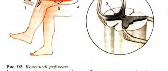
The reflex function is no less important. The concept itself explains what it is. Thanks to the spinal cord, if the hand feels pain or burning when exposed to fire receptors, the person quickly pulls it back reflexively. During the examination, neurologists check the knee reflex and other body reactions to stimuli; if they are absent, this helps to diagnose hidden disorders in a timely manner.
The spinal cord has a rather complex structure, since it is responsible for the most important functions in the body, namely: digestion, urination, blood circulation, breathing, sex life, motor activity and many others. It is thanks to the reflexes for which he is responsible that a person is saved in moments of danger. It consists of many segments that help transmit impulses from the periphery towards the brain. Thanks to their coordinated work, a person’s muscles contract and he feels the world around him.
Reflex Bow
Reflex = stereotypical response
Important Midlife crisis in women: when ladies go crazy
There are afferent fibers that transmit their excitation directly to the motor neurons of the anterior horn cells, which in turn control the muscles through their efferents. This reaction occurs at the level of the spinal cord and is called a simple reflex. The underlying neural circuit is called a reflex arc. The afferent may pass directly to the motor neuron (monosynaptic reflex arc) or involve interneurons (multisynaptic reflex arc) that reach the motor anterior horn cells.
Self-reflex
In self-reflex, the receptor and effector are located in the same organ. The reflex arc proceeds monosynaptically.
Example:
Patellar tendon reflex = PSR
The patellar tendon reflex is a monosynaptic reflex.
A blow to the patellar tendon causes the hamstring muscles to contract. The receptors that sense the stimulus are stretch receptors in the muscle spindle of the quadriceps femoris muscle. Sensory afferents travel to the dorsal horn of the spinal cord and are transmitted to the anterior horn motor neurons at the L2-L4 level by only one synapse. The effects now travel through the lumbar plexus and are then isolated in the femoral nerve back into the muscle and cause it to contract.
What is important for the clinical assessment of a reflex is not the strength of the response to a stimulus, but the fact that the reflex can be evoked on both sides.
External reflex
In an external reflex, the receptor and effector are not located in the same organ. Several synapses are inserted into the reflex arc.
Examples:
polysynaptic flexor reflex
The flexion reflex is a protective reflex. Nociceptors are stimulated, and the excitation spreads to various levels of the spinal cord. The flexors on the irritated side and the extensors on the opposite side contract.
- Cremaster reflex
- Abdominal cutaneous reflex
- Eyelid reflex
Segmental structure
A segment of the spinal cord is a piece of a tourniquet along with associated nerves. There is no morphological separation of one segment of the spinal cord from another. It is extremely functional. Each of the segments innervates a certain region. The designation of spinal cord segments is represented by alphanumeric indices, oriented to a part of the spinal cord and containing segment numbers.
The spinal cord consists of about 33 segments. The spinal cord segments have 4 roots, a pair of anterior and posterior. The spinal column is significantly longer than the cord, so it should be remembered that the segments are not numbered in the same way as the vertebrae. Any nerve consists of motor-sensitive roots. They come out in bunches from this bundle to the openings between the vertebrae.
The nerve ending located behind forms a ganglion and merges with the nerve ending in front. In this case, a mixed nerve is formed, which is divided into branches:
- The meningeal branch innervates in accordance with the nature of the spinal cord membrane and the canal wall.
- Dorsal - the skin in the corresponding areas, as well as deep muscle tissue.
- The connective tissue branch is the connecting link between the tourniquet and the ganglia.
- The abdominal branch is responsible for innervation of the limbs, lateral surfaces of the body and tissue of the abdominal part of the body.
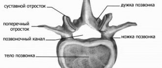
Segment structures
The structure of the segments in the spinal cord is paired, and they are connected to the organs by nerves. The anterior and posterior roots provide the transmission of information, and their main task is to stimulate muscle contraction. Only the dorsal roots are sensitive; they are the ones that are activated by receptor stimuli. The spinal cord is not homogeneous in cross section, but is a substance consisting of white and gray matter.
Segmental structure of the spinal cord with the designation of roots in different sections
White matter
The white matter of the spinal cord is represented by processes of nerve cells that make up the pathways. It is located on the periphery of the gray matter and is formed from nerve bundles. The white matter contains descending ascending fibers that pass into the brain. They transmit information coming from receptors. Their location is natural: on the side of the back there are ascending ones, on the abdominal side there are outgoing ones. The white matter is delimited by the grooves of the spinal cord into:
- anterior - make up the descending pathways.
- lateral - there are both ascending and descending paths.
- posterior cord - make up the ascending pathways.
White matter contains projection, association and commissural nerve tracts. The first provide communication with the brain.
Gray matter
Gray matter consists of interneurons and motor neurons. They provide motor reflexes and communication between neurons. In total, it occupies approximately 18% of the total volume of the spinal cord, and includes approximately 13.5 million neurons. This is a special device that contains some functions of the central nervous system. Thanks to two-way communication, irritation can pass through both ascending and ascending pathways. The response is a contraction of muscle tissue and a motor response.
Do you know that:
- The spinal cord stops growing at the age of 5 years.
- In the process of evolution, it was the spinal cord that appeared first, and only then the brain.
- People with spinal cord injury cannot sweat below the injury site. Therefore, in hot weather they need to be in the shade or otherwise cool their body to avoid overheating.
- The spinal cord relaxes all the muscles of the body during sleep. This function allows a person not to repeat the movements that he performs in his sleep.
No ads 2
What are the segments?
The sections of the spinal cord differ from those used to conventionally divide the areas of the spine. Their length varies; the fewest elements are found in the coccygeal part. The segments are connected to certain areas in the body by nerve conductors. In general, the brain is divided into the following segments:
- Neck – 8.
- Chest – 12.
- Sacral – 5.
- Lumbar – 5.
The main length falls on the thoracic segments of the spinal cord, after which 23.2% is allocated to the neck and only 7.3% to the lower back. They are posterior and anterior nerve roots that alternate regularly. They are located above the vertebrae with the same number. Their main task is to report movements and be responsible for muscle contractions. Therefore, the anterior nerve roots are also called motor, and the posterior - sensitive.
The intervertebral foramen contains the roots of each individual segment. Their direction is not the same, because the spinal column is filled with the brain. In the cervical region they lie horizontally, in the thoracic region they are directed diagonally, and in the lower part they are almost vertical.
The shortest segments are the neck segments, and the longest ones are in the lower back and sacrum. In the lower part they form the so-called “horse tail” - this is a bunch of roots located below the 2nd vertebra.
A visual representation of what a spinal cord segment looks like in cross section
Location Features
The skeletotopy of the areas is individually variable, since the lower part of the lumbar region can be located in adults from the lower third of the body of the 9th thoracic vertebra to the disc between L1-L2. Because of this, a certain feature appears. When the upper processes are directed in the transverse direction, the further down the canal, the higher the exit site is located relative to the intervertebral foramen.
Each segment of the spinal cord is responsible for its part of the periphery. This could be muscle tissue, internal organs or skin. The division into such sections is almost identical in all people, so it is not difficult for doctors to identify the location of the lesion based on the specific sensitivity of a particular area. The relationship can be seen in more detail in the table:
| Department | Number of spines | Related department |
| Cervical | 8 | Skin and muscles in the neck, upper limbs, diaphragm |
| Chest | 12 | Skin on the chest, back and abdomen |
| Lumbar Sacral Coccygeal | 5 5 1 | Lower body, legs, feet, pelvic organs |
For example, when a patient complains of discomfort in the navel area, there is a high probability that the pathology is hidden below the 10th thoracic vertebra. The doctor will check for the presence of a lesion in this particular part of the body.
[node:field_similarlink]
Role and functions in the body
Functionally, the spinal cord performs the following tasks:
- Regulating the functioning of organs and systems through the transmission of nerve impulses to them. In other words, it performs a reflex function.
- Transmission of information to the brain, as well as from it to motor neurons.
The gray matter of this spinal segment contains many pathways that provide motor reactions of the body. The activity of each reflex occurs through a special department of the central nervous system - the nerve center. In the latter, special cells are localized, which occupy a certain section of the organ and ensure the functionality of specific systems in the body. For example, knee reflexes are provided by nerve cells localized in the lumbar region of the spine. The urination process is in the sacrum, dilation of the pupils is in the thoracic.
Important Psychosis
The nervous center processes information sent by skin receptors, as well as other systems and organs in the body. As a response, the brain generates certain impulses, which are subsequently transmitted to the executive organs (for example, skeletal muscles, vascular apparatus, cardiac muscles, etc.). As a result, a change in the functional state of the latter occurs.
Motor neurons carry out the process of contraction of muscles in such parts of the body as limbs, intercostal spaces, etc. Regulation of such a reflex also occurs with the help of higher parts of the central nervous system. Nerve impulses that travel along the spinal cord to the brain transmit information about the dysfunction of any organ or system in the body. Impulses transmitted by various organs to the spinal cord and from there to the region of the dorsal roots of the brain are processed by sensory neurons. From them, information is distributed either to the dorsal horns or to the cerebral hemispheres.
Blood supply
The supply of nutrients is carried out through the blood, which flows through numerous vessels. They extend from the upper part, along the thyroid and vertebral arteries. They start from the aorta and vertebral arteries, of which there are normally about 6–8 in humans. The largest of them provide nutrition to the cervical and lumbar thickening.
The largest radicular-spinal artery is the inferior artery of Adamkiewicz; there are 15–20 posterior arteries in total, but all of them are significantly narrower. Their main task is to provide nutrition to the posterior third of the spinal cord.
All vessels are connected to each other; such joints are called anastomosis. The entire system provides nutrition to different parts of the brain, while being protected from the formation of blood clots. Even if a separate vessel is closed by a plug, the area for which it is responsible will not be left without power; this is what the anastomosis provides. It saves neurons from hypoxia and death.
In addition to the arteries, the spinal cord is also supplied by veins that are connected to the cranial plexuses. This is a whole system of vessels through which blood flows from this organ to the vena cava. Thanks to the presence of special valves, blood is not able to flow back.
Skeletotopy of the spinal cord
[node:field_opros]