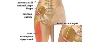
The vestibulocochlear nerve (VCN) is part of the system of the organ of the same name, which is responsible for the functioning of the hearing aid. This is a fiber of special sensitivity, which includes several roots. The vestibular one is responsible for impulses from the static apparatus, and the cochlear one conducts signals from the organ spiral to the labyrinth. In medicine, there are several methods for testing the vestibulocochlear nerve in order to identify its pathologies. After all, they affect many unpleasant symptoms: from dizziness to severe weakness and headaches.
Anatomy and topography of the nerve ending
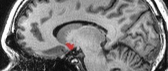
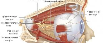
Anatomy of the vestibulocochlear nerve
Topography divides the fiber into two parts. The first is the cochlear part of the vestibular-cochlear nerve fiber, which is also called the vestibular. It integrates with afferent endings and transmits information from them. They are embedded in the semicircular channels of the elliptical and spherical sac. The second part is the cochlea, which transmits impulses from the spiral organ to the brain.
The anatomy of the parts of the vestibulocochlear nerve is as follows:
- The vestibule. The node of the same name is located here, deep in the auditory canal. It is also divided into 2 parts: the upper, directed to the receptive fields, and the lower, forming the spherical-saccular nerve. The ampullary nerve, which arises from the vestibulocochlear nerve, also originates here. The central roots of the cells of this node form the vestibular part, and in the cochlear zone they connect to the auditory canal. Inside, the nerve goes along with the intermediate endings and the branch of the facial nerve, and then diverges into the cranial cavity through the auditory canal. At the cerebellopontine angle the ending passes into the brain stem. The nuclei of the vestibular part of the vestibulocochlear nerve are also formed here. The vestibular part in the region of the vestibular field of the rhomboid fossa ends at the top in the ankylosing spondylitis nucleus, in the middle – in the Schwalbe nucleus, and at the back – in the Deiters nucleus.
- Cochlear zone. This part originates from the spiral ganglion formed by the neurons of the auditory pathway. Then it passes into the spiral canal of the cochlea rod. The peripheral roots end in the spiral organ. And the central ones run through the rod and the opening of the spiral path in the ear canal. The cochlear part of the apparatus is formed there. Then, together with the vestibular nerve ending, it passes into the cranial cavity through the auditory opening and goes to the brain stem. In the cerebellopontine part it enters the trunk and ends in the ventral and dorsal nuclei.
Topographically, the features of PUN do not differ from the characteristics of other sensitive fibers passing in this zone.
Structure [edit]
Path[edit]
The vestibular nerve fibers enter the medulla oblongata on the medial side of the cochlear fibers and pass between the inferior peduncle and the dorsal course of the trigeminal nerve.
They are then divided into ascending and descending fibers. The latter end with branching around the cells of the medial nucleus, located in the acustica region of the rhomboid fossa. The ascending fibers either end in the same way, or in the lateral nucleus, which is located lateral to the acustica region and further from the floor of the ventricle.
Some axons of the cells of the lateral nucleus, and possibly also the medial nucleus, continue upward through the inferior peduncle to the superior nuclei of the opposite side of the cerebellum, to which other fibers of the vestibular root also continue. without interruption in the nuclei of the medulla oblongata.
A second set of fibers from the medial and lateral nuclei partially terminate in the tegmentum, while the rest ascend in the medial longitudinal fasciculus to branch around cells of the oculomotor nerve nuclei.
Fibers from the lateral vestibular nucleus also pass through the vestibulospinal tract to anterior horn cells at many levels of the spinal cord to coordinate movements of the head and trunk.
Subnuclei [edit]
There are 4 sub-cores; they are located at the bottom of the fourth ventricle.
| Name | Location | Notes |
| medial vestibular nucleus (dorsal or main vestibular nucleus) | medulla oblongata (floor of the fourth ventricle) | corresponds to the lower part of the area acustica in the rhomboid fossa; [ citation needed ] The caudal end of this nucleus is sometimes called the descending or spinal vestibular nucleus . |
| lateral vestibular nucleus or Deiters nucleus | medulla (upper) | consists of large cells and is located in the lateral corner of the rhomboid fossa; the dorsolateral part of this nucleus is sometimes called the ankylosing spondylitis nucleus . |
| inferior vestibular nucleus | medulla (lower) | |
| superior vestibular nucleus | bridges |
Functions of the nerve ending
There are several functions of the vestibulocochlear nerve. Violation of even a small segment in the structure of the ending can lead to significant changes in a person’s well-being:
- Auditory function. The human hearing system is a very complex organ consisting of the middle, inner and outer ears. The vestibulocochlear nerve is responsible for the innervation of the internal, most complex structure. First, the sound hits the membrane, and then is transmitted by its vibrations to the hammer. From there it moves to the stapes and anvil, combined with each other. Then it is transmitted to the oval window in front of the labyrinth. Intra-auricular fluid ensures prolongation of vibrational waves, which facilitates transmission through the lymphatic layers. The basilar plate receives sound from the spiral ganglion of the inner ear. And the processes of the nerve endings entering the node go through the opening of the ear canal and penetrate to the nerve. The nerve then sends a signal to the nucleus of the brain stem. This is how the brain receives a signal about incoming sound.
- Balance option. The hearing aid is closely related to the vestibular one. If disturbances occur in it, they also affect the coordination of movements. You may even experience symptoms such as nausea and fainting. The vestibulocochlear nerve provides innervation to important parts of the vestibular system. It allows the nervous system to analyze the position of the head and adjust the tone of the muscles of the whole body. And the organ responsible for balance is located right in the labyrinth of the middle ear and consists of 3 channels. They contain sensitive hairs. These hairs are equipped with neuronal processes that go to the vestibular canal of the bottom of the temporal bone. Further, passing into the medulla, the nerve enters the vestibular nuclei. They are responsible for the body's friendly reactions.
Any pathology of the vestibulocochlear nerve, disruption of its circuit, formation of tumors and rupture of innervation lead to problems with hearing and coordination. And damage to the vestibular apparatus can lead to other diseases.
Brain stem motor systems
The structures of the brain stem provide a higher level of regulation of movements and are classified as structures of direct action. Their activity consists not only in the implementation of action programs launched by higher motor centers.
They are characterized by their own complex reflexes of coordination of the tone of different groups of skeletal muscles. Thus, the structures of the brain stem are involved in the regulation of posture and various motor acts. The stem centers include: the red nucleus, the vestibular nucleus (Deiters nucleus), the nuclei of the reticular formation of the pons and the medulla oblongata (Fig. 38).
The nuclei of the brainstem regulate the tone of antagonistic muscle groups through pathways.
The red nucleus forms the descending rubrospinal tract, activates alpha and gamma flexor neurons, and inhibits extensors.
Deiters' nucleus forms the vestibulospinal tract and excites alpha and gamma extensor neurons.
The reticular formation of the bridge activates alpha and gamma neurons of the extensors and inhibits the flexors. The reticular formation of the medulla oblongata activates alpha and gamma flexor motor neurons and inhibits extensors.
Decerebrate rigidity demonstrates the role of brainstem centers in the regulation of tone and posture. It occurs when the central nervous system is cut below the red nucleus. Consists in an increase in extensor tone, which manifests itself in the characteristic posture of the animal.
This phenomenon is explained by the predominance of the tonic influence of the Deiters nucleus on extensor motor neurons. The proof is the elimination of rigidity after transection of the central nervous system below the medulla oblongata. In the occurrence of decerebrate rigidity, the gamma loop is essential, since deafferentation of the limb eliminates it. Tonic reflexes of the brain stem are divided into static and statokinetic; static ones, in turn, are divided into posnotonic and installation ones.
Posnotonic reflexes are provided mainly by the bulbar region. Associated with a certain redistribution of flexor and extensor tone in the process of maintaining a pose. For the implementation of this group of reflexes, afferentation from skeletal muscles is important.
Setting reflexes are closed at the level of the midbrain. They are more complex and consist of a dynamic redistribution of antagonist muscle tone in the process of taking a pose. Their implementation is very difficult in the absence or disruption of afferent impulses from the receptors of the vestibular apparatus, proprioceptors, and exteroceptors of the skin.
The sequence of chain reflex reactions of the installation reflex is as follows: irritation of the receptors of the vestibular apparatus - turning the head up with the crown of the head - irritation of the proprioceptors of the neck - rotation of the body - irritation of the exteroceptors of the body - taking a position that is comfortable for the animal.
Statokinetic reflexes occur during linear or angular acceleration. These are the most complex reflexes of the brain stem. They are carried out with the participation of all its structures. Thus, the motor reflexes of the brain stem ensure the coordinated work of many muscle groups in the process of maintaining a posture and changing it.
These reflexes are necessarily used during complex motor acts (walking) due to the connections of the brain stem with the cerebellum and basal ganglia. The stem centers are the highest subcortical centers providing direct action. Complex motor acts are associated with the implementation of action programs laid down at the level of higher motor centers.
Possible diseases
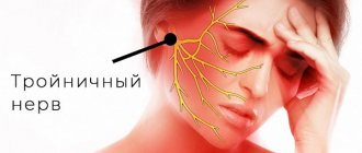
Symptoms of damage to the vestibular-cochlear nerve
Damage to the vestibular-cochlear nerve is accompanied by several symptoms. They are closely related to the part in which the violation is localized:
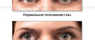
- If the vestibule is damaged, a person experiences dizziness, problems with stability, nausea, and vomiting. The pulse and breathing rhythm change, and blood pressure fluctuations are possible.
- If the cochlear part is damaged, the patient experiences severe tinnitus and hearing loss. Complete deafness is possible.
If both parts are damaged, all symptoms appear simultaneously and can be either severe or mild.
Causes of nerve damage
The most common diseases leading to pathologies of PUN include:
- Infections and viruses. Symptoms can be caused by influenza, colds, as well as measles, scarlet fever, syphilis, mumps and tuberculosis.
- Diseases of the heart and blood vessels. Strokes, heart attacks, atherosclerosis and myocarditis can also cause poor health.
- Intoxication. Symptoms appear when treated with antibiotics and other medications.
There are other diseases that are specific only to the ears, such as labyrinthitis. With this pathology, the inner ear is affected.
Rarely occurs acoustic neuroma involving PUN. It develops very slowly and provokes the gradual onset of all symptoms. Meniere's syndrome is also possible. It is associated with a disease of the same name, in which the production and outflow of fluid in the inner ear changes. From the side of the affected nerve, noise in the ear and a feeling of fullness appear. The person may suffer from attacks of dizziness and hearing loss.
To identify the exact cause, a full diagnosis is required. The patient is prescribed electroencephalography and examination by an ENT specialist or a neurologist. A CT and MRI of the ear area may be required.
Features of the treatment of pathologies
Methods for treating pathology of the vestibulocochlear nerve
Some diseases of this structure require only surgical intervention. Neuroma is considered especially dangerous. It is removed during surgery, since as it develops, especially in the brain part of the PUN, it leads to pressure on the vascular and respiratory centers. After removal, rehabilitation therapy is carried out.
For other disorders, symptomatic treatment is used:
- the patient is provided with complete rest during attacks of dizziness and hearing loss;
- prescribe B vitamins to improve well-being and fiber nutrition;
- take medications to normalize fluid levels in the inner ear;
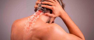
- perform light gymnastics for the cervical spine;
- massage the head and area;
- use means to improve microcirculation.
Treatment is carried out by a neurologist in combination with the help of an otolaryngologist, neurosurgeon and other specialists.