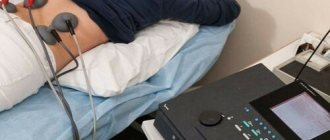
The signs of a neurotic disorder are variable and imprecise; each person’s neurosis can manifest itself differently. Therefore, in order to accurately establish a diagnosis and receive adequate treatment, you need to consult a psychotherapist.
Important
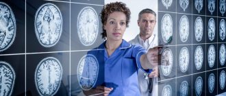
A psychotherapist diagnoses neurosis. The treatment regimen that will help cope with the disorder depends on the type of neurosis and its severity.
Neurosis is a functional disease of the nervous system. This means that the disorder occurs temporarily; there is no infection, tumor, or persistent pathology of blood vessels or internal organs in the body. Negative factors deplete the nervous system:
- stress;
- overvoltage;
- internal conflict;
- traumatic situation.
This causes disturbances in the functioning of the heart and blood vessels, indigestion, strange pain and discomfort in different parts of the body. Somatologists (who deal with diseases of the body - therapists, neurologists) do not find any abnormalities: neither peptic ulcers, nor abnormalities of the endocrine system (for example, the thyroid gland), nor inflammation.
Manifestations of neurosis in adults are often confused with other diseases, so most patients undergo examinations for years, but treatment does not produce results. In such cases, it is recommended to consult a psychotherapist.
The body is healthy with neurosis, most tests are normal. “All diseases come from nerves” - that’s exactly what it’s about.
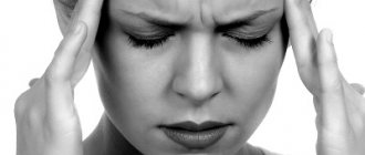
How to recognize neurosis? It is characterized by the following symptoms:
- somatic manifestations (from the body) - vague pain, increased fatigue, changes in blood pressure, fever, sweating;
- emotional instability - frequent unreasonable or causeless anxiety, fear, irritability, tearfulness;
- poor load tolerance - a person cannot concentrate, sometimes cannot sit still. Both work and personal life suffer; basic tasks become tiresome very quickly.
In the next part we will take a closer look at how neurosis manifests itself and what specific symptoms and complaints are possible.
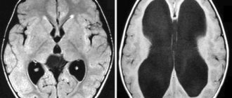
What is organic damage to the nervous system?
Organic damage to the central nervous system
- a pathological condition in which the brain does not function fully. The lesion can be congenital or appear due to trauma, stroke, infectious diseases of the brain, alcoholism and drug addiction.
Lesions are divided into grades 1-3. Stage 1 lesions are diagnosed in many people; they do not manifest themselves in any way and do not require treatment. Lesions of degrees 2 and 3 interfere with normal life and can provoke more dangerous diseases, so they need to be treated.
Nervous diseases: treatment
The choice of treatment method for diseases of the nervous system depends on many factors: the type of pathology, the immune status of the patient, symptoms of the disease, characteristics of the patient’s body, etc. Nervous diseases, the treatment of which gives the desired result in combination with a certain lifestyle, as a rule, go away with changes in psychology person. Optimists, as scientists state, suffer from nervous diseases less often than pessimists. To treat these diseases, exercise therapy, physiotherapy, reflexology, mechanotherapy, and manual therapy are used. Surgical intervention is used for brain tumors, abscesses, aneurysms, intracerebral hematomas, as well as certain cases of Parkinson's disease. To solve psycho-emotional problems, doctors recommend antidepressants. We are against the use of such drugs for the reason that they do not solve the problem, but only “push it aside” for a while, while causing side effects. In the complex treatment of nervous diseases, we recommend taking Transfer Factor. This drug is a component of our immune system, it is an “extract” from cow colostrum and chicken egg yolks of transfer factors - immune molecules - carriers of immune “memory”. Once in the body, these particles: - restore the normal functioning of the nervous and immune systems and metabolic processes of a person; - enhance the therapeutic effect of medications that a person takes, and at the same time neutralizes their side effects on the body (which is important); — transfer factors “record” cases of invasion of foreign bodies into the body, information about these agents and methods for neutralizing them. When these foreign agents invade again, transfer factors “extract” information about them and the immune system, using this information, destroys them. This algorithm of action is available only for this immunomodulator, which, today, has no analogues in the world, either in terms of effectiveness or safety for humans. When treating nervous diseases by any method, it is advisable to use Transfer Factor Advance or Classic - this increases the chance of successfully getting rid of this disease. This immune drug is necessary when using antibiotics or antidepressants.
How does organic damage to the nervous system manifest and what is dangerous?
When the central nervous system is damaged, the following symptoms are observed:
- fast fatiguability;
- impaired coordination, inability to perform simple movements and actions because of this;
- problems with hearing, vision;
- inability to concentrate on a task, the need to constantly be distracted;
- trouble sleeping, insomnia, nightmares or constant awakenings;
- urinary incontinence;
- decreased immunity, which often causes colds and other diseases.
Brain lesions are especially dangerous for children who are lagging behind in physical development and cannot study normally and communicate with peers. Also, lesions lead to oligophrenia, that is, mental retardation, and dementia - loss of skills and knowledge, and the inability to acquire new ones.
Text of the book “Neuropathy”
11.1. Inflammatory diseases of the nervous system
Inflammatory diseases of the nervous system can be divided into two groups: one includes diseases that primarily affect the nervous system and are not associated with inflammatory diseases of other organs;
in the other - diseases that arose as complications of a number of infections and intoxications, such as pneumonia, dysentery, scarlet fever, tuberculosis, food poisoning, etc. Based on the localization of the infectious process in the nervous system, they distinguish: meningitis
– inflammation of the meninges,
encephalitis
– inflammation of the gray matter of the brain, myelitis – inflammation of the spinal cord (white matter),
poliomyelitis
– inflammation of the gray and white matter of the spinal cord,
polyneuritis
– multiple inflammation of the peripheral nerves.
Meningitis.
In neurological clinics, the term “meningitis” is used to refer to inflammation of the soft meninges. Inflammation of the dura mater is called pachymeningitis. If the arachnoid membrane is predominantly affected, then they speak of arachnoiditis.
Meningitis, according to its etiology, can be toxic and infectious. Among the latter, meningitis is meningococcal, pneumococcal, streptococcal, tuberculosis, typhoid, syphilitic, etc. - depending on the microbe that caused the damage to the membranes. Etiological diagnosis of meningitis is not always possible; meningitis of unknown etiology occurs (Fig. 34).
Meningitis most often has an infectious etiology. Like encephalitis, it can be a primary (epidemic and serous meningitis) and secondary (tuberculous, secondary serous and purulent meningitis) disease. Although the clinical manifestations of various meningitis are similar, they differ in symptoms, course, and outcome.
According to pathogenesis, meningitis is divided into primary and secondary. Primary meningitis is an independent disease and occurs when microbes penetrate the subarachnoid space and affect the pia mater of the brain. Secondary meningitis develops as complications of other diseases.
According to the pathological picture, meningitis is distinguished as serous, serous-fibrous, purulent and hemorrhagic.
According to the course, meningitis is divided into acute, subacute and chronic.
The most common are cerebrospinal epidemic (meningococcal), secondary purulent meningitis, tuberculous and acute serous meningitis.
Infection with meningococcus occurs mainly through droplet infection and contact. The entrance gate is the mucous membrane of the pharynx and nasopharynx. The incubation period lasts 1–2 days. Children under 5 years of age suffer from cerebrospinal meningitis, but it often occurs in adults. Epidemic outbreaks are more common in winter and spring.
Rice. 34. Child with meningitis
Clinic.
With cerebrospinal epidemic meningitis, patients experience characteristic symptoms: headache, vomiting, stiff neck, positive meningeal symptoms, changes in the cranial nerves (double image, strabismus, ptosis, limited eye movement, weakness of the facial muscles, hearing and vision impairment , depending on the involvement of the auditory and visual nerves in the process), disorders of tendon reflexes, etc.
The disease develops suddenly, in the midst of complete health. The temperature quickly rises to 39–40°, severe chills, intense headaches, vomiting, neck stiffness appear, and within 1–2 days the entire clinical picture of acute purulent inflammation of the soft meninges develops. The symptoms of meningitis are usually very clear.
There is an increase in leukocytes in the blood. Changes in cerebrospinal fluid (CSF) are especially characteristic. As a rule, the liquid is cloudy and flows out under pressure.
The duration of the disease varies. In most cases, the meningeal symptom complex lasts 3–4 weeks.
Severe atypical forms include fulminant, hyperacute, septic.
The fulminant form of cerebrospinal meningitis is characterized by an extremely violent onset. The patient falls, loses consciousness, the temperature rises sharply, the pulse becomes rapid and weak, and breathing is irregular. Without regaining consciousness, the patient dies within 1-24 hours from the onset of the disease.
The hyperacute form also ends in death, but only 3–5 days after the onset of the disease. With this form, convulsions, bulbar disorders, clouding of consciousness, and impaired sphincter activity are observed.
The septic form gives a variety of clinical manifestations and often goes unrecognized for a long time. Meningeal symptoms are present, but they often remain unrecognized. Lumbar puncture immediately reveals the etiology of an unclear disease. Vigorous, persistent treatment often leads to a favorable outcome.
With cerebral epidemic meningitis, complications are extremely varied.
Long-lasting meningitis is often complicated by hydrocephalus, especially in young children. Complications also include the formation of adhesions in the subarachnoid space of the brain and spinal cord. Sometimes meningitis can turn into meningoencephalitis and cause major complications. In some cases, the inflammatory process can spread to the cranial nerves and cause neuritis of these nerves, which can subsequently cause various complications associated with the innervation of the cranial nerves. Meningitis often ends in complete recovery without any defects.
Acute serous meningitis
may be secondary or primary. Secondary serous meningitis is more common. They complicate common acute infections: pneumonia, sepsis, inflammatory processes in the bones of the skull, middle and inner ear, nose, its accessory cavities, exogenous and endogenous intoxications. Primary serous meningitis is an independent nosological entity.
Acute serous meningitis begins suddenly, in the midst of complete health or against the background of catarrhal symptoms of the upper respiratory tract. It begins with sharp headaches. The temperature is high, up to 39°, sometimes the disease occurs at low-grade fever. There may be a long prodromal period with general malaise, a feeling of weakness, headaches, nasopharyngitis, and low-grade fever.
Sometimes primary serous meningitis occurs particularly violently, accompanied by convulsions and attacks of loss of consciousness. In most cases, this disease ends in recovery.
Acute serous meningitis also includes meningitis caused by the mumps virus (pig meningitis). It is characterized by a violent onset, a benign course and a rapid ending.
Treatment is inpatient, medicinal.
Tuberculous meningitis
develops mainly against the background of general tuberculosis disease. It affects mainly children aged 2–7 years; it is rare in adults. Tuberculous meningitis mostly occurs in the spring, but it can also occur at other times of the year. Measles, whooping cough, influenza, bronchitis, pneumonia predispose to tuberculous meningitis.
Usually children who have had contact with a patient with tuberculosis, weak children, mostly with a tuberculous process in the bronchial glands, become ill. A tuberculosis bacillus is detected in the cerebrospinal fluid.
The disease usually begins with a period of warning signs, which lasts about two weeks. The child loses weight, turns pale, loses appetite, and his character and behavior change. He becomes lethargic, boring, irritable, sits quietly alone for a long time, resting his head on his hand or the back of a chair, and goes to bed during the day. Low-grade fever is often observed. Then vomiting appears, which is characteristic of tuberculous meningitis, headache, constipation, fever (rarely above 38–39°), neck tension, Kernig and Brudzinski symptoms (meningeal symptoms). In severe cases, the patient’s position is typical: the head is thrown back, the legs are bent at the knee joints, the stomach is retracted. All phenomena gradually increase: strabismus, paralysis of facial muscles, and ptosis of the eyelid develop. A frozen, mask-like face with a motionless gaze directed into space and slanting, unblinking eyes is considered characteristic. Tendon reflexes decrease or completely fade away, motor restlessness sets in with a slow pulse and a darkened consciousness. The patient becomes increasingly deaf and has attacks of clonic convulsions.
Possible consequences of tuberculous meningitis - hydrocephalus, hypertension syndrome, epileptiform syndrome, endocrine and autonomic disorders - require special therapy. In advanced cases, death is possible.
Inpatient treatment is based on the use of special anti-tuberculosis drugs.
Epidemic cerebrospinal meningitis
– damage to the lining of the brain and spinal cord, often spreading in the form of epidemics.
The outbreak of epidemics usually occurs in winter and in the first months of spring. Sporadic (isolated) cases occur at any time of the year. Epidemic cerebrospinal meningitis usually affects young children - from several months to 5 years; the disease is less common in adolescence and very rarely in adults.
Epidemic meningitis belongs to the group of purulent meningitis; its specific pathogen is meningococcus - a gram-negative diplococcus living in a cell. Once in the nasopharynx, the infection is transferred from there to the nerve sheaths, spreading by the lympho-hematogenous route.
The existence of septic forms of meningitis
indicates the possible transmission of infection by blood flow. The infection is transmitted through direct contact with the patient, as well as through third parties, carriers of the bacteria, who, in contact with the patient, may not get meningitis, but often infect others with it.
The disease begins violently - a febrile state with a temperature of up to 40°, headache, vomiting. The patient lies in a characteristic position: the occipital muscles are sharply tense, the head is thrown back, the back is arched, the stomach is retracted, the legs are bent and brought to the stomach. The patient becomes hypersensitive to light and sound stimuli. There is often urinary and stool retention.
In the first days of the disease, a rash sometimes briefly appears on the skin, which may resemble the rash of scarlet fever or measles, rubella, or urticaria.
Depending on the severity of the disease, the patient may experience blackouts, unconsciousness, delirium, agitation, and muscle spasms in the limbs and torso. If the course of the disease is unfavorable, at the end of the first week a coma occurs, paralysis of the eye muscles, facial nerve, mono- or hemiplegia comes to the fore; seizures become more frequent, and during one of them death occurs. In cases where the course of the disease takes a favorable turn, the temperature drops, the patient develops an appetite, and he enters the stage of recovery.
In the presence of epidemic meningitis
The causative agent of meningitis is often found in the cerebrospinal fluid.
The duration of epidemic meningococcal meningitis is on average 2–6 weeks. However, there are known cases of fulminant disease, when the patient died within a few hours of the onset of the disease. There are protracted cases when the patient, after a period of improvement, again began to rise in temperature, which was established for a long time, with significant fluctuations between morning and evening, sharply exhausting the patient and sometimes leading him to complete exhaustion, decline in cardiac activity and death. This protracted form represents either the hydrocephalic stage, i.e., the period when the patient develops chronic dropsy of the brain, or the stage when he experiences meningococcal sepsis with the penetration of meningococcus into the blood.
Treatment is inpatient, medicinal: broad-spectrum antibiotics.
Prevention.
The disease is transmitted from person to person by droplets, that is, with splashes of saliva and nasal mucus when coughing, sneezing, spitting, kissing, etc. In people with meningitis, meningococcus can almost always be found in the nasopharynx, from where it disappears by the end of 3 years. th week from the onset of the disease.
Meningococcal carriage in persons in contact with the patient usually lasts 2–3 weeks. The main preventive control measure should be the popularization of information about the ways of spreading epidemic meningitis. The second extremely important preventive measure is hygienic living, working and living conditions. The patient must be isolated and hospitalized. Early diagnosis and isolation are very important. Epidemic meningitis leaves immunity, and recurrent diseases are rare.
Secondary purulent meningitis.
The disease develops in the presence of a purulent focus in the body, for example in the ear, nose, nasopharynx, orbit, or pneumococcal infection. Microorganisms from the purulent focus penetrate through the lymphatic or bloodstream, causing a diffuse purulent process in the soft meninges. Due to the presence of a primary focus that causes headaches, poor general health, the onset of meningitis is not always determined. Increased headaches, a further increase in temperature (up to 40° and above) and a number of meningeal objective symptoms give reason to think about the onset of meningitis. The patient's consciousness soon becomes dark, he becomes delirious, and falls into a drowsy state.
Treatment is inpatient, medicinal.
Prevention is of great importance. Timely surgical treatment of purulent processes in the accessory cavities of the nose and ear is necessary.
Cerebral arachnoiditis
– cerebral arachnoiditis – is a type of serous inflammation of the soft meninges with predominant damage to the arachnoid membrane, characterized by a tendency to be chronic with exacerbations and remissions.
Arachnoiditis occurs in many organic diseases of the central nervous system; it can be based on brain injuries, local (inflammation of the paranasal cavities, ear diseases) and general (influenza, rheumatism, etc.) infections. In childhood, arachnoiditis has a significant prevalence and occurs as complications of many infectious diseases - influenza, respiratory viral infections, measles, mumps, otitis media, sinusitis, etc., as well as traumatic brain injuries. Primary viral arachnoiditis occurs.
The course can be acute, but more often it is subacute with the transition to chronic.
Headache, sometimes very long, sometimes strictly localized. Various degrees of stagnation in the fundus with subsequent loss of vision are observed.
Focal symptoms are varied: damage to certain cranial nerves (with arachnoiditis of the base of the brain); convulsive seizures, Jacksonian (arachnoiditis of the convex surface of the hemispheres); polydipsia, polyuria; neuritis and atrophy of the optic nerve with loss of visual fields, often leading to blindness (optic-chiasmatic arachnoiditis), then disturbances in statics and coordination with severe cerebral symptoms (severe headaches, vomiting), in acute cases (arachnoiditis of the cistern magna).
Despite the chronic and relapsing course of the disease, the prognosis in most cases is favorable. It is serious with arachnoiditis of the cisterna magna, since during one of the acute attacks of increased intracranial pressure death can occur.
Mild inflammatory changes are observed in the cerebrospinal fluid.
Treatment is medicinal: salicylates, antibiotics. X-ray therapy gives good results for arachnoiditis. If conservative treatment is unsuccessful, surgical intervention is resorted to.
Medical and pedagogical activities.
After suffering various forms of meningitis, children may experience complications (neuritis, hearing loss), as well as an asthenic state. The first months after the illness, the child needs special care, a gentle regime is necessary. In preschool age, a child needs dosed time for play and rest. Particularly important is daytime sleep, a strict balanced diet, vitamins, microelements and proteins. It is necessary to monitor the load on the visual sensory system. A child can watch children's programs on TV, but no more than 10–15 minutes. It is necessary to ventilate the room. Children should be outdoors every day. At first, developmental activities with the child should be carried out for a short time. School-age children who have suffered meningitis should be educated in a gentle manner. For the first two to three months, training is conducted according to an individual plan at home or at school, but the school day should be shortened. Physical education lessons should also be individual and gentle. If your child is hearing impaired, it is necessary to have them examined. Detection of hearing impairment requires adequate correction. In some cases, children need education in specialized preschool and school institutions. In case of visual impairment, its correction is provided; in some cases, there is a need for training in specialized institutions for the visually impaired or blind.
Encephalitis
– inflammation of the gray matter of the brain. The Clinic of Nervous Diseases knows a large number of types of encephalitis. The main etiological factor of encephalitis is infection. According to the anatomical distribution, encephalitis can be diffuse or focal, and by nature - purulent and non-purulent. Encephalitis can be a primary or secondary disease; Primary encephalitis is an independent disease, secondary encephalitis is a complication resulting from some other inflammatory process. The primary ones include epidemic, tick-borne, mosquito, enterovirus, herpetic, etc. In childhood, encephalitis may occur due to common childhood infectious diseases, and vaccine encephalitis.
Epidemic encephalitis
belongs to the group of primary encephalitis. Isolated cases of the disease still occur today; It received the name “epidemic” after the epidemic of 1918–1926 that swept across the world. It was described in 1917 by the Viennese neurologist K. Economo, which is why it is also called Economo’s disease. Around 1928, the epidemic ended, leaving a large number of patients in all countries in the chronic stage of lethargic encephalitis.
Epidemic encephalitis most often occurs at the age of 20–30 years; children of school age are less likely to get sick. The causative agent is a filterable virus that enters the body through the mucous membrane of the pharynx and nasopharynx. The incubation period is 1-14 days.
Epidemic encephalitis refers to polioencephalitis, characterized by damage to the gray matter of the brain.
The epidemic encephalitis virus mainly affects the central nodes of the hemispheres and the brain stem. Particularly affected are the striatum, red nuclei, substantia nigra, central gray matter of the aqueduct of Sylvius, the quadrigeminal plate, the optic thalamus and the hypothalamus.
The disease begins acutely, the temperature is often raised to 39–40°. There is a severe headache, general malaise, and vomiting. At the onset of the disease, there are often phenomena of inflammation of the upper respiratory tract, which in some cases leads to an erroneous diagnosis of acute respiratory disease. It is typical that with epidemic encephalitis in the first hours of the disease the child becomes lethargic and drowsy. Pathological drowsiness gave encephalitis the name lethargic. Patients sleep all day long. Less commonly, with encephalitis, pathological wakefulness is observed - insomnia, which sometimes precedes hibernation.
Motor restlessness in the form of general tremors or choreiform twitches may be noted.
Oculomotor disorders – diplopia, ptosis – may develop. Autonomic disorders are also noted - hyperhidrosis, vasomotor lability, tachycardia, changes in breathing rhythm. Symptoms such as “greasy face” and hypersalivation are typical. Some patients experience hiccups. A more severe course of the disease is observed in young children. The duration of the acute period varies, it ranges from 2–4 days to 4 months. In most cases, the acute period lasts 2–3 weeks.
Epidemic encephalitis can result in recovery or become chronic, but death can also occur.
The chronic form of epidemic encephalitis is more often manifested by parkinsonism syndrome, a condition similar to Parkinson's disease, i.e., shaking palsy.
The chronic stage of encephalitis usually occurs at normal temperature, without cerebral and meningeal symptoms. In the chronic phase of the disease, there are autonomic-endocrine disorders caused by progressive damage to the hypothalamic nuclei.
Sick children become slow, their movements are inexpressive, they become “sedate,” which is not characteristic of childhood. The face is amicable, they walk in small steps, write in small letters. A characteristic posture may be noted: a stooped back, half-bent legs, a head tilted forward, and tremor of the head and fingers.
Echolalia gradually develops, speech becomes impoverished. Many patients become aggressive and grumpy. Hypersalivation, greasiness of the skin, disturbances of carbohydrate and fat metabolism, increased thirst and appetite are noted.
Inpatient treatment is medication, anti-inflammatory drugs, vitamin therapy.
Tick-borne encephalitis
also called spring-summer, taiga or Far Eastern. It was described by A.G. Panov in 1935. The scientist isolated a filter virus, the causative agent of encephalitis, and discovered the route of transmission: from rodents, which are the reservoir of the virus, through ixodid ticks, the main carriers of spring-summer encephalitis. The virus, entering the stomach of a tick with the blood of infected animals, penetrates all its organs and is then transmitted to other animals, as well as to the offspring of the tick (transovarial transmission of the virus). The penetration of the virus into the milk of farm animals (goats) has been proven, so nutritional routes of infection of humans through the milk of goats and cows are possible.
The disease is strictly seasonal and is transmitted by tick bites, which multiply and attack humans mainly in May–July. Spring-summer seasonality is associated with the biology of tick vectors. Having sucked on the blood of rodents, the tick bites a person and infects him. Tick-borne encephalitis is observed mainly in persons living or working in wooded and taiga areas. The incidence of tick-borne encephalitis is associated with the habitat of ixodid ticks. Previously, epidemic foci of tick-borne encephalitis in Russia were noted in Siberia, the Urals, and the Far East. Currently, tick-borne encephalitis occurs in Moscow and the Moscow region.
The incubation period lasts from 1 to 3 weeks.
The disease develops acutely, on average 10–14 days after a tick bite or 4–7 days after drinking infected goat milk. In the prodromal period, malaise, pain in the muscles, neck and limbs are observed. The acute period of the disease begins with severe headache, vomiting, chills and fever, which lasts 4-10 days. Against the background of high temperature, meningeal symptoms occur. There may be disturbances in the functioning of the gastrointestinal tract. Consciousness is darkened. The pulse often slows down. The meningeal symptom complex is quickly joined by atrophic paralysis, which occurs on the 3-4th day. Paralysis of the muscles of the neck, shoulder girdle and proximal parts of the upper extremities may be observed. When the nuclei of the cranial nerves of the medulla oblongata are damaged, the patient’s breathing is upset, cardiac activity, speech, phonation, swallowing are disrupted, and the mobility of the soft palate and tongue suffers. Along with this, the patient develops more or less significant pyramidal symptoms: spastic paresis of the lower extremities, pathological reflexes. Sensitivity is usually not impaired.
When contracting tick-borne encephalitis, the following external signs are characteristic: the head hangs on the chest, the shoulders are drooping, the arms hang along the body, the muscles are sharply atrophic. Sometimes spastic hemiparesis occurs. Hyperkinesis and epileptiform seizures are common. The disease sometimes begins with an epileptic seizure.
Tick-borne encephalitis belongs to panencephalitis (meningoencephalomyelitis) and is accompanied by damage to both gray and white matter of the brain.
There are several of its forms: polioencephalomyelitis, meningeal, stem, cerebral and erased.
With polioencephalomyelitis form
the brain (mostly stem structures) and spinal cord (anterior horns) are affected. This form is quite rare.
Meningeal form
caused by serous inflammation. The clinical course is characterized by an acute onset, with meningeal symptoms dominating. The total duration of the meningeal form does not exceed 2–3 weeks. After recovery, symptoms of vegetative-vascular dystonia may be observed. Children adapt slowly to the stress of school and need individual preparation; they experience fatigue, irritability, poor appetite and sleep.
Stem form
– damage to stem structures, cranial nerve nuclei. This is the most severe pathology in tick-borne encephalitis; it impairs breathing and cardiac activity. Bulbar disorders are one of the main causes of high mortality in this form.
Cerebral form
characterized by pyramidal disorders. In some cases, the cerebral form is manifested by hyperkinesis associated with the localization of the process in the subcortical nuclei. Restoration of motor functions in the cerebral form is slow and not always complete.
Preventive
measures include the destruction of ticks, rodents, and specific vaccination of people. Immunization of the population leads to a decrease in the frequency of paralytic forms.
Mosquito encephalitis,
otherwise it is called Japanese, summer-autumn. It is an epidemic disease described after an outbreak in 1924. The causative agent of the disease is a virus that circulates in nature among animals and birds. The virus is transmitted by mosquitoes. A person becomes infected by the bites of virus-carrying mosquitoes; this is due to the reproduction of mosquitoes, which occurs in the summer and autumn. The mosquito encephalitis virus penetrates the central nervous system. The disease is common in Japan and the southern regions of Primorye Russia. Mosquito encephalitis is one of the most severe forms.
The incubation period ranges from 2 to 14 days. In the prodromal period (up to 2 days), headache, pain throughout the body, and chills are observed. The disease begins suddenly - a sharp rise in temperature to 40° within 7-10 days.
Against the background of high temperature, rapid development of symptoms occurs: severe headache, the skin is sharply hyperemic, meningeal symptoms, vomiting, convulsions and impaired consciousness are noted.
Muscle tone is extrapyramidal and pyramidal in nature, paresis and paralysis may appear, in the form of mono- and hemiparesis, damage to the cranial nerves. In some cases, bulbar disorders occur, and in severe cases, bulbar palsy occurs.
In approximately 40–70% of cases, patients die, and in the first week of the disease. The cause of death is respiratory and cardiovascular disorders.
In those recovering, general cerebral symptoms quickly disappear, but disturbances in mental activity, certain focal symptoms and general asthenia may persist.
Preventative
measures include mosquito control. It is necessary to drain swamps and apply individual and collective measures to protect against mosquitoes. Immunization can be carried out.
Treatment is inpatient, medicinal.
Medical and pedagogical activities.
Severe complications often occur after encephalitis. They are expressed in the form of paresis and paralysis of the limbs, impaired speech activity, and lesions of the cranial nerves remain. In some cases, there is a decrease in intelligence and emotional-volitional sphere. Children who have suffered encephalitis often become disabled and confined to a wheelchair. If the musculoskeletal system is damaged, the child can be educated in a comprehensive school inclusively or at home. Such children need an individual approach, the educational load should be gentle, and the daily routine should be strictly regulated. Children need enough time to be outdoors. Adults should strictly monitor the load on the visual sensory system. In case of speech disorders, a session with a speech therapist is necessary. If intelligence decreases, study in specialized correctional educational institutions.
Myelitis. Myelitis
– infectious disease of the spinal cord. It can develop secondary to acute infections (measles, scarlet fever, diphtheria, whooping cough, tonsillitis, influenza, etc.). Primary acute myelitis caused by viruses is very rare. The causative agent of myelitis is introduced into the subarachnoid space through the blood or lymph flow. Possible infectious-toxic origin of the spinal process, when microbes are localized far from the focus, damage to the nervous system is caused by their toxins.
The disease develops very acutely - with fever and chills. Minor pain and paresthesia appear in the back and legs. The legs become weaker, and after 1–3 days movement in them disappears or becomes limited. Most often the entire diameter of the spinal cord is affected. The clinical picture is determined by the level of spinal cord damage. When the thoracic segments of the spinal cord are damaged, spastic paralysis with a sensitivity disorder develops, but when the cervical parts of the spinal cord are damaged, paralysis and sensitivity disorder of the torso and limbs occur, with spastic paralysis in the legs and flaccid paralysis in the arms.
The pain is often of a shingling nature. This is explained by the fact that the pathological process involves the meninges with the roots of the spinal cord. Urinary dysfunction is often observed.
The outcome of myelitis varies, depending on the location of the lesion, severity and accompanying complications. Severe myelitis is often fatal; in mild cases, complete recovery without visible defects is possible.
Inpatient treatment, medication, anti-inflammatory drugs, antibiotics, vitamin therapy, interferon, tissue therapy.
Neuritis. Neuritis
(neuritis) is inflammation of a nerve.
In clinical practice, it is customary to divide diseases of the peripheral nervous system into neuritis and neuralgia. Neuritis is associated with pathological changes in nerve bundles and is manifested by objective clinical symptoms in the motor, sensory and autonomic spheres. Neuralgia
differs from neuritis in the presence of attacks of pain along the course of a particular nerve. For example, trigeminal neuralgia or intercostal neuralgia is observed.
Advantages of treating central nervous system lesions in our clinic
- We accept adults and children
. We enroll adult patients and preschool children. We send young patients to a doctor who specializes in brain damage in children. - We treat children in the presence of parents
. If a child experiences fear or anxiety, we conduct a consultation in the presence of the parents. If necessary, we carry out further treatment in front of the parents to reduce stress for the child. - We select therapy individually
. We assess the degree of damage, the presence of concomitant diseases and disorders. We select treatment taking into account the patient’s age, general health, and response to previous therapy. - We warn you about the duration of treatment and possible results
. We will tell you how long the treatment can last, since organic lesions require long-term therapy. We predict how effective the therapy will be in a particular case, and discuss this with the patient.
To make an appointment with a doctor, call or use the feedback form. The administrator will call you back, answer questions and help you choose a convenient appointment time.
Herpetic encephalitis
The most common sporadic encephalitis in Western Europe is herpes simplex encephalitis (HSVE). The main symptoms of this deadly disease include headaches, fever, as well as various quantitative and qualitative disturbances of consciousness. If a patient is suspected of having herpetic encephalitis, antiviral therapy with Acyclovir should be carried out. Without treatment, the disease can be fatal, and any delay in starting antiviral therapy can cost the patient his life.
Prevention of central nervous system diseases
The consequences of diseases of the nervous system can be mild or severe. Most severe complications develop against the background of weak immunity, chronic lack of vitamins, minerals, and enzymes. Hence the conclusion - for the proper prevention of nervous pathologies, especially in the age group over 50, daily intake of vitamin and mineral complexes is necessary.
Among the drugs that have proven themselves is the therapeutic and prophylactic product Lamifaren . What does it contain and why is it effective for the prevention of nervous pathologies?
Lamifaren - a means of preventing diseases of the nervous system
The drug contains a large complex of minerals and vitamins of natural origin. It is based on an extract from brown algae Laminaria. Thanks to this plant, the product provides the body with vital substances.
- Salts of calcium, sodium, macro - micro - elements in digestible bioorganic form. The absorption of minerals in Lamifaren reaches 98%. The number of macro-micro components reaches forty - selenium, silver, phosphorus, titanium. Essential zinc, sodium, manganese. Metals – magnesium, copper, iron in the correct daily doses.
- Sorbents necessary for cleaning the digestive tract, blood, and body cells. From heavy metals, chemical toxins, cholesterol plaques, fats. And also to normalize intestinal flora and metabolic processes.
- Full supply of vitamins. The drug contains vitamins A, B, C, D, E, K, PP, polysaccharides, unsaturated fatty acids (omega 3).
Lamifaren is the prevention of diseases of the nervous system. Your restful sleep , good rest, the opportunity for an active life, playing sports at any age. Your healthy children and active, sensible parents.
Get a free consultation
Classification of diseases
Demyelinating diseases (DD) are divided into primary (acute disseminated encephalomyelitis, clinical forms - polyencephalitis, encephalomyelopolyradiculoneuritis, optomyelitis, disseminated myelitis, optoencephalomyelitis) and secondary (vaccinal - develop during vaccination with DPT, DTP, atirabiic vaccine; parainfectious - encephalomyelitis with influenza, coc Lushe, Corey , chicken pox, other diseases).
Subacute forms of the disease manifest themselves in the form of diseases:
- multiple sclerosis. Clinical forms – cerebrospinal and spinal, optical, cerebral, brainstem, cerebellar;
- chronic forms of ID – Dawson, Doering, Pette, Van Bogart encephalitis, Schilder’s periaxial diffuse leukoencephalitis;
- Disorders affecting predominantly peripheral nerves - infectious polyradiculoneuropathy, infectious-allergic primary Guillain-Barré polyradiculoneuritis, toxic polyneuropathy, diabetic and dismetabolic polyneuropathy.
Genetic diseases
When damage to nerve tissue occurs, the body responds by destroying myelin. Diseases that are accompanied by the destruction of myelin are divided into two groups - myelinoclastics and myelinopathy. Myelinoclasty is the destruction of the membrane under the influence of external factors. Myelinopathy is a genetically determined destruction of myelin associated with a biochemical defect in the structure of the neuron membrane. At the same time, this distribution into groups is considered conditional - the first manifestations of myelinoclasty may indicate a person’s predisposition to the disease, and the first manifestations of myelinopathy may be associated with damage caused by external factors. Multiple sclerosis is considered a disease of people with a genetic predisposition to the destruction of the membrane of neurons, metabolic disorders, a deficient immune system and the presence of a slow infection. Genetic demyelinating diseases include: neural amyotrophy of Charcot-Marie-Tooth, hypertrophic neuropathy of Dejerine-Sottas, diffuse periaxial leukoencephalitis, Canavan disease and many other diseases. Genetic demyelinating diseases are less common than autoimmune demyelinating diseases.
Meningitis
Most often, bacterial meningitis is caused by streptococci (Streptococcus pneumoniae), Listeria (Listeria monocytogenes) and meningococci (Neisseria meningitidis). Symptoms of the disease include headaches, fever, nausea and vomiting, as well as so-called meningism, or stiff neck muscles. In very young as well as elderly patients, the symptom of meningism may be absent. Like viral diseases, bacterial meningitis requires immediate treatment, rapid diagnosis and qualified treatment of the patient in the intensive care unit.
Myelitis
Another example of inflammatory diseases of the central nervous system is myelitis - inflammation of the spinal cord. It can diffusely affect an entire section of the spinal cord (transverse myelitis) or be focal (disseminated myelitis). The main symptoms of the disease are:
- muscle weakness;
- paresis, including spastic;
- sensitivity disorders;
- gait disturbances;
- pain throughout the body;
- depression;
- exhaustion;
- as well as bowel and/or bladder dysfunction.
Both transverse and disseminated myelitis can also be accompanied by transverse paralysis with sensory or motor impairments, depending on the region of the lesion.
Treatment of demyelinating diseases of the nervous system
Treatment of demyelinating disease depends on the type of disease and its severity, and is a long-term and complex process. In addition to drug treatment, the patient is prescribed a diet, recommended to follow a strict daily routine, sleep and wakefulness, regularly undergo massage, and engage in physical therapy. Each patient with demyelinating disease requires an individual approach; maintenance therapy for patients with demyelinating disease takes many years.