Oculomotor nerve (n. oculomotorius, III pair of cranial nerves)
Motor neurons of the oculomotor nerves (n. oculomotorius, III pair of cranial nerves) are located on both sides of the midline in the rostral part of the midbrain. These nuclei of the oculomotor nerve innervate the five extrinsic muscles of the eyeball, including the levator palpebral muscle. The nuclei of the oculomotor nerve also contain parasympathetic neurons (Edinger-Westphal nucleus), which are involved in the processes of pupil constriction and accommodation.
The normal position of the eyeballs and divergent strabismus are shown with weakness of the medial (internal) rectus muscle of the eye on the right (n. oculomotorius, III pair of cranial nerves).
There is a division of supranuclear groups of motor neurons for each individual eye muscle. The fibers of the oculomotor nerve innervating the medial rectus, inferior oblique and inferior rectus muscles of the eye are located on the side of the same name. The subnucleus of the oculomotor nerve for the superior rectus muscle is located on the contralateral side. The levator palpebrae superioris muscle is innervated by the central group of cells of the oculomotor nerve.
Trochlear nerve (n. trochlearis, IV pair of cranial nerves)
The motor neurons of the trochlear nerve (n. trochlearis, IV pair of cranial nerves) are closely adjacent to the main part of the complex of nuclei of the oculomotor nerve. The left nucleus of the trochlear nerve innervates the right superior oblique muscle of the eye, the right nucleus innervates the left superior oblique muscle of the eye.
Abducens nerve (n. abducens, VI pair of cranial nerves)
Motor neurons of the abducens nerve (n. abducens, VI pair of cranial nerves), innervating the lateral (external) rectus muscle of the eye on the side of the same name, are located in the nucleus of the abducens nerve in the caudal part of the pons. All three oculomotor nerves, leaving the brain stem, pass through the cavernous sinus and enter the orbit through the superior orbital fissure.
Clear binocular vision is ensured precisely by the joint activity of individual muscles of the eye (oculomotor muscles). Conjugate movements of the eyeballs are controlled by the supranuclear gaze centers and their connections. Functionally, there are five different supranuclear systems. These systems provide various types of eyeball movements. Among them there are centers that control:
- saccadic (rapid) eye movements
- purposeful eye movements
- convergent eye movements
- holding the gaze in a certain position
- vestibular centers
Weakness of the lateral (external) rectus muscle of the right eye (n. abducens, VI pair of cranial nerves) was shown before and after treatment.
Saccadic (rapid) eye movements
Saccadic (fast) movements of the eyeball occur as a command in the opposite visual field of the cortex of the frontal region of the brain (field 8). The exception is fast (saccadic) movements that occur when the central fovea of the retina is irritated, which originate from the occipital-parietal region of the brain. These frontal and occipital control centers in the brain have projections on both sides in the supranuclear brainstem centers. The activity of these supranuclear brain stem centers of vision is also influenced by the cerebellum and the vestibular nuclei complex. The paracentral sections of the reticular formation of the bridge are the stem center, providing friendly rapid (saccadic) movements of the eyeballs. Simultaneous innervation of the internal (medial) rectus and opposite external (lateral) rectus muscles when moving the eyeballs horizontally is provided by the medial longitudinal fasciculus. This medial longitudinal fasciculus connects the nucleus of the abducens nerve with the subnucleus of the complex of oculomotor nuclei, which are responsible for innervation of the opposite internal (medial) rectus muscle of the eye. To initiate vertical rapid (saccadic) eye movements, bilateral stimulation of the paracentral sections of the pontine reticular formation is required from the cortical structures of the brain. The paracentral sections of the pontine reticular formation transmit signals from the brain stem to the supranuclear centers that control the vertical movements of the eyeballs. This supranuclear eye movement center includes the rostral interstitial nucleus of the medial longitudinal fasciculus, located in the midbrain.
Diagnosis of abducens neuropathy
The level of development of medical technologies does not cause problems with the diagnosis of pathology. Abducens neuropathy does not occur overnight. Its development occurs gradually. Diagnosis of the disease is usually carried out not only by an ophthalmologist, but also by a neurologist. First of all, the doctor interviews the patient, collects a detailed history, and inquires about the presence of other diseases.
The first thing the doctor must do is to determine the cause of the neuropathy. Usually, an ophthalmoscopy is performed by an ophthalmologist for this purpose. This procedure allows you to analyze in detail the condition of the fundus. Ophthalmoscopy is performed to diagnose retinal breaks, identify thinned areas of the eye nerve, and analyze the condition of the fundus vessels. To carry out the procedure, oculists use an ophthalmoscope or fundus lenses.
If neuropathy of the abducens ophthalmic nerve is suspected, doctors often prescribe angiography of the ocular vessels to patients. Using a fluorescent dye and a special camera, the doctor takes a series of pictures called angiograms. They depict the vessels of the retina. Angiography allows you to identify pathologies of the optic nerve, a predisposition to blockage of blood vessels, and inflammatory processes occurring in the tissues of the eyeball.
If the patient's condition is severe, doctors may recommend magnetic resonance imaging or computed tomography. This study allows you to obtain accurate data on the condition of the tissues and blood vessels of the eyeball. MRI and SCT diagnostics can be performed on adults and children. The list of contraindications to these procedures is quite small, which is why doctors often prescribe these studies to their patients.
Purposeful eye movements
The cortical center for smooth targeted or tracking movements of the eyeballs is located in the occipital-parietal region of the brain. Control is carried out from the side of the same name, i.e. the right occipital-parietal region of the brain controls smooth, targeted eye movements to the right.
Testing of targeted eye movements is carried out by tracking an object from the center to the periphery with the patient's head motionless.
Which doctors treat atrophy?
Not everyone knows which doctors treat atrophy. Ophthalmologists are involved in restoring vision and getting rid of optic nerve atrophy. They are the ones who identify the disease and help restore vision. But sometimes the services of a neurologist and neurosurgeon are required. These specialists perform a number of procedures necessary for the patient's recovery.
Our clinic employs highly professional specialists with extensive experience who will help get rid of optic nerve atrophy. All that remains is to see a doctor and get medical help.
Eye movement with changes in gravity and acceleration
Coordination of the movements of the eyeballs in response to changes in gravity and acceleration is carried out by the vestibular system (vestibular-ocular reflex). When the coordination of movements of both eyes is disturbed, double vision develops, since images are projected onto disparate (inappropriate) areas of the retina. In congenital strabismus, or strabismus, a muscle imbalance that causes the eyeballs to be misaligned (nonparalytic strabismus) may cause the brain to suppress one of the images. This decrease in visual acuity in the non-fixing eye is called amblyopia without anopia. In paralytic strabismus, double vision occurs as a result of paralysis of the muscles of the eyeball, usually due to damage to the oculomotor (III), trochlear (IV) or abducens (VI) cranial nerves.
Clinic JSC "Medicine" will help cure optic nerve atrophy
Atrophy can be eliminated if the process of restoring health is carried out by professionals. Clinic JSC "Medicine" offers vision restoration services. We employ experienced ophthalmologists and other specialists who will take care of your health. Our medical center has the following advantages:
- Experienced specialists with extensive work experience. The center employs doctors of the highest category with extensive experience in the field of ophthalmology. If the doctor doubts the correctness of the diagnosis, he has any suspicions, he consults with foreign colleagues from the best leading medical centers in the world.
- The doctor is always in touch. You can contact us at any time and assistance will be provided to you.
- The complex is equipped with modern equipment. This is our main advantage, because we use modern technology that allows us to accurately diagnose various diseases, determine the degree of its development and prescribe the most effective method for eliminating pathologies.
- All rooms are equipped with the necessary devices to ensure maximum comfort. Patients will feel at home.
- Convenient location. We are located in Moscow, near the Mayakovskaya metro station. Convenient to reach from almost any part of the city.
- The center is open even on holidays and weekends. You don’t need to take time off from work to see a specialist. We adapt to you, sign up for any convenient time, even if it’s a weekend or holiday.
- Polite and responsive staff. Tired of long lines at clinics, constant quarrels and shouting in the corridors? With us this is impossible, the staff is always polite, there are no queues, assistants will guide you through the center to the desired office. We guarantee international level service.
Get rid of health problems. There is only one step left: make an appointment with a specialist. Call us and a consultant will tell you more about the provision of services and answer all questions. We will be happy to help you improve your health.
Eyeball muscles and gaze palsies
There are three types of paralysis of the external muscles of the eyeball:
- paralysis of individual eye muscles
- paralysis of friendly movements (gaze)
- mixed paralysis
Paralysis of individual eye muscles
Characteristic clinical manifestations occur with isolated damage to the oculomotor (III), trochlear (IV) or abducens (VI) nerve.
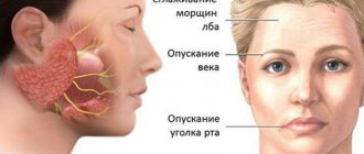
Complete damage to the oculomotor (III) nerve leads to ptosis. Ptosis manifests itself in the form of weakening (paresis) of the muscle that lifts the upper eyelid and disruption of voluntary movements of the eyeball upward, downward and inward, as well as divergent strabismus due to the preservation of the functions of the lateral (lateral) rectus muscle. When the oculomotor (III) nerve is damaged, pupil dilation and lack of reaction to light (iridoplegia) and paralysis of accommodation (cycloplegia) also occur. Isolated paralysis of the muscles of the iris and ciliary body is called internal ophthalmoplegia.
The normal position of the eyeballs and convergent strabismus are shown with weakness of the lateral (external) rectus muscle of the eye on the right (n. abducens, VI pair of cranial nerves).
Injuries to the trochlear (IV) nerve cause paralysis of the superior oblique muscle of the eye. Such damage to the trochlear (IV) nerve leads to outward deviation of the eyeball and difficulty moving (paresis) downward gaze. Paresis of downward gaze is most clearly manifested when turning the eyes inward. Diplopia (double vision) disappears when the head is tilted to the opposite shoulder, which causes a compensatory inward deviation of the intact eyeball.
Damage to the abducens (VI) nerve leads to paralysis of the muscles that abduct the eyeball to the side. When the abducens (VI) nerve is damaged, convergent strabismus develops due to the predominance of the influence of the tone of the normally working internal (medial) rectus muscle of the eye. With incomplete paralysis of the abducens (VI) nerve, the patient can turn his head towards the affected abductor muscle of the eye in order to eliminate the existing double vision using a compensatory effect on the weakened lateral rectus muscle of the eye.
The severity of the above symptoms in cases of damage to the oculomotor (III), trochlear (IV) or abducens (VI) nerve will depend on the severity of the lesion and its location in the patient.
Friendly gaze paralysis
Companionate gaze is the simultaneous movement of both eyes in the same direction. Acute damage to one of the frontal lobes, for example, during cerebral infarction (ischemic stroke), can lead to transient paralysis of voluntary conjugate movements of the eyeballs in the horizontal direction. At the same time, independent eye movements in all directions will be completely preserved. Paralysis of voluntary conjugate movements of the eyeballs in the horizontal direction is detected using the doll eye phenomenon when passively turning the head of a horizontally lying person or using caloric stimulation (infusion of cold water into the external auditory canal).
Unilateral damage to the inferiorly located paracentral section of the reticular formation of the pons at the level of the nucleus of the abducens nerve causes persistent paralysis of gaze in the direction of the lesion and loss of the oculocephalic reflex. The oculocephalic reflex is a motor reaction of the eyes to irritation of the vestibular apparatus, as with the phenomenon of the head and eyes of a doll or caloric stimulation of the walls of the external auditory canal with cold water.
Damage to the rostral interstitial nucleus of the medial longitudinal fasciculus in the anterior midbrain and/or damage to the posterior commissure causes supranuclear upward gaze palsy. Added to this focal neurological symptom is the dissociated reaction of the patient’s pupils to light:
- sluggish pupil reaction to light
- rapid reaction of the pupils to accommodation (changing the focal length of the eye) and looking at nearby objects
In some cases, the patient also develops convergence paralysis (movement of the eyes towards each other, in which the gaze will focus on the bridge of the nose). This symptom complex is called Parinaud's syndrome. Parinaud's syndrome occurs with tumors in the pineal gland, in some cases with cerebral infarction (ischemic stroke), multiple sclerosis and hydrocephalus.
Isolated downward gaze palsy is rare in patients. When this occurs, the cause is most often blockage (occlusion) of the penetrating arteries in the midline and bilateral infarctions (ischemic strokes) of the midbrain. Some hereditary extrapyramidal diseases (Huntington's chorea, progressive supranuclear palsy) can cause restrictions in the movement of the eyeballs in all directions, especially upward.
Mixed paralysis of gaze and individual muscles of the eyeball
The simultaneous combination of gaze paralysis and paralysis of individual muscles that move the eyeball in a patient is usually a sign of damage to the midbrain or pons. Damage to the lower parts of the pons with destruction of the abducens nerve nucleus located there can lead to paralysis of rapid (saccadic) horizontal movements of the eyeballs and paralysis of the lateral (external) rectus muscle of the eye (abducens nerve, VI) on the affected side.
With lesions of the medial longitudinal fasciculus, various gaze disturbances occur in the horizontal direction (internuclear ophthalmoplegia).
Unilateral damage to the medial longitudinal fasciculus caused by infarction (ischemic stroke) or demyelination leads to disruption of the inward adduction of the eyeball (to the bridge of the nose). This can manifest clinically as complete paralysis with the inability to move the eyeball inward from the midline, or as a moderate paresis, which will manifest itself as a decrease in the speed of adducting rapid (saccadic) eye movements to the bridge of the nose (adductive delay). On the side opposite to the lesion of the medial longitudinal fasciculus, as a rule, abduction nystagmus is observed: nystagmus that occurs when the eyeballs are abducted outward with a slow phase directed towards the midline and fast horizontal saccadic movements. An asymmetrical arrangement of the eyeballs relative to the vertical line often develops with unilateral internuclear ophthalmoplegia. On the affected side, the eye will be positioned higher (hypertropia).
Bilateral internuclear ophthalmoplegia occurs with demyelinating processes, tumors, infarction, or arteriovenous malformations. Bilateral internuclear ophthalmoplegia leads to a more complete syndrome of eyeball movement disorders, which are manifested by bilateral paresis of the muscles that lead the eyeball to the bridge of the nose, impaired vertical movements, purposeful tracking movements and movements caused by the influence of the vestibular system. There is a disturbance of gaze along a vertical line, upward nystagmus when looking up and downward nystagmus when looking down. Lesions of the medial longitudinal fasciculus in the overlying (rostral) parts of the midbrain are accompanied by a violation of convergence (convergent movement of the eyes towards each other, towards the bridge of the nose).
Causes of abducens neuropathy
The disease rarely develops on its own. It is usually a consequence of other diseases. Most often, neuropathy of the abducens nerve occurs against the background of pathologies of the cardiovascular system, after traumatic brain injury and neurosurgical operations. Much less often, the disease is congenital. Cases of a hereditary form of pathology are not excluded, but they are not so common in medical practice. Doctors consider the main causes of the development of abducens nerve neuropathy
- endocrine disorders (diabetes mellitus, hypothyroidism, hyperthyroidism);
- neurological pathologies (osteochondrosis of the cervical spine, multiple sclerosis, stroke);
- complications of infectious diseases (encephalitis, diphtheria, meningitis);
- intoxication of the body (poisoning with ethyl alcohol, chemicals, alcohol);
- chronic arterial pathologies (atherosclerosis, arteritis, phlebitis);
- cardiovascular diseases (varicose veins, arrhythmia, angina).
Doctors cannot always immediately determine what exactly caused the development of neuropathy of the abducens nerve of the eye. Diagnosis of this disease is often carried out by several specialists simultaneously, including: neurologists, cardiologists, endocrinologists, and traumatologists.
The disease is easier to detect in children than in adults. One of the causes of abducens neuropathy is hydrocephalus. This disease allows doctors to immediately carry out procedures that will allow them to diagnose the pathology.
Symptoms
Normally, in a healthy person, the edge of the cornea is in contact with the outer edge of the eyelid junction.
In cases where this is not observed, nerve pathology is present. Symptoms of the pathology are:
- limited mobility of the eyeball
- secondary eye deviation
- dizziness
- spatial orientation disorder
- unsure gait
- diplopia (double images of one object)
- forced, involuntary position of the head.
With a mild form of paresis, the symptoms are mild and practically do not cause any discomfort. These signs are characteristic of both the right and left eyes.