Stories that even very small structures in the brain perform important functions have not surprised anyone for a long time. But still, the realization that even smaller areas in the same structures also work differently and are responsible for many things is admirable. We have previously talked about the small but important hypothalamus and its numerous nuclei. Now it’s the turn to talk about the thalamus and its nuclei.
Thalamus. Credit: Wikimedia Commons
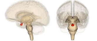
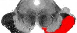
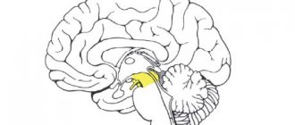
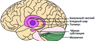
The thalamus, or, in other words, the visual thalamus, is a paired organ that is located between the anterior and middle sections of the brain and on the sides of the third ventricle. It is shaped like a chicken egg, 3-4 centimeters in size. Its anterior end is pointed and is sometimes called the “anterior tubercle”, and the rear end is thickened and is called the “cushion”. The thalamus is small, but occupies approximately 80% of the total volume of the diencephalon. In each of the thalami, internal, external, superior and inferior surfaces can be distinguished. The visual thalamus is associated with a large number of structures of the nervous system, including the hypothalamus, cerebellum, basal ganglia, hippocampus, cerebral cortex, and spinal cord.
The thalamus collects information from all the body's senses (except smell) and sends it to the cerebral cortex. It turns out that each sensory system has its own “representative” there - a special core. In essence, the thalamus can be imagined as a hub into which a lot of information is concentrated and then transmitted to the right recipients. It is interesting that, according to available information, not all the information that comes to the visual thalamus is sent to the cortex, but only some. The other part probably takes part in the formation of unconditioned and, possibly, conditioned reflexes.
In addition to collecting and distributing sensory information, the thalamus controls the cycles of sleep and wakefulness, is also involved in the process of memorization, and exercises conscious control over automatic movements. Regarding the latter, let us explain that this organ is the most important link in the system that provides control over our usual movements (running, walking, jumping, swimming). The thalamus also regulates consciousness because it connects the areas of the cortex that are responsible for perception with other parts of the brain and spinal cord.
As with the hypothalamus, the thalamus also has nuclei. These are clusters of nerve cells that form gray matter and have a specific function. The rest of the thalamus is filled with white matter.
Cerebellum
Has 3 pairs of legs . One of the most important structures of the brain, taking part in ensuring the accuracy of targeted movements and regulating the coordinated action of antagonist muscles.
The cerebellum is a corrective and controlling structure. By itself it does not cause any movement. If affected , movements will be inaccurate.
Thanks to the activity of the cerebellum, namely its hemispheres, all voluntary and involuntary movements of the upper and lower extremities become smooth.
Functions of the cerebellum:
- They coordinate and regulate voluntary and involuntary movements.
- Participates in maintaining balance and posture (anti-gravity function).
- Takes part in maintaining muscle tone.
- Performing precise, targeted movements.
These functions are performed by 3 zones: the vermis, intermediate and lateral zones.
1) Vermis (medial zone) - responsible for the correlation of the simplest trunk movements (balance, posture, maintaining muscle tone).
Receives impulses from:
- proprioceptors along the spinocerebellar tracts,
- vestibular nuclei of the trunk.
Directs impulses to:
- reticular formation (RF),
- Deiters nucleus.
The cerebellar vermis receives impulses about the position of the head and torso in space. It receives impulses about the position of the head in space from the vestibule of the cochlea.
When a person moves, impulses are transmitted from the receptors of the semicircular canals. This allows you to maintain balance regardless of body position and movement.
When a worm is damaged, gross static disturbances occur, that is, is lost .
In the most severe cases, the patient cannot sit or stand even with his legs spread wide apart and leans back and forth.
Violations:
- Astasia is the inability to stand without support.
- Ataxia is a disorder of balance and stability when walking, a “drunk gait.”
- Nystagmus - “shifting eyes”.
2) Intermediate zone (cork-shaped and spherical nuclei) - corrects more complex trunk movements.
Receives impulses from:
- proprioceptors along the spinal cerebellar tracts,
- vestibular nuclei of the trunk,
- cortex through the pontine nuclei.
Directs impulses to the red nucleus .
Violations:
- tremor (shaking) of the limbs,
- violation of locomotor tests (finger-nose test).
3) Lateral zone (dentate nuclei) - the youngest area of the cerebellum (corrects the most complex fast and precise movements).
Violations:
- Adiadochokinesis is a violation of supination - pronation of outstretched arms.
- Dysarthria - speech in syllables.
The consequences of removal of the cerebellum and loss of its functions were characterized by the Italian physiologist Luciani (1907) as a triad :
- Muscular asthenia or hyposthenia is a decrease in muscle performance.
- Atony is a decrease in muscle tension at rest.
- Astasia is a decrease in the stability of voluntary movements.
- Ataxia (as a result of 1, 2 and 3).
Thus, the cerebellum, having received information about the upcoming movements, adjusts the program of this movement and at the same time prepares the muscle tone for the implementation of this movement by the spinal cord. Also involved in the regulation of autonomic functions.
Reticular formation (RF)
The reticular formation is a nonspecific section of the central nervous system, a network of neurons in the thickness of the gray matter of the medulla oblongata, pons, midbrain and diencephalon, associated with all structures of the central nervous system.
Features of neurons:
- long dendrites and short, well-branched axons,
- a huge number of synapses (up to 25 thousand) between one RF neurons and neighboring ones,
- polysensory, i.e. the ability of one neuron to perceive afferent impulses from various receptors,
- high excitability and lability,
- sensitivity to certain substances (carbon dioxide, adrenaline, etc.).
RF neurons are in a state of constant tone due to:
- High sensitivity and humoral factors (neurons of the vasomotor center, sensitive to the content of carbon dioxide in the blood, hydrogen ions).
- Collaterals from ascending pathways.
There is also an internal organization of the RF - the axons of reticular neurons have a large number of collaterals and synaptic structures.
Functions of RF neurons:
- Coordinating influence - brain stem reflexes are combined into complex motor acts.
- Complex forms of motor behavior are carried out: chewing, swallowing, singing, friendly eye movements, etc.
- Regulation of muscle tone by influencing motor neurons.
- The RF and medulla oblongata cause inhibition of neurons in the cerebral cortex.
- Midbrain RF increases cortical activity (wake-up effect).
- Regulates the activity of breathing, cardiovascular and other centers of the trunk.
Introductory part
Exam questions:
1.19. Visual thalamus: anatomy, physiology, symptoms of damage.
1.20. Hypothalamus: anatomy, physiology, symptoms of damage.
1.22. Internal capsule: anatomy, physiology, symptoms of damage.
1.23. White matter of the cerebral hemispheres, corpus callosum, commissural and associative fibers: anatomy, physiology, symptoms of damage.
1.26. Olfactory and taste analyzers: structure, research methods, symptoms of damage.
1.27. Visual analyzer: structure, research methods, symptoms of damage.
2.16. Hypothalamic syndrome: etiology, clinical picture, treatment.
Practical skills:
1. Taking anamnesis in patients with diseases of the nervous system.
Thalamus (visual thalamus)
- large symmetrical formation,
- has the shape of an egg
- occupies 4/5 of the diencephalon,
- contains about 120 nuclei (a cluster of neuron bodies).
3 groups of cores:
- anterior - projects neuron axons into the cingulate gyrus,
- lateral (largest) - in the parietal, temporal and occipital cortex,
- medial - to any part of the cerebral hemispheres.
Classification by functional features:
- specific,
- nonspecific,
- associative.
Specific nuclei of the thalamus:
Function: rapid transmission of information from afferent systems to local areas of the cortex.
The lateral nuclei provide the transmission of impulses to the somatosensory areas of the 3-4 layers of the cerebral cortex (CBC) to the posterior central gyrus from the receptors of superficial and deep sensitivity of the limbs, trunk and head.
Dysfunction of specific nuclei of the thalamus leads to loss of specific types of sensitivity.
The nuclei of the thalamus themselves have a somatotropic localization (like the cortex).
They receive signals from receptors of the skin, eyes, ear, muscular system, as well as from interoceptors n. vagus and splanchnic nerves and hypothalamus.
Nonspecific nuclei of the thalamus form diffuse connections with all layers of the cortex.
The nonspecific nuclei of the thalamus receive impulses from:
- RF barrel,
- hypothalamus,
- limbic system,
- basal ganglia,
- specific nuclei of the thalamus.
Dysfunction of nonspecific nuclei leads to disruption of sleep onset.
Associative nuclei - do not receive direct projections from the periphery; they form connections with other nuclei of the thalamus and CBP.
Function: integrative activity of the KBP.
Functions of the thalamus:
- regulation of the functional states of the body,
- processing of all signals coming from the periphery to higher-lying centers.
The thalamus is called the “gate of consciousness”: the implementation of primary processing of information with the formation of primitive sensations.
Anatomical and physiological features and syndromes of damage to the olfactory analyzer
I pair - N. Olfactorius
1. Anatomy of the olfactory analyzer:
— Pathway for olfactory information:
1) receptor for odorant substances - the mucous membrane of the superior turbinate,
2) olfactory bipolar cells (body I) (~10 million), peripheral processes ending in club-shaped thickenings with olfactory hairs à filae olfactoriae (unmyelinated, through lamina cribrosa) à
3) mitral cells bulbus olfactoria (body II) à tractus olfactorius à substantia perforata anterior rostralis à
- stria olfactoria medialis (medial bundle) à area subcallosa (body III) and bulbus olfactorius;
- stria olfactoria intermedia (intermediate bundle)à trigonum olfactorium (body III)
- stria olfactoria lateralis (lateral fasciculus) à circle of Peipetz [arched gyrus (cingulum) à uncus of the parahippocampal gyrus ( uncus) à Ammon's horn à hippocampus à fornix (fornix) à corpus mamillare] à
- amygdala;
- transparent septum (diagonal Broca's bundle - to the tonsil);
- tr.mamillotegmentalis à superior colliculi
— bundle of Vic d'Azir à anterior nucleus of the thalamus à posterior thigh of the internal capsule à ventral surface of the frontal lobe
— The cortical olfactory center is located in the medio-basal parts of the temporal lobe and in the hippocampus. The primary olfactory centers have bilateral cortical connections. The centers and connections of the olfactory analyzer are part of the limbic-reticular system.
- The vomeronasal organ (vomeronasal organ, Jacobson's organ) is a peripheral section of the additional olfactory system of some vertebrates, the receptor surface is directly behind the area of the olfactory epithelium in the projection of the vomer. A connection between the vomeronasal system and the functions of the genital organs, sex-role behavior and the emotional sphere has been discovered. Reacts to volatile pheromones and other volatile aromatic substances (VAS), most of which are not perceived as odor or are poorly perceived by smell; in some mammals there is a characteristic movement of the lips (phlehmen) associated with the capture of VAS in the area of the Jacobson's organ:
1) releaser pheromones - induce an individual to take some immediate action and are used to attract mating partners, signal danger and induce other immediate actions.
2) pheromone primers - the formation of some specific behavior and influence on the development of individuals: for example, a pheromone secreted by a queen bee prevents the sexual development of other female bees.
2. Theories of smell - the olfactory epithelium is covered with a fluid produced in special glands; molecules of odorous substances dissolve in this liquid, and then reach the olfactory receptors and irritate the endings of the olfactory nerve.
— Adsorption of odorous substances during breathing;
— Enzymatic theory – 4 groups of enzymes that form electrical potential;
— Wave theory – high-frequency waves
— Electronic theory – electrochemical energy
- Stereochemical theory - the shape of the odorant molecule - 7 primary odors = 7 types of cells (according to Eimur), complex odors are made up of primary ones: 1) camphor (eucalyptus), 2) acrid (vinegar), 3) ethereal (pears), 4) floral (roses), 5) mint (menthol), 6) musky (musk deer glands), 7) putrid (rotten eggs).
3. Olfactory pathway syndromes:
— Symptoms of loss:
1) hypo(an)osmia - decrease (absence) of smell (rhinogenic lesions, damage to the olfactory nerves and bulbs, damage to the olfactory triangle, bulb, tract of the anterior perforated substance).
2) olfactory agnosia - failure to recognize familiar odors (damage to the limbic system and temporal lobe). A. Kitzer (1978) believes that when the olfactory pathways are damaged, anosmia to olfactory substances develops; when the cortical centers are damaged, the recognition of odors to olfactory, trigeminal, and glossopharyngeal substances is impaired.
— Symptoms of irritation:
1) hyperosmia – increased sensitivity to odors,
2) parosmia – qualitative change in odors, distortion of odors, inadequate perception of odors (cacosmia)
3) olfactory hallucinations – a feeling of a non-existent smell.
4. Olfaction research methods
— It must be remembered that some strong odors can be perceived by other sensory nerves (trigeminal, glossopharyngeal).
— The olfactory set (W. Bornstein) consists of 8 substances: 1) washing soap, 2) rose water, 3) bitter almond water, 4) tar, 5) turpentine, 6) ammonia (V), 7) acetic acid (V ), chloroform (IX).
— Zwaardemaker olfactometer.
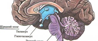
Hypothalamus
This is the structure of the diencephalon that organizes the emotional, behavioral and homeostatic reactions of the body.
50 pairs of nuclei are isolated. Functionally divided into front, middle and rear.
Features of the hypothalamus:
- It is richly vascularized and has a permeable blood-brain barrier (BBB) to various substances.
- The nuclei of the hypothalamus are highly sensitive to shifts in the constants of the internal environment of the body: hormones, glucose, ions, etc.
- Neurons secrete peptides, neurotransmitters and other substances.
Connections of the hypothalamic nuclei:
- together,
- with the above and underlying structures of the central nervous system.
Functions:
- Impact on the vegetative functions of the body through humoral and nervous pathways .
- The hypothalamus is located centers:
- homeostasis,
- thermoregulation,
- hunger-satiation,
- thirst,
- sexual behavior,
- fear,
- rage,
- sleep-wake.
- Regulation of the pituitary gland:
- Neurons of the hypothalamus produce releasing factors (liberins) and inhibitory factors (statins) , which regulate the activity of the anterior pituitary gland (adenohypophysis).
- The hypothalamus secretes vasopressin (antidiuretic hormone - ADH) and oxytocin, which enter the neurohypophysis.
Anatomical and physiological features and syndromes of damage to the taste analyzer
1. Anatomy of a taste analyzer:
— Path of taste information:
1) receptor - mucous membrane of the tongue,
- receptor on the anterior 2/3 of the tongue - facial nerve (VII) - gang. inf. nn. glossopharingei (body I) ->
- receptor on the posterior 1/3 of the tongue - glossopharyngeal (IX) - gang. inf. nn. vagi (body I) ->
- receptor on the epiglottis - vagus (X) - gang. geniculi (body I) ->
2) nucl. solitarius (body II) => tractus solitarius =>
3) ventrolateral nucleus of the thalamus (body III) => posterior 1/3 of the posterior limb of the internal capsule =>
4) insula and base of the postcentral gyrus (body IV) .
2. Types of taste sensitivity:
- salty - lateral surfaces of the tongue (concentration of sodium ions, less often potassium),
– sour – lateral surfaces of the tongue (concentration of hydrogen ions),
- sweet - tip of the tongue (specific receptor),
- bitter - root of the tongue (specific receptor),
- “umami” - the root of the tongue (a specific receptor for glutamate),
- “chili” – root and back of the tongue (pain receptor).
3. Syndromes of damage to the taste analyzer
— Symptoms of loss:
1) hypo(a)geusia - decrease (absence) of taste.
— Symptoms of irritation:
1) hypergeusia – increased sensitivity to smell and taste,
2) taste hallucinations - a feeling of a non-existent smell or taste.
4. Methods for studying taste sensitivity
- drip method (applying standard solutions with a volume of 10 ml (temperature 250C) using pipettes to different parts of the tongue, with rinsing the mouth for 3-5 seconds, at intervals for bitter irritants of 3 minutes, and for other irritants 2 minutes):
1) 20% sugar solution - sweet,
2) 10% solution of table salt - salty,
3) 0.2% hydrochloric acid solution is acidic,
4) 0.1% solution of quinine sulfate is bitter.
Basal ganglia. Striopallidar system
-Improvement of movements in their gradual economization and automation is ensured by the basal ganglia.
This system activates the necessary components of movement and inhibits unnecessary ones.
Basal ganglia:
- caudate nucleus,
- shell,
- pale ball,
- fence.
The globus pallidus, together with the substantia nigra, red nucleus, and subthalamic nucleus, constitute the pallidal system .
Extrapyramidal system:
- caudate nucleus,
- shell,
- pale ball,
- subthalamic nucleus
- black substance.
Information processed in the basal ganglia enters the nuclei of the anterior thalamus. It is then combined with information coming from the cerebellum. The impulses then go to the frontal motor cortex. From there to the neurons of the spinal cord.
Thus, the basal ganglia are integrative centers for organizing established types of motor activity of the body associated with learning.
Violations of the extrapyramidal system manifest themselves in the form of changes in motor function, muscle tone, autonomic functions, and emotional disorders.