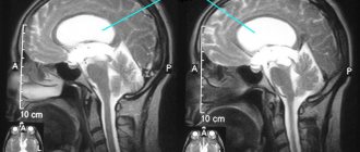
| Insula, insular cortex | |
| Right hemisphere insula | |
| Coronal section of the brain, insula marked top right | |
| Part | Cerebral cortex |
| Catalogs | |
| |
| Media files on Wikimedia Commons | |
Ostrovkovaya
, or
central lobe
(lat. lobus insularis), or
insula
(lat. insula), in a number of sources -
insular cortex
(lat. cortex insularis) - part of the cerebral cortex located in the depths of the lateral sulcus. The insular cortex is considered responsible for the formation of consciousness and also plays a role in the formation of emotions and the maintenance of homeostasis.
Content
- 1 Anatomy 1.1 Pathways
- 1.2 Cytoarchitecture
- 1.3 Brodmann fields
- 3.1 Processing of multimodal sensory information
- 4.1 Progressive expressive aphasia
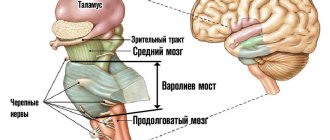
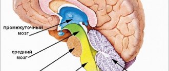
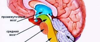
Diencephalon
This department is part of the front part of the organ and controls and switches all incoming information. The functions of the forebrain are the adaptive capabilities of the human body (external negative factors) and the regulation of the autonomic nervous system.
The diencephalon includes:
- Thalamic region;
- Hypothalamic-pituitary system (hypothalamus and posterior pituitary gland);
- Epithalamus.
The hypothalamus regulates the functioning of internal organs and systems and is the center of pleasure. This part is presented in the form of a small cluster of neurons that transmit signals to the pituitary gland.
The thalamus processes all signals coming from sensory receptors, redistributing them to the appropriate parts of the central nervous system.
The epithalamus synthesizes the hormone melatonin, which is involved in the regulation of human biorhythms and emotional background.
The hypothalamus is part of an important system of the central nervous system - the limbic system. This system performs a motivational-emotional function (adapts when habitual conditions change). The system is closely connected with memory and smell, evoking clear memories of a vivid event or reproducing a favorite smell (food, perfume).
Anatomy
The central insular groove (lat. sulcus centralis insulae) divides the lobe into two parts: the larger anterior and smaller posterior. A large area of cortex covering the insula superiorly and laterally forms the operculum (lat. pars opercularis) and is formed from parts of the adjacent frontal, temporal and parietal lobes. The anterior part of the central lobe is divided into three or four short convolutions, and the posterior part is formed by long convolutions.
Pathways
The central lobe communicates through the white matter tracts with the nuclei of the thalamus, located ventrally at the base, and the central nucleus of the amygdala.
Studies in rhesus monkeys have established bidirectional connections between the insula and the small nuclei of the amygdala. Its posterior sections predominantly communicate with the central and dorsolateral part of the amygdala. The anterior sections of the insula contain the anterior, medial, cortical and other nuclei of the amygdala.
The posterior part of the insula is interconnected with the secondary somatosensory cortex. Thus, it receives signals from the ventral basal ganglia of the thalamus, which receive afferent information from the spinothalamic tract. This area also receives signals from the medial ventral nucleus of the thalamus, specialized in transmitting homeostatic information - pain, tactile, temperature sensitivity, local oxygen status, irritation and others.
Neuroimaging studies [ what?
] using diffusion MRI showed that the anterior part of the insula is interconnected with areas in the temporal and occipital lobes, the opercular and fronto-orbital cortex, and the triangular and tegmental parts of the frontal lobe. The same study found differences in anatomical connectivity patterns between the left and right hemispheres.
Cytoarchitecture
In the insular cortex, areas with different cellular structures or cytoarchitecture were found, in particular, granular cell in the posterior part and agranular cell in the anterior. John Allman and his colleagues showed that the anterior cortex contains a population of spindle neurons. They are also called von Economo neurons.
Paul Brodmann
According to the classification of cytoarchitectonic Brodmann areas of the cortex, the insular region of the cerebral cortex contains Brodmann areas 13, 14, 16, as well as areas 44 and 55.
Main parts of the brain
The human nervous system has been studied quite well, which has made it possible to describe in detail what parts the brain consists of and their relationship with various organs, as well as their influence on behavioral reactions. The CNS organ contains billions of neurons through which electrical impulses pass, transmitting information to brain cells from internal organs and systems.
Brain structures are firmly protected from the effects of negative external factors:
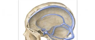
- Cerebrospinal fluid (CSF) is located between the membranes and the surface of the organ. Cerebrospinal fluid acts as a shock absorber, protecting structures from damage and friction. The fluid continuously circulates in the ventricles of the brain, in the subarachnoid space and the spinal canal. In addition to mechanical protection, it also maintains stable intracranial pressure and metabolic processes;
- The arachnoid membrane (arachnoid membrane) is the middle membrane, the deepest and softest. It is formed from connective tissue and contains a large number of collagen fibers. Participates in the exchange of cerebrospinal fluid. The arachnoid membrane contains very thin thread-like strands that are woven into the soft shell;
- The inner shell (soft) fits tightly to the structures, filling all spaces (cracks, grooves). It consists of loose connective tissue penetrated by a vascular network that delivers nutrients to the cells of the organ;
- Superficial shell (hard) - formed from dense connective tissue and has two surfaces. The outer surface contains a large number of vessels and has a rough surface. The inner surface is smooth and fits tightly to the bones - it fuses with the periosteum of the skull and the sutures of the vault;
- The cranium - forms a protective frame for the structures of the brain and its membranes, consists of 23 bones connected to each other. The skull serves as a site for attachment of the soft tissues of the brain.
The cells of the brain structures are formed from the cell bodies of neurons (gray matter, the main component of the nervous system) and the myelin sheath (white matter). Each functionally active cell of an organ has a long process (axon), which branches and connects to another neuron (synapse).
Thus, a kind of circuit is obtained for transmitting and receiving an electrical impulse from one neuron to another. Signals to the brain structures enter through the spinal cord and cranial nerves extending from the brainstem. In some parts of the brain, neurons are transformed due to the synthesis of hormones.
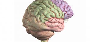
The human brain consists of: anterior, middle and posterior sections. Scientific works of researchers describe the brain after opening the skull as two cerebral hemispheres and an extended formation (trunk), therefore the brain is usually divided into three sections. The hemispheres are separated by a longitudinal groove - an interweaving of nerve fibers (corpus callosum), which looks like a wide strip, consists of axons.
The functions of these parts of the brain are the formation of thought processes and the possibility of sensory perception. Each hemisphere has different functionality and is responsible for the opposite half of the body (the left for the right half and vice versa). The main parts of the brain are formed by dividing the organ using grooves and convolutions.
Brain structures are divided into 5 sections:
- Hindbrain (diamond-shaped);
- Average;
- Front;
- Finite;
- Olfactory.
The organ of the central nervous system has high plasticity - when one of the departments is damaged, compensatory capabilities are temporarily activated, allowing it to perform the functions of the damaged department. Conventionally, the brain is divided into: the right hemisphere and the left hemisphere, the cerebellum, the medulla oblongata. These three departments are connected into a single network, but differ in functionality.
Functions
Processing multimodal sensory information
Functional imaging studies show activation of the insular cortex throughout the performance of audio-visual integration tasks.
Interoceptive self-awareness
There is evidence that in addition to its basic function, the insula may play a role in some higher mental functions. Functional imaging studies have shown that activity in the right anterior insula correlates with a person's ability to sense their own heartbeat or empathize with the pain of others. It is believed that these functions do not differ from the basic functions of the insula, since they arise as a result of the insula's perception of homeostatic information from the thalamus. Thus, the insula is involved in the perception of heat and cold (without pain) on the skin. This includes a feeling of fullness in the stomach and bladder.
Insula activity has been found to be involved in blood pressure control, particularly during and after exercise; In addition, its activity depends on the amount of conscious effort.
The central lobe stands out as the center for evaluating the sensations that arise, which is also expressed in empathy, for example, when a person experiences painful sensations when looking at someone else's pain.
One tomography study showed that the sensation of shortness of breath is processed in the insula and amygdala.
Cortical processing of the vestibular sensation (balance) also involves the central lobe cortex, so with minor damage to the anterior insula the patient may experience dizziness.
Other interoceptive perceptions that are processed in the insular cortex are passive listening to music, laughing and crying, compassion and empathy, and language.
Motor control
The work of the central lobe cortex is involved in hand and eye movements, swallowing, gastric motility and language articulation. Studies of the insular cortex during conversation have shown its connection with the ability to speak for a long time and complex phrases. The insular cortex is also involved in motor learning and has been identified as playing a significant role in recovery and restoration of motor function after stroke.
Social emotions
In the central lobe, processes of processing sensations of disgust take place, both to smells, to the sight of dirt and mutilation - even imaginary ones. In the social aspect, the insular cortex is involved in processing information about violation of generally accepted norms of behavior, emotional processes, empathy and orgasm. The activity of the lobe was discovered when making social decisions made as a result of passing various tests.
Medulla
The posterior section of the central organ of the central nervous system includes the bulb (medulla oblongata), which is included in the stem part. The bulb is responsible for coordinating movements and maintaining balance in an upright position.
Anatomically, the structure is located between the exit of the first spinal nerve (region of the foramen of the occipital bone) and the bridge (upper border). This department regulates the respiratory center - a vital department; if it is damaged, instant death occurs.
Main functions of the medulla oblongata:
- Regulation of blood circulation (work of the heart muscle, stabilization of blood pressure);
- Regulation of the digestive system (production of digestive enzymes, salivation);
- Regulation of muscle tone (rectifying, postural and labyrinthine reflexes);
- Control of unconditioned reflexes (sneezing, vomiting, blinking, swallowing);
- Regulation of the respiratory center (condition of the lung tissue and its stretching, gas composition).
The medulla oblongata has an internal and external structure. On the outer surface there is a midline that divides the pyramids (the connection of the cortex with the nuclei of the cranial nerves and motor horns).
The line crosses the nerve fiber and forms the corticospinal tract. On the side of the pyramid is an olive (an oval extension). The pyramidal system allows a person to perform complex coordination of movements.
Internal structure (nuclei of gray matter):
- Olive nucleus (plate of gray matter);
- Nerve cells with complex connections (reticular formation);
- Nuclei of the cranial nerves (glossopharyngeal, hypoglossal, accessory and vagus);
- Communication between vital centers and the nucleus of the vagus nerve.
Bundles of axons in the bulb provide communication between the spinal cord and other parts of the central nervous system (conducting tracts - long and short). Autonomic functions are regulated in the medulla oblongata.
The vasomotor center and the nuclei of the vagus nerve invert the signals necessary to maintain tone - the arteries and arterioles are always slightly narrowed, and the activity of the heart is slowed down. The bulb contains active poles that stimulate the production of various secretions: salivary, lacrimal, gastric enzymes, bile formation, pancreatic enzymes.
Clinical significance
The central lobe is believed to be involved in the functioning of consciousness and plays an important role in various functions, usually related to the regulation of homeostasis and emotions. Functions of the insula include, but are not limited to: perception, motor control, self-awareness, cognition, and interpersonal experience. This gives rise to a role in the corresponding psychopathological processes.
Progressive expressive aphasia
Type of semantic aphasia. In progressive expressive aphasia, there is a deterioration of normal language function, resulting in loss of fluent speech with preserved ability to understand single words and unaffected nonlinguistic cognitive functions. Occurs in a variety of degenerative neurological diseases, including Pick's disease, motor neuron disease, corticobasal degeneration, frontotemporal dementia, and Alzheimer's disease. This is due to hypometabolism and atrophy of the left anterior central lobe.
Addiction
A number of functional brain imaging studies have shown that the central lobe cortex is activated when drug abusers are exposed to the environment and cues that trigger cravings to use. This has been shown for a variety of drugs, including cocaine, alcohol, opiates and nicotine. Despite these findings, the role of lobe has been ignored in the addiction literature.
A 2007 study showed that smokers who have central lobe damage after a stroke are able to quit tobacco addiction. This has been confirmed by newer studies, making the central lobe a promising site for new research and a target for new anti-drug drugs.
Other clinical conditions
It is believed that the central lobe plays a role in the occurrence and course of such painful conditions as anxiety disorders, emotional dysfunctions, and anorexia.
Midbrain
The middle section of the organ performs quite a lot of physiologically significant functions.
Anatomical structure:
- Four hillocks (two upper and two lower) - these hillocks form the upper surface of the middle part of the organ;
- Sylvian aqueduct – is a cavity;
- The cerebral peduncles are paired parts that connect to the tegmentum of the midbrain.
This section belongs to the stem structure of the organ and has a complex structure, despite its small size. The midbrain is a subcortical section of the brain, part of the motor center of the extrapyramidal system.
Functions of the internal brain:
- Responsible for vision;
- Controls movements;
- Regulates biorhythms (sleep and wakefulness patterns);
- Responsible for concentration;
- Regulates pain;
- Responsible for hearing;
- Regulates protective reflexes;
- Supports thermoregulation in the body.
In the thickness of the cerebral peduncles there are nerve fibers that concentrate almost all the pathways of general sensitivity. Various lesions of the internal structure of the organ lead to impaired vision and hearing. Movements of the eyeballs become impossible, severe strabismus is noted along with hearing loss (bilateral). Hallucinations, both auditory and visual, often occur.