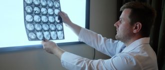
Electroencephalography (EEG) is a method that is used to study the state of the human brain and is based on recording its electrical activity. This examination allows you to detect the spread of pathological processes and signs of epilepsy.
The average duration of the procedure is about an hour, but it is highly informative and makes it possible to track functional changes in the brain, the dynamics of the disease, and evaluate the impact of the therapy.
Since the procedure does not cause any pain or discomfort, EEG can be called one of the most accurate and gentlest methods of examining the brain.
EEG principle
The human brain consists of millions of special cells called neurons. Each of them generates its own electrical impulse. Within individual areas of the brain, impulses must be consistent. They can also strengthen each other or make each other weaker. Their strength and amplitude are not stable and are constantly changing.
Content:
- How EEG works
- Historical reference
- Significance of EEG
- Benefits of an Electroencephalogram
- Indications for the procedure
- Contraindications
- How to prepare for an EEG
- Performing an EEG
- Purpose of EEG video monitoring
- Conclusion of electroencephalography
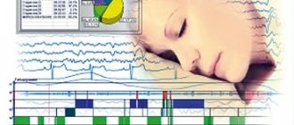
This is the bioelectrical activity of the brain. To register it, special electrodes are applied to the intact scalp, which will pick up vibrations, amplify them and record them in the form of special curves, so-called waves. The latter, depending on their shape, frequency and amplitude, are divided into five types: α- (alpha), β- (beta), δ- (delta), θ- (theta) and μ- (mu) waves. Each of the waves reflects the work of a specific part of the brain and is named by the first letter of its Latin name.
Their registration in real time is the essence of encephalography.
Parameters of normal beta rhythm
Frequency 12-25 Hz (some recognize 13-25 Hz) Beta1 rhythm 25Hz Beta2 rhythm -40Hz
The amplitude is small - 10-15 µV.
Zoning - normally distributed in the anterior-central and temporal regions. According to Zhirmunskaya E.A. The beta 1 rhythm is not purely physiological and is not typical for the norm. Temporal beta rhythm is often the result of muscle artifact.
C-rhythm is a variant of the normal rhythm with a frequency of 8 - 13 Hz and is detected in the central sections. It has the following features: it disappears with the contralateral active clenching of the hand into a fist, it is narrowly localized in the central sections. Slow rhythms that occur normally. Theta rhythm - frequency 4-8 Hz, amplitude up to 30-40 µV. Delta rhythm - frequency 0.5-4 Hz, amplitude up to 30-40 µV.
Historical reference
One of the founders of the electroencephalography method is considered to be the physiologist and psychiatrist from Germany Hans Berger. In 1924, using a device for measuring small currents called a galvanometer, he was the first to perform a procedure resembling an EEG recording.
Later, a special device called an encephalograph was created to conduct electroencephalography. Today, there are stationary encephalographs, which make it possible to conduct research exclusively in a special room, and portable ones, which are designed for long-term monitoring and can be moved.
It is noteworthy that initially EEG was considered exclusively as a method for identifying mental disorders in a person. Only over time it became clear that the technique also makes it possible to detect deviations not related to psychiatry.
Significance of EEG
EEG is a highly informative diagnostic method that allows:
- assess the nature of brain dysfunction and how severe they are;
- determine the localization of the pathological focus;
- clarify information obtained during other diagnostic procedures (for example, computed tomography);
- monitor how effective the therapy is;
- identify areas of the brain in which epileptic activity is present;
- assess brain function in the periods between attacks;
- identify the causes of panic attacks and fainting;
- study the sleep-wake cycle.
It should be taken into account that if a person has seizures, the study will be informative only if it is carried out after about a week.
The essence of the diagnostic method
Back in the 19th century, it was established that the human brain is capable of generating electrical impulses, but more than eighty years passed before doctors were able to use this ability in medical practice. This happened after the creation of the first electroencephalograph apparatus, and the graphic image that was obtained by recording the biocurrents of the human brain was called an electroencephalogram.
Signals emanating from brain neurons change under the influence of various factors, while different parts of the brain give coordinated impulses, mutually weakening or enhancing each other’s activity. An encephalogram records various changes in impulses and helps to identify abnormalities in the functioning of the brain or its individual parts. Electroencephalography allows you to track the activity of each part of the brain separately, study neural impulses and the coordination of the actions of brain parts (rhythm).
Benefits of Electroencephalography
Today, electroencephalography is widely used in neuropathological practice. It makes it possible to clarify a huge number of problematic situations that are associated with the diagnosis and differentiation of neurological diseases. One of the undeniable advantages of encephalography is the fact that it not only helps to identify certain problems, but also helps to distinguish true disorders from hysterical manifestations or simulation.
In addition, the procedure is not as expensive as examination using a tomograph or other similar devices. EEG equipment is available in most hospitals.
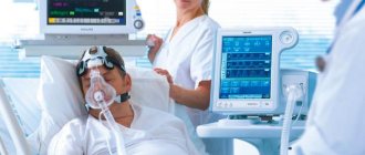
The procedure does not have any negative impact on human health and condition. The patient remains fully functional. The study can be carried out even on patients in serious condition, children and adults of any age, since it does not cause deterioration of the condition, discomfort or pain.
Indications for the procedure
Today, electroencephalography is widely used in the practice of neurologists to solve a number of problems.
EEG is recommended:
- for prolonged insomnia and other sleep disorders, including apnea, somnambulism and talking during sleep;
- during a convulsive attack;
- for diseases of the thyroid gland;
- with signs of developmental disorders in children or mental disorders in adults;
- after recently experienced traumatic brain injury;
- in case of pathological changes in the vessels of the neck and head, brain tumors detected during ultrasound;
- with frequent migraines, complaints of regular dizziness, feeling of constant fatigue;
- for panic attacks, autism, Asperger's syndrome, stuttering, nervous tics, delayed speech and mental development in children;
- for meningitis, encephalitis, after a stroke and micro-stroke;
- after neurosurgical operations.
Contraindications
It is noteworthy that there are no absolute contraindications for electroencephalography. If a person suffers from convulsive attacks, is diagnosed with coronary heart disease, hypertension, or mental disorders, an anesthesiologist must be present during the diagnosis.
The procedure should be postponed if there are open wounds, traumatic injuries, postoperative sutures or any signs of an inflammatory process in the area where it is necessary to install electrodes. Also, the study is not carried out for patients with ARVI.
How to prepare for an EEG
It should be noted that there are no special restrictions that must precede the procedure. However, there are a number of rules that are recommended to be followed to ensure that the examination is successful and informative.
First of all, inform your doctor if you are taking any medications. You may need to stop taking them for a while or change the dosage.
At least twelve hours before the procedure, or even better, a day before, eliminate from your diet foods that contain caffeine, carbonated drinks, chocolate and cocoa, as well as foods that contain them, foods with energy components, for example, taurine. You should also avoid products with sedative effects.
Wash your hair before the procedure. It is not recommended to use additional styling products (oils, gels, balm, varnishes, etc.) as this may affect the quality of contact of the electrodes with the skin.
If the main purpose of the procedure is to detect seizure activity, you need to sleep before the study.
In order for the result to be as reliable as possible, the patient should avoid stressful situations and refrain from driving for twelve hours before the procedure.
Eating is recommended two hours before the procedure.
If the patient is prescribed EEG sleep monitoring, the night before should be sleepless. Immediately before the procedure, the subject will receive a special sedative, which will enable him to fall asleep during the electroencephalography.
If a child is to undergo an examination, parents should first psychologically prepare him for the manipulations, explaining that there will be no pain or discomfort. You can practice putting on a pool cap under the pretext of playing pilots or astronauts, teach your child to breathe deeply, showing him how to do it by personal example. On the eve of the procedure, the child should wash his hair without using any additional styling products. Before leaving the house, the baby should be fed and calmed down. Just in case, parents are advised to take with them tasty food and drink, and a favorite toy that will help calm and distract the baby.
Please note that if the above rules are not followed, the EEG result may not be very accurate or very informative. In this case, the procedure will have to be repeated.
International system of electrode placement “10-20%” when conducting EEG
➥ Main article: System 10-20
Layout of electrodes 10-20
Bioelectrical activity of the brain can be recorded from any point on the convexital surface. The repeatability of the results and their comparability with the data of other studies is achieved only if all specialists use a single standard system of electrode placement.
In 1958, Henry Jasper proposed an original electrode placement scheme. The “coordinate system” proposed by Jasper is based on a strict ratio of the distances between the electrodes in a “coordinate grid” on the convexital surface. The system of “meridians and parallels” is built relative to the line “occipital protuberance - bridge of the nose” and the interauricular “equator” passing through the crown. Based on the selected ratio of the distances between the electrodes, Jasper’s scheme is called the “10-20% system.” Currently, the 10–20% electrode placement system is recommended by the International Federation of Clinical Neurophysiologists (IFCN) as standard.
Letter symbols indicate the main areas of the brain and landmarks on the head: O - occipitalis, P - parietalis, C - centralis, F - frontalis, T - temporalis, A - auricularis. Odd digital indices correspond to electrodes above the left, and even ones - above the right hemisphere of the brain. Electrodes located along the sagittal line are assigned the index “ z ”.
The location points of the electrodes in the “10-20%” lead system are determined as follows. Measure the distance along the sagittal line from the occipital protuberance (inion) to the bridge of the nose (nasion) and take it as 100%. At 10% of this distance from the reference points (inion and nasion), the lower frontal (Fpz) and occipital (Oz) sagittal electrodes are installed, respectively. The remaining sagittal electrodes (Fz, Cz and Pz) are placed between these two at equal distances, constituting 20% of the inion-nasion distance. The second main line runs between the two ear canals through the vertex (top of the head). The inferior temporal electrodes (TZ and T4) are located, respectively, 10% of this distance above the auditory canals, and the remaining electrodes of this line (SZ, Cz, C4) are located at equal distances, constituting 20% of the length of the biauricular line. Lines are drawn through points T3, SZ, C4, T4 from inion to nasion and the remaining electrodes are placed along them. Electrodes Oz, Pz, Fz are placed along the midsagittal line (through Cz). Along the lines passing through SZ and C4, electrodes 01, RZ, F3, Fpl are placed on the left and 02, P4, F4, Fp2 on the right. Along the lower lines passing through the electrodes T3 and T4, electrodes F7 and T5, F8 and Tb are placed. Electrode clips are placed on the earlobes: A1 - on the left ear and A2 - on the right.
The advantage of the “10-20%” scheme is a large number of electrodes, which allows one to obtain a detailed picture of the potential distribution over the convexital surface and perform mapping procedures.
American Neurophysiological Society in the early 1990s. (1991) proposed a “10-10” lead system. Additional electrodes in this system are installed at a distance equal to half the distance between the electrodes in the “10-20” system. This system, as well as systems with an even larger number of electrodes (up to 64-128), represent an attempt to increase the “resolution power of the EEG”. By “EEG resolution” we can conditionally assume the ability to identify two sources of bioelectrical activity of the brain as independent independent sources.
Performing an EEG
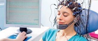
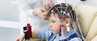
Electroencephalography is performed in a special room, which is completely isolated from light and sound. The patient sits in a chair or is asked to lie on the couch. A special cap with electrodes is first placed on his head. During the procedure, the patient is alone in the room, contact with doctors is maintained using a camera and microphone. If the diagnosis is performed on a child, one of the parents remains in the office.
Before starting the procedure, the patient is asked to close and open his eyes several times to adjust the equipment. During the diagnosis, the eyes must be closed. If during the procedure the patient needs to change position or visit the restroom, he can inform the doctors about this, after which the diagnosis will be suspended.
It is extremely important that the patient lies with his eyes closed and does not move during the procedure. In the event that a person opens his eyes slightly or moves, the doctor makes a corresponding note, since these actions must be taken into account when deciphering the electroencephalogram.
After the resting EEG is recorded, so-called “stress tests” are performed. Their goal is to test how the brain will react to situations that are stressful for it.
So, a hyperventilation test can be performed. The patient is asked to breathe frequently and deeply for three minutes. Photostimulation with a stroboscopic light source can also be used. It flashes frequently, and this allows you to evaluate how the brain reacts to bright light.
Stress tests can trigger convulsions or an epileptic seizure. The doctors who conduct the study have the appropriate skills to provide emergency care to the patient if necessary.
After the study is completed, the physician should remind the patient to resume taking medications that were stopped the day before the EEG.
The total duration of the procedure is from forty minutes to two hours.
Why is electroencephalography needed? Complete patient guide
Frequent awakenings at night, insomnia, enuresis, the need to detect epilepsy - these are just some of the pathologies that electroencephalography will help in diagnosing.
The neurologist at the Expert Kursk Clinic, Olesya Olegovna Bratchikova, told us about her.
— Olesya Olegovna, what is electroencephalography and how often is this research method prescribed?
Electroencephalography is a non-invasive method for studying brain function by recording its bioelectrical activity. “Non-invasive” means that there are no punctures, incisions, insertion of instruments into body cavities and organs, etc.
It is used everywhere, all over the world. It is widely used to diagnose and monitor the effectiveness of treatment for various conditions. Among them: loss of consciousness, differentiation of epileptic syndromes, various neurological conditions.
Why is MRI performed for epilepsy? Radiologist at MRI Expert Sochi, Zarema Bardudinovna Tseeva, tells
— What is the difference between daytime and nighttime EEG?
Daytime (otherwise known as routine) electroencephalography is performed during the daytime. Its duration does not exceed 20 minutes. Used to identify obvious disorders - for example, to diagnose epileptic syndrome.
It is used for medical examinations (screening studies), and for the delimitation of paroxysmal (accompanied by certain attacks) conditions. Daytime EEG provides no more than 30% of all possible information.
You can sign up for electroencephalography in your city here
Please note: the service is not available in all cities
Night EEG is the “gold standard” of this examination. It is used for a deep search for a problem: separating epileptic syndrome from non-epileptic syndrome, sleep disorders (walking and talking in sleep, periodic short-term pauses in breathing during sleep - for example, with snoring, etc.), enuresis, etc.
— What are the indications for electroencephalography?
An EEG can detect signs of epilepsy. This is her main indication. In this case, diagnosis is prescribed when epilepsy is suspected for the first time, when the treatment regimen is changed and its control.
Read material on the topic: Epilepsy: symptoms, causes, diagnosis and treatment
Electroencephalography is prescribed for enuresis; paroxysmal states; neuroses, anxiety, irritability; hyperactivity and learning disorders in children; delayed mental development, speech, stuttering; autism; frequent awakenings and excessive motor activity during sleep; episodes of walking and talking in a dream, etc.
— At what age is EEG monitoring performed for children?
Starting from 4 weeks.
— Are the indications for daytime and nighttime electroencephalography different or the same?
For a medical examination/screening of a healthy person (without complaints, burdened anamnesis), a daylong study is sufficient. If a person has a history of, for example, an episode of epilepsy, then an overnight study is necessary.
— In what cases does a patient need to undergo both daytime and nighttime electroencephalography?
In the presence of paroxysmal conditions, suspected epilepsy. Suppose a person had a history of an epileptic seizure, or the doctor himself observed it. In this case, a day examination is carried out. If epileptic activity is not detected during the daytime study, a night study must also be prescribed.
— Is it necessary to undergo EEG monitoring to obtain a certificate from the traffic police?
Yes, but not everyone. Since June 2015, according to the order of the Ministry of Health of the Russian Federation No. 344n, an EEG must be performed on any road user applying for a driving license of categories C, D, E. An EEG for drivers of other categories is performed according to indications determined by a neurologist at an appointment.
— How is electroencephalography performed? How long does diagnosis take?
A special cap with electrodes is placed on the subject’s head. A special gel is applied to the scalp at the point of contact with the electrode (for better contact and conductivity of the recorded impulses). Signals from the brain are transmitted through electrodes to a device called an electroencephalograph. It amplifies them and transfers them to a computer for further processing.
As a result, a special “curve” is formed - an electroencephalogram. Based on it, a conclusion is made about the state of the functional activity of the brain.
The daytime session lasts no more than 20 minutes. The study takes place in the so-called state of “relaxed wakefulness”. The man is relaxed, lying with his eyes closed, but not sleeping.
What are the reasons for insomnia? Olga Ivanovna Kuyantseva, a neurologist at Clinic Expert Voronezh, talks about the treatment of insomnia.
During diagnosis, functional tests are carried out to change the functional activity of the brain. The doctor asks the patient to close and open his eyes and breathe actively (3-5 minutes). Photostimulation is also performed: the subject looks at light flashes.
Night research is best done at home. It starts between 20 and 21 pm. Technical nuances are the same as during a daytime examination.
Two complete sleep cycles are recorded, which is approximately 2-3 hours after falling asleep.
— How to properly prepare for an electroencephalogram?
No special preparation is required for an EEG of the brain. The head must be clean; styling products must not be used when drying. On the eve of the study, avoid stress, overwork, and maintain a measured regimen. It is necessary to refrain from taking nervous system stimulants, strong tea, coffee, and alcohol.
Noisy games, watching TV, cartoons, working or playing on the computer are also undesirable for a child.
The presence of metal objects and jewelry on the body during diagnosis is not allowed.
In some cases, when conducting a sleep EEG to establish or exclude a diagnosis of epilepsy, as well as for the differential diagnosis of paroxysms, so-called sleep deprivation is recommended to the patient in preparation for the study. This means that on the eve of the procedure, the patient reduces the duration of night sleep, wakes up early - 3-4 hours earlier than his usual awakening time.
— Olesya Olegovna, how often can you undergo electroencephalography? Is this diagnostic method safe?
EEG is completely safe. You can undergo EEG monitoring as often as necessary for the doctor to make a diagnosis or monitor the patient’s condition.
— Does EEG have any limitations or contraindications?
No.
— In order to undergo an EEG in your clinic, do you need a doctor’s referral?
If a person undergoes a medical examination or is examined on his own initiative, in principle he can undergo the diagnosis independently, without a referral. It is enough to have an identity document.
If we are purposefully looking for some kind of pathology, determining the type of paroxysms, etc., then in this case it is advisable to have a doctor’s referral. From it we can understand the purpose of the survey, i.e. what the doctor needs.
Other questions:
Educational program on medical professions. When to contact a neurologist?
Holter (24-hour) ECG monitoring – complete instructions for the patient
Tension headache: causes, symptoms, diagnosis and treatment
For reference:
Bratchikova Olesya Olegovna
Graduate of the Faculty of General Medicine of Kursk State Medical University in 2004.
From 2004 to 2005, she completed an internship, and from 2005 to 2007, a clinical residency in the specialty “Neurology”.
I took a course in epileptology and certification courses.
Currently working as a neurologist at Clinic Expert Kursk.
Purpose of EEG video monitoring
One of the types of electroencephalography is EEG video monitoring. This is a long-term recording of electroencephalography, usually lasting several hours, which can be performed during sleep. The duration of the procedure in each specific case is determined by the attending physician and the personnel of the laboratory that conducts the examination.
EEG video monitoring is prescribed if a short standard procedure does not reveal pathologies, but they are present.
Also, this type of examination allows you to evaluate the EEG during wakefulness and sleep.
Many patients are interested in the question of whether they should necessarily sleep during the study. The answer to this question cannot be unambiguous, because it depends on the specific situation. So, for example, if the reason for the examination is a tic that bothers the patient while awake, there is no need to sleep during the examination.
At the same time, EEG video monitoring during sleep sometimes helps to identify conditions that neither the patient nor his loved ones may even be aware of.
The peculiarity of this procedure is that it can be carried out not only during the day, but also at night. In the event that sleep EEG is required, night monitoring is more rational. Not everyone can fall asleep without problems during the daytime.
At the same time, we should not forget that carrying out a multi-hour procedure in a completely isolated room can be extremely tiring for the patient, especially if we are talking about a child. Most pathologies can be detected during a relatively short recording of a conventional EEG.
Best materials of the month
- Coronaviruses: SARS-CoV-2 (COVID-19)
- Antibiotics for the prevention and treatment of COVID-19: how effective are they?
- The most common "office" diseases
- Does vodka kill coronavirus?
- How to stay alive on our roads?
Also, overnight EEG video monitoring is much more expensive.
What is EEG and its features
Electroencephalography is an examination of the brain and its electrical activity. The procedure is called EEG in short. Investigations help to detect inflammatory processes, vascular anomalies, tumors, epilepsy, and other serious pathologies in a timely manner.
EEG of the brain is the only method that allows us to examine and diagnose the patient’s condition, even if he has lost consciousness. The research is absolutely safe for the body. The procedure takes no more than 30 minutes.
Using electroencephalography, the doctor traces the dynamics of the pathology, adjusts therapy, and evaluates the effect of medications already used on the body. EEG is able to monitor all changes in the brain, and this distinguishes the method from MRI.
The activity of the organ is determined using a special map. This scheme traces the degree of manifestation of pathology, problems with the central nervous system, to which a specific rhythm corresponds. The doctor determines the synchronicity of the parts of the brain and how it uses its capabilities.