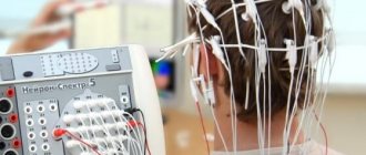
Our center performs EEG (electroencephalogram of the brain), including a full neurological protocol and with sleep deprivation. The study allows you to diagnose pathology in the functioning of the vascular system of the brain and nervous system. The interpretation of the study results is carried out by the doctor immediately after the completion of the EEG, and does not take more than 15–20 minutes.
In particular, the method allows you to determine:
- epileptic activity characteristic of different lobes of the brain;
- probable cause provoking panic attacks and loss of consciousness;
- location of pathological foci in certain lobes of the brain;
- the degree of change in the electrical activity of the brain before attacks.
Also, an electroencephalogram of the brain allows you to find the cause of neurology in a person (is it a functional disorder or caused by an organic lesion), assess how effective treatment and rehabilitation are - then an EEG is done 3 times: before treatment, during therapy and after its completion.
| Service | Price |
| Electroencephalogram (EEG) | 2000 |
| EEG with sleep deprivation | 4000 |
| Urgent EEG interpretation | 300 |
Our address: St. Petersburg, st. Marata, house 78.
Our phone: +7 (812) 385-55-82.
Opening hours: from 9:00 to 21:00.
We work by appointment.
EEG is performed on modern medical equipment that meets international (ISO) and European certification - Mitsar and Neurosoft.
When is it necessary to do an EEG?
Before doing an electroencephalogram, the doctor examines the patient and listens to complaints. The basis for the study may be:
- sleep disorders - for example, insomnia;
- frequent awakenings and sleepwalking;
- frequent attacks of dizziness and fainting;
- fatigue and feeling tired;
- headaches that occur for no reason.
Even a seemingly insignificant deterioration in a person’s well-being may indicate irreversible negative processes in the GM. Based on this, it is extremely important to do an EEG in the following cases:
- vascular diseases of the cervical region of the head;
- VSD;
- heart rhythm disturbances;
- post-stroke condition;
- speech delays;
- logoneurosis;
- autism;
- inflammation of the brain;
- hormonal disorders;
- suspicion of cancer.
Brain EEG is mandatory for patients with TBI, epilepsy, or who have undergone neurosurgery.
Video-EEG monitoring and neurology
— Are you worried about headaches?
-Have you started sleeping poorly?
— Has your child started to study poorly?
- Do you know what epilepsy is?
In practical neurology, night and daytime video-EEG monitoring (VEM) is the method of choice in the differential diagnosis of diseases of the central nervous system in adults and children, non-epileptic and epileptic paroxysmal conditions:
- allows you to record a video image of the patient synchronously with changes in the Electroencephalogram (EEG), myography, and Electrocardiogram (ECG). Synchronization of video recordings with EEG and ECG makes it possible to interpret certain pathological conditions of the patient with a high degree of reliability. Using this method, it is possible to identify non-epileptic conditions masquerading as epileptic seizures, and vice versa, to make a diagnosis of epilepsy in cases where routine electroencephalography is uninformative.
- Compared to routine EEG, video-EEG monitoring allows one to obtain a significantly larger amount of useful diagnostic information. Video EEG monitoring in various functional states (passive and active wakefulness, sleep, awakening, specific tests) also helps to increase the information content of the research.
- Long-term EEG recording makes it possible to detect rarely occurring pathological changes in the bioelectrical activity of the brain.
- the likelihood of recording pathological forms of electrical activity in the brain increases sharply when falling asleep and during sleep. Moreover, in some pathologies, the bioelectrical activity of the brain is characterized by maximum representation only in certain stages of sleep.
The method has no age restrictions or contraindications, is painless and safe.
How is an electroencephalogram performed?
According to global practice, EEG of the brain in St. Petersburg is carried out using an encephalograph. It includes a processing unit, a monitor and a set of electrodes mounted in a rubber “cap” worn on the head. EEG is carried out in a compact room, protected from light and noise from outside. The procedure is quick and consists of several stages.
Preparation
First, the subject chooses a comfortable position - reclining or sitting. Next, you put on a cap with electrodes that record the electronic activity of the brain.
Study
During an EEG of the brain, the device reads data from the electrodes, forming a graph based on it that is displayed on the monitor. In some cases, a series of functional tests is carried out: the subject must blink, look at flashes of light, change the breathing rhythm, listen to certain sounds.
Completion of the study
At this stage, the electrodes are removed, the results of the study are printed, and a transcript is made.
Duration of the study
An electroencephalogram in St. Petersburg lasts depending on the need for functional tests and the area of the brain being studied. On average, an EEG is performed in 20–30 minutes using:
- rhythmic photostimulation of various frequencies;
- hyperventilation (the subject will have to breathe deeply and rarely);
- loads in the format of slow blinking (opening and closing the eyes on command).
In addition, at this time, functional changes that occur covertly are detected.
Indications for Video-EEG monitoring (VEM) in children aged from 4 weeks to 18 YEARS
Indications for 1-3 hour VEM in children aged 4 weeks to 2 years
- Children who have suffered intrauterine hypoxia:
- from mothers with various somatic pathologies during pregnancy
- from mothers with hormonal disorders, threat of miscarriage
- Children who suffered hypoxia during childbirth
- Children born prematurely
- Children born with low gestational age and intrauterine hypotrophy
- Children with central nervous system disorders at birth, of varying degrees
- Children from mothers with epilepsy
- Children with epilepsy
- Children with delayed psychomotor development
- Children who have suffered traumatic brain injury (TBI) and neuroinfections
- Children with sleep disorders
Indications for 2-4 hour and overnight VEM in children aged 2 to 7 years
- Children with delayed psychomotor development
- Children with speech delay
- Children with borderline behavioral disorders
- Tics, hyperkinesis
- Children with post-vaccination complications from the central nervous system
- Children with sleep disorders
- Syncope
- Children suffering from epilepsy (primary clarification of the form of epilepsy and confirmation of the effectiveness of the therapy)
- Children who have undergone brain surgery
- Children with hypertension and hydrocephalic syndromes
- Children who have suffered TBI of varying severity and neuroinfections
- Children suffering from enuresis, encopresis
Indications for 4-hour and overnight VEM in children aged 7 to 18 years
- Children with attention deficit disorder with hyperreactivity, decreased academic performance
- Children with sleep disorders (sleeping, sleep talking, night terrors, startling, unexpected awakenings)
- Children with borderline behavioral disorders
- Children with delayed speech and psychomotor development
- Children suffering from paroxysmal headaches
- Children who have suffered a TBI of varying severity, neuroinfection, poisoning, coma or clinical death.
- Children suffering from epilepsy (primary clarification of the form of epilepsy and confirmation of the effectiveness of the therapy).
- Adolescents of military age with a history of consciousness disorders and fainting.
- Girls with disorders of consciousness during the perimenstrual period.
EEG with sleep deprivation
EEG with sleep deprivation is a type of electroencephalography in which sleep time is limited or interrupted multiple times at night followed by falling asleep. This is used in cases where the patient’s EEG in a normal state does not reveal pathology in brain activity. This type of study is 30% more accurate than traditional EEG and is more effective in relation to the following types of pathologies:
- epilepsy;
- episyndromes;
- syncope;
- sleep disorders;
- insomnia;
- sleepwalking and dream-talking.
EEG with sleep deprivation can last from 1.5 to 3 hours.
Indications for Video-EEG monitoring
Non-epileptic
- Headaches, dizziness.
- Unexplained speech delay, minimal cerebral dysfunction.
- Progressive decline in cognitive function.
- Affective respiratory paroxysms.
- Febrile seizures.
- Disturbance of daytime and night sleep.
- Enuresis, encopresis.
- Asthenic syndrome.
- Non-epileptic paroxysms:
- Psychogenic attacks (conversion)
- Syncope: Cardiogenic syncope
- vasovagal syncopation
- the vague syncopations of small children
- night terrors
- myoclonus in sleep
Epileptic
- Confirmation of the diagnosis of epilepsy.
- Clarification of the form of epilepsy and localization of epileptic foci.
- Confirmation of drug remission of epileptic disease and the adequacy of antiepileptic therapy (absence of paroxysms and epileptiform discharges during night and daytime Video-EEG monitoring).
- Resolving the issue of discontinuing antiepileptic therapy.
- Paroxysmal disorders of consciousness.
- Paroxysmal movement disorders.
- Clarification of the cause of a single epileptic seizure.
- Clarification of the cause of rare attacks (incomplete compensation, incorrect diagnosis).
- Epileptic seizures in the presence of a history of traumatic brain injury.
- Recurrence of attacks due to continuous therapy.
If your child has problems with behavior, learning, memory, concentration, if there is a speech delay of unknown origin or brain dysfunction - call and come to us for examination. Our staff will help you understand the current situation and offer you solutions to problems in the comfortable conditions of our center.
What you need to know about preparing for an EEG
To do an EEG in St. Petersburg, the patient does not need to prepare for a long time. The main thing is to strictly follow a number of recommendations necessary to obtain reliable results:
- Avoid taking anticonvulsants, sedatives or stimulants at least 3-4 days before the procedure;
- limit your consumption of tea, coffee, chocolate and energy drinks;
- eat a heavy meal 2 hours before the EEG, since hunger and a decrease in blood sugar against its background distorts the diagnostic results;
- When conducting an EEG during sleep, the night before the study, you are prohibited from sleeping.
The patient's head must be washed and free of cosmetics, the hair must be dry, and if it is long, it must be loose. Metal jewelry must be removed. The patient should be calm and not react to sounds or flashes of light. An electroencephalogram of the brain must be carried out during periods of mental calm - any experiences, nervous shocks and breakdowns a few days before the study should be excluded.
A complete medical examination includes the collection of information about the patient’s life, the development of the disease and, most importantly, a very detailed description of the attacks, as well as the conditions preceding them, by the patient themselves and eyewitnesses of the attacks. If seizures occur in a child, the doctor will be interested in the course of pregnancy and childbirth in the mother. A general and neurological examination and electroencephalography are required. Special neurological studies include nuclear magnetic resonance imaging and computed tomography. The main task of the examination is to identify current diseases of the body or brain that could cause the attacks.
What is electroencephalography (EEG)? Using this method, the electrical activity of brain cells is recorded. This is the most important test in diagnosing epilepsy. An EEG is performed immediately after the first seizures appear. In epilepsy, specific changes (epileptic activity) appear on the EEG in the form of discharges of sharp waves and peaks of higher amplitude than normal waves. During generalized seizures, the EEG shows groups of generalized peak-wave complexes in all areas of the brain. In focal epilepsy, changes are detected only in certain, limited areas of the brain. Based on EEG data, a specialist can determine what changes have occurred in the brain, clarify the type of seizures, and, based on this, determine which drugs will be preferable for treatment. Also, with the help of EEG, the effectiveness of the treatment is monitored (especially important for absence seizures), and the issue of stopping treatment is decided.
How is an EEG performed? EEG is a completely harmless and painless study. To carry it out, small electrodes are applied to the head and secured to it using a rubber helmet. The electrodes are connected via wires to an electroencephalograph, which amplifies the electrical signals of brain cells received from them by 100 thousand times, records them on paper or enters the readings into a computer. During the examination, the patient lies or sits in a comfortable diagnostic chair, relaxed, with his eyes closed. Usually, when taking an EEG, so-called functional tests (photostimulation and hyperventilation) are performed, which are provocative loads on the brain through bright flashing light and increased respiratory activity. If an attack begins during an EEG (this happens very rarely), then the quality of the examination increases significantly, since in this case it is possible to more accurately determine the area of impaired electrical activity of the brain.
Are changes in the EEG grounds for identifying or excluding epilepsy? Many EEG changes are nonspecific and provide only supporting information for the epileptologist. Only on the basis of the identified changes in the electrical activity of brain cells, one cannot speak of epilepsy, and, conversely, this diagnosis cannot be excluded with a normal EEG if epileptic seizures occur. Epileptic activity on the EEG is regularly detected in only 20-30% of people with epilepsy.
It is necessary to constantly keep in mind that the interpretation of changes in the bioelectrical activity of the brain is, to some extent, an art. Changes similar to epileptic activity may be caused by eye movement, swallowing, vascular pulsation, respiration, electrode movement, electrostatic discharge, and other extracerebral causes. In addition, the electroencephalographer must take into account the patient's age, since the EEG of children and adolescents differs significantly from the electroencephalogram of adults.
What is a hyperventilation test? This is frequent and deep breathing for 1-3 minutes. Hyperventilation causes pronounced metabolic changes in the brain due to intensive removal of carbon dioxide (alkalosis), which, in turn, contribute to the appearance of epileptic activity on the EEG in people with seizures. Hyperventilation during EEG recording makes it possible to identify hidden epileptic changes and clarify the nature of epileptic seizures.
What is EEG with photostimulation? This test is based on the fact that flashing lights can trigger seizures in some people with epilepsy. During EEG recording, a bright light flashes rhythmically (10-20 times per second) in front of the eyes of the patient being studied. Detection of epileptic activity during photostimulation (photosensitive epileptic activity) allows the doctor to choose the most appropriate treatment tactics.
Why is an EEG with sleep deprivation performed? Sleep deprivation for 24-48 hours before EEG is carried out to identify hidden epileptic activity in difficult to recognize cases of epilepsy.
Sleep deprivation is a fairly strong trigger for attacks. This test should only be used under the guidance of an experienced physician.
What is EEG during sleep? As is known, in certain forms of epilepsy, changes in the EEG are more pronounced, and sometimes can only be perceptible during a study during sleep. Recording an EEG during sleep makes it possible to detect epileptic activity in the majority of those patients in whom it was not detected during the daytime, even under the influence of ordinary provocative tests. But, unfortunately, such a study requires special conditions and trained medical personnel, which limits the widespread use of this method. It is especially difficult to carry out in children.
Is it right not to take antiepileptic drugs before an EEG? This should not be done. Abruptly stopping the medication provokes seizures and can even cause status epilepticus.
When is video EEG used? This very complex study is carried out in cases where it is difficult to determine the type of epileptic seizure, as well as in the differential diagnosis of pseudo-seizures. Video-EEG is a video recording of an attack, often during sleep, with simultaneous EEG recording. This study is carried out only in specialized medical centers.
Why is brain mapping done? This type of EEG with computer analysis of the electrical activity of brain cells is usually carried out for scientific purposes. The use of this method in epilepsy is limited to identifying only focal changes.
Is EEG harmful to health? Electroencephalography is an absolutely harmless and painless study. EEG is not associated with any effect on the brain. This study can be carried out as often as necessary. Carrying out an EEG causes only minor inconvenience associated with putting a helmet on the head and slight dizziness that may occur during hyperventilation.
Do EEG results depend on what device the study is used on? Equipment for conducting EEG – electroencephalographs, produced by different companies, are not fundamentally different from each other. Their difference lies only in the level of technical service for specialists and in the number of recording channels (electrodes used). EEG results largely depend on the qualifications and experience of the specialist conducting the study and analyzing the data obtained.
How to prepare a child for an EEG? The child must be explained what awaits him during the examination and convinced that it is painless. The child should not feel hungry before the test. The head should be washed clean. With small children, the day before you need to practice putting on a helmet and staying still with your eyes closed (you can pretend to play as an astronaut or tank driver), and also teach them to breathe deeply and often under the commands “inhale” and “exhale.”
Computed tomography Computed tomography (CT) is a method of examining the brain using radioactive (X-ray) radiation. During the study, a series of images of the brain are taken in different planes, which allows, unlike conventional radiography, to obtain an image of the brain in three dimensions. CT allows you to detect structural changes in the brain (tumors, calcifications, atrophy, hydrocephalus, cysts, etc.).
However, CT data may not have informative value for certain types of attacks, which include, in particular:
any epileptic seizures for a long time, especially in children; generalized epileptic seizures with the absence of focal changes in the EEG and indications of brain damage during a neurological examination.
Magnetic resonance imaging Magnetic resonance imaging is one of the most accurate methods for diagnosing structural changes in the brain.
Nuclear magnetic resonance (NMR) is a physical phenomenon based on the properties of certain atomic nuclei, when placed in a strong magnetic field, to absorb radio frequency energy and emit it after the exposure to the radio frequency pulse ceases. In its diagnostic capabilities, NMR is superior to computed tomography.
The main disadvantages usually include:
low reliability of calcification detection; high price; impossibility of examining patients with claustrophobia (fear of closed spaces), artificial pacemakers (pacemakers), large metal implants made of non-medical metals.
Is a medical examination necessary in cases where there are no more attacks? If a person with epilepsy has stopped having seizures, but the medications have not yet been stopped, then it is recommended that he undergo a control general and neurological examination at least once every six months. This is especially important for monitoring the side effects of antiepileptic drugs. Usually the condition of the liver, lymph nodes, gums, hair is checked, as well as laboratory blood tests and liver tests. In addition, it is sometimes necessary to monitor the amount of anticonvulsant drugs in the blood. A neurological examination includes a traditional examination by a neurologist and an EEG.