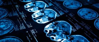
Cerebral palsy (cerebral palsy) is a disease that primarily affects the musculoskeletal system. It develops against the background of pathological damage to certain areas of the brain or their incomplete development. Many women carrying a child are afraid that this could happen to their baby, so quite often the question is raised among expectant mothers: is cerebral palsy visible on an ultrasound during pregnancy?
First of all, you need to clearly find out what this pathology is, what factors provoke it, and also what role ultrasound plays in prenatal diagnosis. Quite often, it is precisely because of ignorance of these concepts that a pregnant woman may have such a question.
General information about cerebral palsy
Cerebral palsy is characterized by a wide variety of disorders of the spinal column and limbs. First of all, a noticeable failure is observed in the coordination of movements, muscle structures are especially affected. Motor ability is lost due to damage to brain structures. The degree of muscle pathologies, their shape and nature directly depend on the volume and area of brain damage.
Musculoskeletal problems are not the only thing that makes cerebral palsy different. In addition, the following pathologies may develop:
- disturbances in the functioning of the organs of hearing and vision;
- decreased speech ability;
- various forms of epileptic seizures and convulsions;
- retardation in mental and mental development;
- impaired perception of the surrounding world;
- spontaneous excretion of feces and urine, breathing problems.
Cerebral palsy does not get worse with age. This is due to localized damage to brain structures and limited spread to new tissue.
Quite often, the causes of cerebral palsy in children are not related either to the mother herself or to the qualifications of obstetricians
Drug treatment
Medicines for cerebral palsy are used to suppress certain symptoms of the disease and play a supporting role. For concomitant epilepsy, anticonvulsants (anticonvulsants) may be prescribed. Increased muscle tone is reduced with the help of antispastic agents. In order to improve the functioning of the central nervous system, nootropics, metabolic drugs (ATP, amino acids, glycine), antidepressants, tranquilizers, antipsychotics, and vascular drugs are used.
Increased salivation and nutrition of a child with cerebral palsy
Shemyatovskaya Olga Dmitrievna, neurologist at the DocMed clinic:
“Approaches to reducing salivation usually include behavioral therapy, medications first and, if not effective, botulinum toxin injections into the salivary glands and surgery.
In some children, drooling improves with age. Children with cerebral palsy are at high risk of food entering the respiratory tract (aspiration). Therefore, you need to feed little by little, choose the right consistency and make sure you don’t choke. If a child constantly chokes, coughs, or vomits immediately, after or during meals, moreover, he hardly gains weight or gains too much - this is a reason to discuss changing your feeding strategy with your pediatrician. Sometimes the solution is feeding through a tube with special mixtures.”
Botulinum therapy
For local hypertonicity and deformation of the limbs, an injection of botulinum toxin, the same one that is widely used in cosmetology to get rid of wrinkles, is considered an effective approach. It blocks nerve signals traveling to the muscles, thereby relaxing them. Due to this, muscle tone decreases. The duration of the effect is 3-6 months or more, after which the injection is repeated.
Botulinum therapy can be used in combination with exercise therapy, helping to reduce pain during exercise, optimize movements and facilitate the use of orthoses.
Intrathecal administration of baclofen
For severe motor disorders and severe spasticity in cerebral palsy, intrathecal (under the spinal cord) administration of baclofen using a pump can be used¹.
The purposes of using this drug are:
- Decreased muscle tone.
- Reducing pain.
- Improved positioning in sitting and lying positions.
- Facilitation of care and use of orthoses.
- Prevention of contractures and dislocations.
- Improving quality of life.
Orthopedic surgery
Orthopedic surgeries are indicated for all types of movement disorders. The more pronounced the spasticity and defects, the earlier surgical intervention is performed. As a rule, operations are performed starting from the age of three. The purpose of this treatment is to eliminate and prevent deformities, contractures and dislocations, stabilize posture, and optimize movements.
Physiotherapy
Physiotherapeutic procedures are widely used as an additional treatment for cerebral palsy. Most often used:
- Oxygen barotherapy.
- Electrical stimulation of nerves and muscles.
- Medicinal electrophoresis.
- Mud therapy.
- Thermal procedures.
- Hydrotherapy.
- Underwater massage shower and whirlpool baths.
- Laser therapy (infrared laser).
Causes of cerebral palsy
Among the possible causes of cerebral palsy are the following:
4 D ultrasound during pregnancy
- Problematic pregnancy. The occurrence of oxygen starvation in the fetus can be caused by impaired fetal-placental circulation, umbilical cord pathology, fetoplacental insufficiency and other complications of pregnancy. In response to hypoxia, the fetus primarily suffers from its nervous system and reflexes. The child loses the ability to maintain body balance, muscles begin to work incorrectly and problems arise with adequate motor activity.
- Injuries received during childbirth. They can be triggered by various labor disturbances: prolonged or rapid labor, weak or discoordinated labor. Pathologies on the part of the fetus: breech presentation, oligohydramnios, large size or prematurity. Problems on the mother's side - the woman's age, post-term pregnancy, narrow pelvis, small uterus, late toxicosis.
- Premature birth. The smaller the baby, the greater the risk of developing postpartum pathologies such as cerebral palsy. In premature babies, the internal organs are still poorly developed, so oxygen starvation and periventricular leukomalacia (damage to the white matter of the cerebral hemispheres) quite often develop.
- Chronic diseases of the mother. A special risk group includes women with hypertension; also, existing heart defects, endocrine diseases, acute infectious diseases, and excess fat deposits strongly influence pregnancy and the birth of a healthy child. The drugs used against this background can greatly affect the condition of the fetus.
- Lifestyle of future parents. If a woman experiences constant stress, physical trauma, drinks alcohol, takes drugs or smokes, then the baby automatically falls into the risk zone and the likelihood of developing cerebral palsy increases.
- Hemolytic disease of infants or erythroblastosis. It develops against the background of incompatibility between the blood of mother and baby due to the Rh factor or due to an ABO conflict. In the case of kernicterus, convulsions, vomiting, frequent regurgitation are observed, the newborn is lethargic, and the sucking reflex is poorly developed. With this pathology, the central nervous system suffers, which subsequently affects the mental development of the baby.
- Hereditary factor. A number of studies have suggested that there is a relationship between cerebral palsy and family history. If there are patients with cerebral palsy in the family tree, then the descendants are at increased risk.
Currently, there are over 400 possible causes of cerebral palsy. They are divided into prenatal, associated with childbirth, as well as the postpartum period (the first 4 weeks).
The final diagnosis - cerebral palsy is made closer to the second year of life, since motor disorders in newborns can be temporary.
Complications of cerebral palsy
Complications develop mainly in the late residual stage and include, first of all, orthopedic pathology - the formation of joint-muscular contractures, deformities and shortening of the limbs, subluxations and dislocations of joints, scoliosis. All of them lead to restriction of normal life activities and the inability to self-care.
Increased load and abnormal position of body parts against the background of constant muscle tension can lead to degenerative changes in bones and the formation of osteoarthritis. Also, these changes, in combination with medications often used for cerebral palsy, cause a decrease in bone density and increased fragility (osteopenia).
With pronounced defects and a predominantly sedentary lifestyle, the likelihood of developing a number of diseases increases, including:
- Pneumonia.
- Inhalation of food and saliva.
- Urinary system infections.
- Neurogenic constipation and chronic dysfunction of the gastrointestinal tract.
- Bedsores.
In the presence of speech defects and intellectual deficits, the child’s education process is complicated. Also, children with cerebral palsy, due to existing physical and mental differences, may be prone to depressive disorders caused by social isolation.
Diagnosis of cerebral palsy during pregnancy
Considering the versatility of factors that can trigger the development of cerebral palsy, no comprehensive diagnosis guarantees 100% exclusion of the disease. It is impossible to reliably detect the presence of this pathology prenatally, but there are diagnostics for other diseases that can provoke this disease.
Determining probable risk factors for the development of cerebral palsy in a child forces a woman to undergo the following diagnostic measures:
- ultrasonography;
- Fetal CTG - continuous recording of fetal heart rate and contractile activity of the pregnant uterus;
- test to determine cardiac activity in accordance with movements;
- ultrasound study of blood flow in the fetal vessels;
- electrocardiography.
These techniques allow you to see whether the fetus is in a state of hypoxia, which significantly increases the risk of cerebral palsy. But at the same time, hypoxia can lead to other serious complications not related to the development of paralysis.
If the fetus has any serious abnormalities in the structure of the brain, then an ultrasound will be able to reveal this, but if there are no obvious deviations from the norm, then the disease may appear only after the birth of the child. After a series of examinations, such a diagnosis can be made by a neonatologist and a neurologist.
So, is it possible to detect cerebral palsy during routine ultrasound scans of a pregnant woman? The answer is obvious - it is impossible to do this. Diagnosis of this particular pathology is not carried out in the womb. In many cases, cerebral palsy appears during childbirth, and cerebral palsy can also develop in a completely healthy fetus if there was a difficult delivery.
Requirements for conducting the study
Ultrasound for Down syndrome during pregnancy is a mandatory screening procedure. If the pregnancy proceeds without complications and other test results do not raise suspicions with the doctor, an ultrasound examination is prescribed once in each trimester.
A complicated pregnancy requires more careful monitoring of the condition of the expectant mother and child, so the gynecologist may prescribe additional ultrasound procedures to carefully monitor the dynamics. Expectant mothers should not worry that ultrasound will harm the fetus. Ultrasound is an absolutely safe procedure that does not expose the body to radiation and does not affect the baby’s development. Diagnosis is carried out using the transabdominal method, so the woman will not experience any discomfort during the examination.
Decoding the results
Signs indicating that the fetus has chromosomal abnormalities:
- The thickness of the collar space exceeds 2.8 mm. This indicates that the volume of fluid accumulating in the subcutaneous fold in the neck area exceeds normal values. On ultrasound, the fold appears white, and the fluid underneath appears darker.
- Absence or small size of the nasal bone. This sign indicates Down syndrome.
- Short upper jaw.
- Heart rate exceeds permissible norms, heart defects.
Anomalies in the development of the musculoskeletal system, pathologies of internal organs, unformed ears - all these signs are characteristic of a chromosomal developmental abnormality.


