The cranium performs a vital function in the human body. This bone structure is the protective shell of the brain, and therefore has a certain strength. However, there are situations when the integrity of the skull, and, accordingly, the safety of brain tissue, may be at risk. Injuries, diseases and abnormalities in the development of the cranium can directly threaten not only human health, but also human life. Considering the structural features of the skull, as well as the density of its structure, the value of non-invasive methods for examining this bone structure cannot be overestimated. One of the most common and accessible diagnostic methods is skull radiography. It is this that doctors often prescribe as the first stage of patient examination, preceding the more complex and expensive one - computed tomography and magnetic resonance imaging.
X-ray of the skull: when is it needed and how is it performed?
Radiography is a quick and reliable way to determine pathologies that are located within dense tissues.
The advantages of a skull x-ray are as follows:
- high information content of images taken in various projections;
- high efficiency;
- non-invasiveness and relatively simple technology for performing the procedure;
- accessibility - today head x-rays
can be taken in almost any clinic; - low radiation exposure.
Indications
X-rays are prescribed to examine the skull for the following symptoms:
- Recent traumatic brain injury.
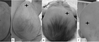
- Congenital developmental anomalies, emerging or acquired asymmetry.
- Headaches and dizziness, fainting.
- Deterioration of vision, hearing, smell.
- Trembling in fingers and limbs.
- Malfunctions in the functioning of various endocrine glands (adrenal glands, thyroid gland and others) caused by a pituitary tumor.
- Frequent ENT diseases, nosebleeds.
- Pain in the jaw joints when chewing.
Doctors can prescribe an x-ray of the skull: traumatologist-orthopedist, oncologist, ENT doctor, endocrinologist.
When is a skull x-ray prescribed?
Survey X-ray of the skull
in different settings can be prescribed to patients who are concerned about:
- cephalgia, or, in other words, headache, of varying localization and intensity;
- trembling in the limbs;
- the appearance of a veil before the eyes or darkness;
- nosebleeds;
- painful chewing of food;
- decreased visual and hearing acuity;
- cases of fainting for no apparent reason;
- the appearance of asymmetry of the facial bones.
X-rays of the head are also indicated for mechanical injuries - bruises, blows, falls from a height, and so on.
Various specialists can prescribe an X-ray examination of the skull: a neurologist, a surgeon, an oncologist, an ophthalmologist and others.
What does the procedure show?
X-ray images clearly visualize:
- cheek bones;
- lower jaw bones;
- bony pyramid of the nose;
- sphenoid bone;
- eye sockets;
- temporomandibular joints;
- mastoid processes of the temporal bones.
If a more accurate and detailed diagnosis of the condition of the bone tissues of the skull is necessary, targeted images are taken, which can show the following pathological conditions:
- formed calcifications - can provoke pathological development of the cranial bones;
- partial calcification of tumors;
- hemorrhages and hematomas;
- fluid in the paranasal sinuses;
- fractures of the skull bones.
Using the X-ray method, it is possible to identify congenital pathologies of the skull, as well as increased intracranial pressure. The latter may be indicated by so-called impressions - marks similar to fingerprints that are located on the inside of the bone tissue.
Advantages of X-ray examinations of the head at the Alfa Health Center clinic
- Gentle dose of radiation. We use a modern digital device – Brivo DR-F manufactured by General Electric, a world leader in the production of medical equipment. With the Brivo DR-F, the radiation dose from x-rays is reduced by thirty percent compared to other x-ray units. The high quality of the equipment allows you to get a clear image the first time, avoiding repeated examinations and thus reducing the impact of radiation.
- Low cost. Thanks to the constant flow of clients and the low cost of the examination, we were able to reduce the price of skull radiography. X-rays at the Alpha Health Center clinic are available to everyone.
- All inclusive. The cost of reading and describing the x-ray is included in the total cost of the x-ray. You don't need to pay extra for decryption.
- Save time. The procedure time is minimal. No need to wait for the film to be developed. The image will be printed on film and recorded on a CD within five minutes after the examination.
- Comfort, convenience, safety. Table and counter surfaces are thoroughly disinfected after each use. Each patient receives a sterile disposable wipe. The room is maintained at an optimal temperature - warm in winter and cool in summer.
Preparation for the procedure
Special preparation x-ray of the head
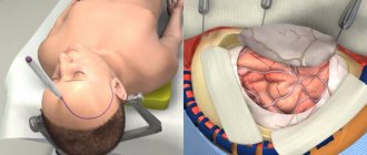
does not require. Before the procedure, the patient must remove all metal jewelry, glasses, and, if possible, dentures. If the dentures are fixed or a metal implant is installed, the radiologist must be warned about this in advance. Then, depending on the configuration of the X-ray machine, the patient takes a lying, sitting or standing position.
A lead vest or apron is placed on the patient's body to prevent exposure below the neck level. The head is secured with special clamps, since the first condition for obtaining a high-quality image is immobility.
Features of radiography of the skull in children
In order for the child not to be afraid of an incomprehensible and unfamiliar procedure, he should be explained in simple and understandable words how radiography is performed, that this process does not cause pain at all, that parents may be nearby, so there is no reason for fear, and you just need to listen to the doctor. Very young children are allowed a pacifier.
The child is seated or laid down and carefully secured so that he does not move. All metal clips, jewelry and hair accessories must be removed. The body is covered with a lead apron; a lead collar can be additionally used to protect the thyroid gland.
After x-rays, the baby should be given plenty of fluids - fruit drinks, teas, juices with pulp, milk and fermented milk drinks to neutralize the effect of the received radiation dose.
Technique
To take an X-ray, the patient must stand up, sit near the X-ray machine, or lie down on its work table. It is important to remain still and not breathe while filming. If you need to take a picture in several projections, the doctor will tell you how to change the position.
X-ray of the skull in 2 projections
To obtain the most detailed and complete information about the condition of the nasal bones, photographs can be taken in two projections - frontal and lateral. In the first case, the patient faces the X-ray machine, in the second - sideways (left or right).
Headshot during pregnancy
A pregnant woman may need an X-ray of the head bones:
- To provide dental care;
- With dislocations;
- For fractures to determine the location of bone fragments.
When deciding on the need for diagnostics, you should weigh the risks and benefits of the procedure. Conditions in which this issue is resolved already pose a danger to the health of the mother. Refusal to take an x-ray can have serious consequences - lack of medical care will lead to the development of dangerous diseases. Then there can be no question of normal childbirth. In addition, precautions effectively reflect radiation.
According to scientific research, the effect of X-ray radiation is especially unfavorable in the early stages of pregnancy. The result may be termination of pregnancy or pathology of fetal development.
How dangerous is the research?
X-ray is a non-invasive and painless procedure. It can also be called relatively safe, since the radiation exposure is minimal. At the same time, of course, x-rays are not a procedure that can be repeated many times in a row. There are certain norms and frequency that must be observed.
Contraindications for
An absolute contraindication for head x-rays is pregnancy. There are also relative restrictions. These are children under 15 years of age, mental illness, serious condition.
Contraindications for head x-rays
X-ray of the head is strictly contraindicated for pregnant women and, with a reservation, for nursing mothers (for health reasons). There are no other contraindications to head x-rays.
If you have previously had an X-ray of your head, it is advisable to have the results with you - this will help us correctly track the dynamics of the changes that have occurred. We guarantee high quality diagnostics, attentive attitude of a specialist to your problem, fast processing of results and affordable prices for x-rays. In our Center you can also find out the price for an appointment with a neurologist.
X-ray of the skull: decoding
When interpreting an X-ray of the skull in 2 projections, specialists evaluate the dimensions and features of the location of the bones, as well as the structure of the nasal sinuses. These indicators must correspond to the norm for the age category of the subject.
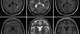
X-rays also make it possible to partially analyze the condition of the soft tissues of the brain (although for an accurate diagnosis of this organ it is better to use MRI or CT). The images can visualize tumor growths, and their location and size can be assessed. The main sign of a malignant neoplasm will be the presence of darkening of an uneven structure. If the tumor is benign, its contours will be smooth and clear.
Normal indicators
Let's figure out what a skull x-ray
in cases where there are no pathologies. When describing the images, the radiologist evaluates the size, shape, thickness and location of the skull bones, as well as the vascular system, the condition of the nasal sinuses and cranial sutures. All of the listed characteristics must correspond to the patient’s age.
X-ray of the skull for head trauma
The main questions that a specialist should answer based on an x-ray for a head injury:
- Is the integrity of the bones of the skull damaged?
- If there is a fracture, is it accompanied by the entry of bone fragments into the cranial cavity?
- Are the eye sockets, sinuses and ears damaged?
- Is there damage to the brain due to compression by the deformed bones of the skull?
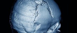
The most common injuries to the calvarium are linear fractures (cracks) of its bones. In most cases, they appear in the place where the force was applied. By the way, this fact greatly facilitates the process of identifying a fracture/crack. The fracture is visualized as a sharp strip, in some places diverging in different directions, with uneven edges. Depending on the complexity, the fracture may have a different position, direction, and size. Multiple fractures can affect one or both halves of the skull. The most unfavorable situation is when the fracture passes to the cranial suture and causes its divergence.
How does the skull work and what functions does it perform?
The cranium is part of the human skeleton. Essentially, it forms the bone frame of the head.
Content:
- How does the skull work and what functions does it perform?
- What does a skull x-ray show and why is it prescribed?
- Indications and contraindications for x-rays of the skull
- Preparation requirements, procedure for performing skull x-rays
- Types of skull radiography
- Features of radiography of the skull in children
- How are skull x-rays interpreted?
This part of the skeleton has its own characteristics, for example, the growth and development of the bones of the skull occurs before a person reaches the age of 30-32 years. In addition, as a person grows older, the proportions of the relationship between the brain and facial parts change, the cartilage located between the bones of the base of the skull disappears, and the fontanelles (non-ossified areas of the cranial vault connecting its parts) become overgrown.
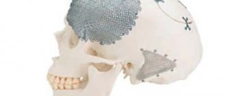
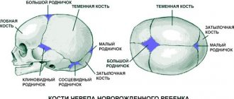
The anatomical structure of the skull includes 23 bones, two sections - the brain and the face, while the first is significantly larger in volume than the second.
In the facial part of the skull there are paired and unpaired bones: the vomer, the ethmoid and hyoid bones, the lower jaw, the inferior nasal concha, the upper jaw, the nasal, palatine, zygomatic and lacrimal bones.
The brain part of the skull is divided into a vault and a base, and is formed by the frontal, occipital, sphenoid, parietal and temporal bones. In the area of the crown there are the parietal bones and parietal tubercles - characteristic convex parts of bone tissue. The temporal bones contain pyramidal processes containing the vestibular apparatus and auditory receptors.
All the bones of the skull are connected by sutures - fixed formations of a fibrous structure. The exception is the lower jaw - it is mobile, and is connected to the main part of the skull by ligaments and paired temporomandibular joints.
What is the purpose of the skull in the human body? First of all, it is a protective box for the brain. The skull is the bony frame of the head and determines its shape. It can be argued that the protective function is the main function of this bone structure.
In the area of the skull are the original openings of the respiratory and digestive tract, as well as the human sensory organs; facial muscles are attached to his bones, which, together with the bones, determine the facial features of a person.
Thanks to the mobility of the lower jaw, a person has the ability to perform the chewing function. The bones of the skull are part of the speech apparatus, allowing communication through articulate speech, and the bones of the jaws themselves represent the base of the teeth.
The occipital bone of the brain part of the skull connects it to the spine; it provides an opening for the transition of the brain into the spinal cord.
Respiratory and speech activity, food absorption, and the work of almost all sense organs and the brain are practically impossible if the cranium cannot fully perform its functions.
Possible complications
The danger of x-rays for human health, and in particular for children, is sometimes significantly exaggerated, but it is also wrong to completely deny it. Harm is ionizing radiation that affects the human (child) body with a certain intensity. This radiation, passing through tissues, ionizes molecules, causes a temporary change in the composition of the blood, and changes the structure of proteins. Also, under the influence of radiation, cells can undergo premature aging. The main negative aspect of X-rays is that radiation can provoke malignant degeneration of cells, and therefore cause further development of oncology. However, such cases are extremely rare in medical practice, but the high diagnostic benefit of x-rays is sometimes priceless.
Contraindications for
If we consider that childhood is already a contraindication for x-rays, it is almost pointless to talk about other prohibitions. The main thing you need to know is head x-ray
child is a last resort measure that is used if other types of diagnostics turn out to be uninformative.
How is the procedure carried out?
Children undergo the examination lying or sitting. There should be no metal objects on the head. Sometimes you need to stand while taking a photo; this is discussed separately. Below the head, the child’s body is covered with personal protective equipment. Pictures are taken in frontal or lateral projection. It is important not to move during the examination. Therefore, soft restraints are used for the smallest children, and sedation is sometimes used. Such measures allow the procedure to be carried out quickly and almost without harm. Sometimes a child calms down thanks to the presence of one of the parents in the office.
Contraindications
Radiography has no significant contraindications unless alternative diagnostic techniques are found with similar information content but less radiation exposure. It is worth discussing the restrictions with the doctor if the child has recently had an x-ray.
Is the procedure harmful?
Ionizing radiation is harmful, especially for a growing organism. Therefore, it is better to do the procedure for a fee and in a clinic with modern equipment. The device used in the SM-Clinic provides a minimum radiation dose: from 0.03 to 0.1 mSv (for children, the permissible radiation exposure is 1 mSv per year). It is also important that the digital technologies used now make it possible to take a picture very quickly, that is, to influence the body with radio radiation for the shortest possible time - up to 1 second. The procedure is practically harmless if it is carried out all the time in the same medical center with the radiation doses received by the child throughout the year recorded in the outpatient card.