Double hemiplegia is the rarest form of cerebral palsy. It occurs in 1.9% of patients.
- The most common form is spastic diplegia. Most often, motor function suffers; in some children, intellectual abilities are affected.
- The hyperkinetic form has a characteristic symptom – hyperkinesis, that is, involuntary movements.
- The hemiparalytic form is characterized by damage to one hemisphere of the brain. Consequently, muscle paresis occurs on one side of the body.
- The atonic-astatic form has the following symptoms: muscle hypotonia, musculoskeletal disorders, imbalance. This is the second most complex form of cerebral palsy after double hemiplegia.
Sometimes the patient has symptoms that are inherent in several forms. In this case, a diagnosis is made – a mixed form of cerebral palsy.
Double hemiplegia is the most complex form of cerebral palsy. The disease is characterized by motor disturbances in the limbs and mental disorders. More often the arms are affected more than the legs, but there are cases of identical damage. Double hemiplegia can be diagnosed immediately after the baby is born.
The disease is caused by damage to the subcortical and cortical structures of the cerebral hemispheres. As a rule, this happens due to circulatory problems.
Why does hemiplegia occur?
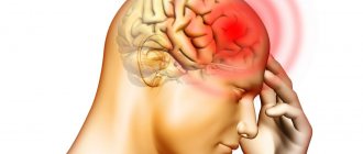
The cause of hemiplegia is either damage to the motor parts of the cerebral hemisphere opposite the paralyzed side of the body, or damage to the spinal cord on the same side where symptoms are observed. It would seem that there are few possible reasons, but, in fact, completely different processes can lead to the occurrence of these lesions. The most common of them, oddly enough, is strokes. In addition, injuries, infectious diseases and even tumors can lead to the appearance of symptoms of hemiplegia.
However, in this whole series of reasons, childhood hemiplegia stands apart, since it is usually congenital. Birth injuries can lead to it, but in about 3/4 of cases the cause is a violation of intrauterine development. In both cases, such hemiplegia in children is a form of cerebral palsy.
Differential diagnosis of alternating hemiplegia in childhood
Symptoms of the following disorders may be similar to those of AHD.
Familial hemiplegic migraine (FHM) is a rare genetic form of migraine headache. The disorder is characterized by recurrent episodes of migraine and additional symptoms. Onset usually occurs during the first or second decade of life. The frequency of these episodes can vary from one per day to less than a few in a lifetime.
The duration of the episode also varies from several hours to several days. Triggers that typically cause migraines, such as certain smells and foods, minor head injuries, and emotional situations that cause stress or anxiety, can also trigger an episode of FMS.
Additional symptoms may vary from one person to another. Those affected may also develop vision problems, sensory problems such as numbness or a tingling or burning sensation in the limbs or face, and movement problems such as temporary weakness or paralysis on one side of the body (hemiplegia). Confusion, drowsiness, and difficulty speaking (dysphasia) have also been noted. Approximately 40-50% of patients may develop irregular, involuntary eye movements (nystagmus) or an inability to coordinate voluntary movements (ataxia).
Cerebral palsy is a relatively rare (approximately 3 in 1000 live births) non-progressive neuromuscular disorder that causes movement impairment. Children with cerebral palsy may have developmental delays until the third year of life and may have abnormally low (hypotonia) or high (hypertension) muscle tone. Affected infants and children often have some combination of movement problems, including tremors, inability to coordinate voluntary movements (ataxia), unsteadiness or inability to walk, hyperreflexia, involuntary muscle spasms (spasticity) that lead to slow movements.
Additional characteristics may include difficulty chewing and swallowing, problems with bladder or bowel control, seizures, and/or poor vision and hearing. Because cerebral palsy is primarily related to motor function, intelligence may not be affected.
Cerebral palsy can be caused by damage to the brain early in fetal development, at birth or after birth. Injury may result from maternal/fetal infection, lack of oxygen (hypoxia), or bleeding into the brain.
Epilepsy is a general term for a group of neurological disorders characterized by recurrent seizures and associated with abnormal electrical discharges in the brain. Signs of an epileptic seizure may include loss of consciousness, convulsions, sensory symptoms, and disturbances in the autonomic nervous system. The attacks may be preceded by an “aura,” a feeling of restlessness or sensory discomfort; the aura marks the onset of a seizure in the brain.
There are many different causes of epilepsy, including head injuries, brain malformations, and metabolic syndromes, in which the body is unable to properly use certain energy sources, making it easier for seizures to occur; however, in many cases of epilepsy the exact cause is unknown.
Epilepsy can also occur as part of larger syndromes. Types of epilepsy or epilepsy-related disorders include Lennox-Guesteau syndrome, Rett syndrome, Angelman syndrome, Landau-Kleffner syndrome, Dravet syndrome, and neuronal ceroid lipofuscinoses.
The symptoms of many neurological disorders are similar.
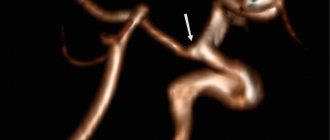
Therefore, it is important to make a differential diagnosis. A wide range of additional disorders may have signs or symptoms similar to those observed in some people with childhood hemiplegia alternating hemiplegia, including vascular disorders such as moyamoya disease and arteriovenous malformations; certain metabolic disorders such as glucose transporter deficiency syndrome type 1, aromatic l-amino acid decarboxylase (AADC) deficiency, homocystinuria, congenital disorders of glycosylation, and mitochondrial disorders such as MELAS or pyruvate dehydrogenase deficiency; and neurotransmitter deficiency disorders such as aromatic L-amino acid decarboxylase deficiency.
Types of hemiplegia
There are several ways to classify hemiplegia, each of which serves a specific purpose: classification by cause (organic and functional); by localization (cerebral hemiplegia and spinal hemiplegia); according to the relative location of the lesion and symptoms (contralateral hemiplegia or homolateral hemiplegia); on the side of the body on which paralysis is observed (right-sided hemiplegia and left-sided hemiplegia); according to symptoms (spastic hemiplegia and non-spastic hemiplegia). Let's look at each option in more detail.
Classification of hemiplegia by cause is the first thing you need to pay attention to. The fact is that in addition to organic hemiplegia - the one in which real damage to nerve cells leads to the development of muscle paralysis, there is also functional hemiplegia, which is also called alternating. And with it, symptoms can appear and disappear spontaneously, usually during times of increased nervous excitement. Depending on what type of hemiplegia was identified, treatment will differ radically.
Classification by location is used to figure out where exactly the lesion is located. As we have already written, damage to the brain - cerebral hemiplegia - leads to the appearance of symptoms of hemiplegia on the opposite side of the body, and damage to the spinal cord - spinal hemiplegia - on the same side. This is “contralateral” and “homolateral” hemiplegia. It is important to clarify that when the spinal cord is affected, the term “central hemiplegia” is sometimes used. The concepts of “right-sided hemiplegia” and “left-sided hemiplegia” speak for themselves.
The diagnosis of “spastic hemiplegia” implies that in addition to the loss of the ability to make voluntary movements, there is also noticeable overstrain in the muscles. This is what childhood hemiplegia usually is - it is cerebral hemiplegia and in most cases it occurs in patients with cerebral palsy. The non-spastic form is very rare, and, as the name implies, muscle spasms are not observed with it.
ALTERNATING SYNDROMES
Accent placement: ALTERNATING SYNDROMESALTERNATING SYNDROMES (lat. alternus - alternating, changing; synonym: alternating paralysis, cross paralysis) - symptom complexes characterized by dysfunction of the cranial nerves on the side of the lesion (paralysis or paresis) and central paralysis or paresis of the limbs or conduction sensitivity disorders on the opposite side . To A. s. refers to cross hemiplegia
(see) - paralysis of one arm and the opposite leg and cross hemianesthesia - sensitivity disorder on one side of the face and hemianesthesia on the opposite half of the torso and limbs.
Rice. 1-6. Localization of lesions in the brain stem that cause the appearance of alternating syndromes. Rice. 1. Weber syndrome. (The lesions are indicated in yellow; the nuclei of the cranial nerves are in orange; the dotted line is the course of the cranial nerves.)
Rice. 1-6. Localization of lesions in the brain stem that cause the appearance of alternating syndromes. Rice. 2. Benedict's syndrome. (The lesions are indicated in yellow; the nuclei of the cranial nerves are in orange; the dotted line is the course of the cranial nerves.)
Rice. 1-6. Localization of lesions in the brain stem that cause the appearance of alternating syndromes. Rice. 3. Foville's syndrome. Rice. (The lesions are indicated in yellow; the nuclei of the cranial nerves are in orange; the dotted line is the course of the cranial nerves.)
Rice. 1-6. Localization of lesions in the brain stem that cause the appearance of alternating syndromes. Rice. 4. Millard-Gubler syndrome. (The lesions are indicated in yellow; the nuclei of the cranial nerves are in orange; the dotted line is the course of the cranial nerves.)
Rice. 1-6. Localization of lesions in the brain stem that cause the appearance of alternating syndromes. Rice. 5. Wallenberg-Zakharchenko syndrome. (The lesions are indicated in yellow; the nuclei of the cranial nerves are in orange; the dotted line is the course of the cranial nerves.)
Rice. 1-6. Localization of lesions in the brain stem that cause the appearance of alternating syndromes. Rice. 6. Jackson syndrome. (The lesions are indicated in yellow; the nuclei of the cranial nerves are in orange; the dotted line is the course of the cranial nerves.)
A. s. are distinguished: 1) according to the topic of the lesion - cerebral peduncles, pons, medulla oblongata, extracerebral localization is also possible; 2) according to clinical syndromes - motor, sensory, combined with dysfunction of cranial nerves, hyperkinesis, etc.; 3) according to the etiology of the disease - circulatory disorders, tumors, injuries, etc.; 4) along the flow - progredient, regredient.
Localization of the lesion in the brain stem is manifested by symptoms of damage to the cranial nerves (on the side of the lesion).
Paralysis or paresis of the limbs on the side opposite to the lesion develops due to damage to the corticospinal (pyramidal) tract. Cross hemianesthesia on the trunk and limbs opposite the pathological focus occurs when sensitive pathways are damaged (median lemniscus, spinothalamic tract). Hemiplegia or hemiparesis and hemianesthesia occur on the side opposite the lesion because the pyramidal tract and sensory pathways intersect in the lower part of the brain stem.
A. s. divided according to the localization of the lesion in the brain stem: a) bulbar - with damage to the medulla oblongata; b) pontine - when the bridge is damaged; c) peduncular - with damage to the cerebral peduncle (color table, see above, Fig. 1-6).
Bulbar alternating syndromes
. Jackson syndrome, or hemiplegia alternans hypoglossica, is characterized by symptoms of damage to the hypoglossal nerve on the side of the lesion and hemiplegia or hemiparesis of the limbs on the opposite side. On the side of the lesion, symptoms of peripheral paralysis of the hypoglossal nerve are found: deviation of the tongue towards the affected side, atrophy of half the tongue, sometimes fibrillary twitching in the tongue, degeneration reaction when studying the electrical excitability of the muscles of the tongue.
Avellis syndrome is characterized by symptoms of damage to the glossopharyngeal and vagus nerves on the side of the lesion and hemiplegia or hemiparesis of the limbs on the opposite side. There is paralysis or paresis of the soft palate and vocal fold on the side of the lesion, a swallowing disorder (liquid food gets into the nose, the patient chokes when eating), dysarthria and dysphonia.
Babinski-Nageotte syndrome consists of cerebellar symptoms in the form of hemiataxia, hemiasynergia, lateropulsion (as a result of damage to the inferior cerebellar peduncle, olivo-cerebellar tract), miosis or Bernard-Gorier syndrome on the side of the lesion and hemiplegia and hemianesthesia on the opposite limbs.
Schmidt syndrome consists of symptoms of damage to the glossopharyngeal, vagus and accessory nerves. Paralysis or paresis of the vocal fold, soft palate, trapezius and sternocleidomastoid muscles on the side of the lesion, as well as hemiparesis of the limbs on the opposite side, is determined.
Wallenberg-Zakharchenko syndrome is characterized by the appearance on the side of the lesion of symptoms of damage to the vagus nerve (paralysis or paresis of the soft palate and vocal fold), the trigeminal nerve (sensitivity disorder on the face of a segmental type, Fig. 1), sympathetic fibers of the eye (Bernard-Horner syndrome), spinocerebellar tracts (hemiataxia, decreased muscle tone, asynergy of movements), and with a large lesion in the reticular formation of the medulla oblongata - respiratory disorder, cardiovascular activity on the opposite side (due to damage to the spinothalamic tract in the lateral cord of the medulla oblongata) hemianalgesia and hemitermanesthesia from level CIII or lower are detected; with damage to the pyramidal tract - hemiplegia (rare).
Rice. 1. A patient with Wallenberg-Zakharchenko syndrome (the dots indicate areas of surface sensitivity disorder)
Tapia syndrome - manifested by symptoms of damage to the accessory and hypoglossal nerves on the side of the lesion. Paralysis of the sternocleidomastoid and trapezius muscles, peripheral paralysis of the hypoglossal nerve, and hemiplegia on the opposite side are determined.
Glick syndrome is characterized by the appearance of symptoms of damage to the facial nerve - peripheral paralysis of the facial muscles with their spasm (Fig. 2); vagus nerve with difficulty swallowing; the orbital branch of the trigeminal nerve (pain in the supraorbital region) and the optic nerve (amaurosis or decreased vision) on the side of the lesion, and on the opposite side hemiplegia develops as a result of damage to the pyramidal tract.
Rice. 2. Glick syndrome (spasm of facial muscles on the right and hemiplegia on the left)
Volestein syndrome manifests itself as transient paralysis of the vocal fold, and on the opposite side - hemianesthesia.
Pontine alternating syndromes
. Millard-Hubler syndrome, or hemiplegia alternans facialis, is manifested by peripheral paralysis of the facial nerve on the side of the lesion (Fig. 3) and spastic hemiplegia on the opposite side.
Rice. 3. Facial nerve paralysis and spastic hemiplegia in Millard-Hübler syndrome
Brissot-Sicard syndrome is characterized by spasm of the facial muscles (irritation of the cells of the nucleus of the facial nerve) on the side of the lesion and spastic hemiparesis or hemiplegia of the limbs on the opposite side.
Foville syndrome - abducent-facial alternating hemiplegia - is expressed by damage to the facial (peripheral paralysis of facial muscles on the same side) and abducens (convergent strabismus) nerves in combination with gaze paralysis on the side of the pathological focus (Fig. 4) and hemiplegia, and sometimes hemianesthesia ( lesion of the median loop) of the limbs on the opposite side.
Rice. 4. Abducentofacial paralysis on the left with Foville syndrome
Raymond-Sestan syndrome is manifested by paralysis or paresis of combined movements of the eyeballs towards the lesion, ataxia and choreoathetoid movements, hemianesthesia (impaired sensitivity according to the hemitype) and hemiparesis on the opposite side (Fig. 5).
Rice. 5. Combined eye movement palsy and choreoathetoid movements in Raymond-Sestan syndrome
Peduncular alternating syndromes
. Weber's syndrome is characterized by paralysis of the oculomotor nerve on the side of the lesion (ptosis, divergent strabismus, mydriasis) and hemiplegia with paresis of the muscles of the tongue and face of the central type (lesion of the corticonuclear pathway) on the opposite side. The syndrome develops due to pathological processes at the base of the cerebral peduncle.
Benedict's syndrome consists of paralysis of the oculomotor nerve on the side of the lesion and choreoathetosis and intention tremor of the opposite limbs (damage to the red nucleus and cerebellar-red nuclear tract).
Nothnagel syndrome includes a triad of symptoms: cerebellar ataxia, oculomotor nerve palsy, hearing impairment (unilateral or bilateral deafness of central origin). Sometimes hyperkinesis (choreiform or athetoid), paresis or paralysis of the limbs, central paralysis of the facial and hypoglossal nerves can be observed.
Claude's syndrome is characterized by paralysis of the oculomotor nerve on the side of the lesion and cerebellar phenomena (impaired coordination and conjugal movements), as well as decreased muscle tone on the opposite side. Sometimes dysarthria and swallowing disorders are observed.
Foix syndrome consists of cerebellar symptoms, intention tremor, choreoathetoid movements, sensitivity disorders and changes in visual fields on the side opposite the lesion.
AS, characteristic of the intrastem process, can also occur when the brain stem is compressed. Thus, Weber syndrome develops not only in pathological processes (hemorrhage, intrastem tumor) in the midbrain (Fig. 6, 1
), but also when the cerebral peduncle is compressed (Fig. 6,
2
). Compression dislocation syndrome of compression of the cerebral peduncle, which occurs in the presence of a tumor of the temporal lobe or pituitary gland, can manifest itself as damage to the oculomotor nerve (mydriasis, ptosis, strabismus, etc.) on the side of the compression and hemiplegia on the opposite side.
Sometimes A. s. are manifested mainly by cross-sensitivity disorder (Fig. 7). Thus, Raymond-Sestan syndrome is manifested by paralysis of gaze towards the lesion and an alternating nature of sensory impairment. On the face, sensitivity is impaired in a segmental manner due to damage to the sensitive nucleus of the trigeminal nerve on the side of the lesion, and on the trunk and limbs - on the opposite side (damage to the median lemniscus and spinothalamic tract).
Rice. 6. Pathological changes in the brain with Weber syndrome: 1 - hemorrhage in the cerebral peduncle
Rice. 6. Pathological changes in the brain in Weber syndrome: 2 - compression of the midbrain by an extensive hematoma
Extracerebral alternating syndromes
. Optical-hemiplogical syndrome [Radovici and Lasco (A. Radovici, F. Lasco, 1948)] is manifested by dysfunction of the optic nerve (blindness) and spastic hemiplegia on the opposite side. The syndrome is pathognomonic for thrombosis of the internal carotid artery and its branches. In this case, due to circulatory disorders in the ophthalmic artery, visual impairment occurs, in the middle cerebral artery - hemiplegia or hemiparesis.
Vertigo-hemiplegic syndrome with dyscirculation in the subclavian artery system is characterized by dizziness and noise in the ear as a result of circulatory disorders in the internal auditory artery (a branch of the anterior inferior cerebellar artery) on the side of the lesion and hemiplegia and hemiparesis on the opposite side (due to circulatory disorders in the branches of the carotid artery) .
Rice. 7. Hemianesthesia scheme: 1 - complete cross hemianesthesia with softening in the area of the left posterior inferior cerebellar artery; 2 - hemianesthesia with a dissociated disorder of pain and temperature sensitivity (syringomyelic type with a limited focus of softening of the medulla oblongata behind the olive). Areas with disorders of pain and temperature sensitivity are shaded
Asphygmo-hemiplegic syndrome occurs when the common carotid artery or brachiocephalic trunk is blocked. In this case, on the affected side there is no pulsation in these vessels and their branches, and on the opposite side there is hemiplegia or hemiparesis.
Topical diagnostic value
. Study of signs of damage to cranial nerves and other focal symptoms in A. s. allows you to determine localization and boundaries; focus, i.e. establish a topical diagnosis. Thus, Jackson syndrome occurs with thrombosis of the anterior spinal artery or its branches; Avellis syndrome - with damage to the branches of the artery of the lateral fossa of the medulla oblongata; Babinsky-Nageotte syndrome - the artery of the lateral fossa or the inferior posterior cerebellar artery, as well as Wallenberg-Zakharchenko syndrome, which develops with damage to the vertebral arteries (Fig. 8. 1-3) (with a large lesion in the dorsolateral medulla oblongata); Foville syndrome - with thrombosis of the basilar artery; cross hemiplegia syndrome - with a focus in the area of the intersection of the pyramidal tracts due to thrombosis of the spinobulbar arterioles. Claude and Foix syndromes develop when the anterior and posterior arterioles of the red nucleus (branches of the posterior cerebral artery) are damaged; Claude's syndrome - with damage to the oculomotor nerve and the posterior part of the red nucleus (inferior red nucleus syndrome), and Foix's syndrome - with an isolated lesion of the anterior part of the red nucleus (superior red nucleus syndrome) without involvement of the oculomotor nerve. Benedict's syndrome occurs when the lesion is localized in the medial dorsal part of the midbrain (the pyramidal tract remains unaffected) - damage to the interpeduncular or central branches. Nothnagel syndrome is caused by damage to the tegmentum of the midbrain - circulatory disturbance in the circumferential and retromamillary arteries; Raymond-Sestan syndrome - with damage to the bridge tire.
Rice. 8. Pathological changes in the system of vertebral arteries: 1 - tortuosity of the inferior posterior cerebellar artery
Rice. 8. Pathological changes in the system of vertebral arteries: 2 - atherosclerosis of the vertebral and basilar arteries with uneven caliber and thrombosis of the right vertebral artery
Rice. 8. Pathological changes in the system of vertebral arteries: 3 - thrombosis of the left posterior inferior cerebellar artery
Studying the dynamics of symptoms allows us to determine the nature of the pathological process. Thus, with ischemic softening of the brain stem as a result of thrombosis of the branches of the vertebral arteries, the main or posterior cerebellar artery (BC). A.s. develop gradually, not accompanied by loss of consciousness, and the boundaries of the lesion correspond to the zone of impaired vascularization. Hemiplegia or hemiparesis is usually spastic In case of hemorrhage into the trunk of the apex, the site may be atypical, since the boundaries of the lesion do not correspond to the vascularization zone and increase due to edema and reactive phenomena in the hemorrhage circumference. The
main diagnostic signs of some alternating syndromes
| Avellisa | Paresis or paralysis of the soft palate and vocal fold | Hemiplegia or hemiparesis | Medulla oblongata (lateral-basal section): nuclei and roots of the glossopharyngeal and vagus nerves, pyramid | Circulatory disorders in the vertebral artery system (artery of the lateral fossa of the medulla oblongata) or cervical tumor |
| Babinsky-Najotta | Hemiataxia, hemiasynergia, lateropulsion, Bernard-Horner syndrome | Hemiplegia (hemiparesis) and hemianesthesia | Medulla oblongata (dorsolateral section): inferior cerebellar peduncle and olivocerebellar fibers, pyramid | Circulatory disorders in the vertebral artery system (artery of the lateral fossa of the medulla oblongata or inferior posterior cerebellar artery) |
| Wallenberg-Zakharchenko | Paresis or paralysis of the soft palate and vocal fold, impaired superficial sensitivity of the segmental type on the face, Bernard-Horner syndrome, hemiataxia | Impairment of surface sensitivity according to hemitype from level CII and below, rarely hemiparesis | Medulla oblongata (dorsolateral section): nuclei of the glossopharyngeal and vagus nerves, spinothalamic tract, oculosympathetic fibers, trigeminal nerve root, rope body, rarely pyramid | Circulatory disorders in the vertebral artery system (inferior posterior cerebellar artery) |
| Voleshteina | Transient paresis or paralysis of the vocal fold | Hemianesthesia | Medulla oblongata (oral-lateral section): oral section n. ambiguus, spinothalamic tract | Circulatory disorders in the vertebral artery system |
| Jackson | Peripheral paresis or hypoglossal nerve palsy | Hemiplegia or hemiparesis | Medulla oblongata (midsection): nucleus or root of the hypoglossal nerve, pyramid | Circulatory disturbance in the vertebral artery system or intrastem tumor |
| Tapia | Paresis or paralysis of the sternocleidomastoid muscles, peripheral paralysis of the hypoglossal nerve | Hemiplegia or hemiparesis | Medulla oblongata (dorsolateral section): nuclei or roots of the accessory and hypoglossal nerves, pyramid | Circulatory disorders in the vertebral artery system |
| Schmidt | Paresis or paralysis of the soft palate. vocal fold, atrophy and paresis of the sternocleidomastoid and trapezius muscles | Same | Medulla oblongata (caudal-lateral section): nuclei and roots of the glossopharyngeal, vagus and accessory nerves, pyramid | Circulatory disorders in the vertebral artery system |
| Pontine syndromes: | ||||
| Brissot-Sicara | Spasm of facial muscles | » | The pons of the brain: irritation of the nucleus of the facial nerve, pyramidal tract | Poor circulation in the main artery or its branches |
| Millard-Hübler | Peripheral paresis or facial paralysis | » | The pons of the brain: nucleus or root of the facial nerve, pyramidal tract | Circulatory disturbance in the main artery or its branches or a space-occupying process |
| Ramona-Sestan | Paresis or paralysis of combined eye movements, ataxia, choreoathetoid movements | Hemiplegia or hemiparesis and hemianesthesia | The pons of the brain: posterior longitudinal fasciculus, middle cerebellar peduncle, median lemniscus, pyramidal tract | Poor circulation in the main artery or its branches |
| Fauville | Peripheral paresis or facial paralysis, divergent strabismus | Same | The pons of the brain: nuclei or roots of the facial and abducens nerves, median lemniscus, pyramidal tract | Poor circulation in the main artery or its branches |
| Peduncular syndromes: | ||||
| Benedicta | Ptosis, strabismus, mydriasis | Choreoathetosis, intention tremor | Cerebral peduncle (mediodorsal division): red nucleus, dentato-rubral tract, oculomotor nerve nuclei | Circulatory disorders in the main artery or its branches (interpeduncular or central) |
| Weber | Ptosis, strabismus, mydriasis | Hemiplegia or hemiparesis and hemiaesthesia | Cerebral peduncle (base): oculomotor nerve roots, pyramidal tract | Poor circulation in the basilar artery or a space-occupying process in the temporal lobe of the brain, inflammation of the meninges at the base of the brain or hemorrhage under the membranes in the middle cranial fossa |
| Claude | Same | Ataxia, decreased muscle tone | Cerebral peduncle (tegmentum): nuclei of the oculomotor nerve, superior cerebellar peduncle | Circulatory disorders in the branches of the posterior cerebral artery (anterior and posterior arterioles of the red nucleus) |
| Nothnagel | Cerebellar ataxia, ptosis, strabismus, mydriasis, deafness (unilateral or bilateral) | Choreiform or athetoid movements, hemiparesis, central paresis of the facial and hypoglossal nerves | Cerebral peduncle (tegmentum): oculomotor nerve nuclei, superior cerebellar peduncle, lateral lemniscus, pyramidal and corticonuclear tract | Circulatory disorders in the main artery or its branches (circumferential or retromamillary) |
| Extracerebral syndromes: | ||||
| Vertigo-hemiplegic | Noise in the ear, dizziness | Hemiplegia or hemiparesis | Brain stem: vestibular apparatus, cerebral hemisphere, motor analyzer | Poor circulation in the branches of the aortic arch (carotid and subclavian) with discirculation in the auditory and middle cerebral arteries |
| Optical-hemiplegic | Blindness | Same | Eye: retina and optic nerve, cerebral hemisphere, motor analyzer | Circulatory disorders in the internal carotid artery system (ophthalmic and middle cerebral) |
In case of acute outbreaks in the bridge of A. s. usually combined with respiratory distress, vomiting, disruption of the heart and vascular tone, hemiplegia - with muscle hypotension as a result of diaschisis
(cm.).
A. s. may also occur with damage to the spinal cord (see Brown-Séquard syndrome
).
Identification of symptoms of A. s. helps the clinician in making a differential diagnosis (see table). In cases of A. caused by damage to the great vessels, surgical treatment (thrombinthymectomy, vascular plastic surgery, etc.) is indicated.
See also Paralysis, paresis
.
Bibliography
.: Bogolepov N.K. Impaired motor functions in vascular lesions of the brain, M., 1953, bibliogr.; aka, Comatose States, M., 1962; aka, Some neurological syndromes, Multivolume. Guide to neurol., ed. S. N. Davidenkova, vol. 2, p. 308, Zh., 1962, bibliogr.; Zakharchenko M. A. Vascular diseases of the brain stem, Tashkent, 1930; Avellis G. Klinische Bciträge zur halbseitigen Kehlkopflähmung, Berl. Klin., Hft 40, S. 1, 1891; Babinski et Nageotte, Syndrome caractérisé par une hémiplégie, une hémianesthésie du côté opposé à la lésion, une hémiasynergie, une latéropulsion, une myosis homonyme, Ann. Oculist. (Paris), no. 127, p. 380, 1902; Brissaud E. Lecons sur les maladies nerveuses, p. 385, P., 1899; Crouzon O. Troubles de la motilite, hemmlegie, in the book: Prat, neurol., publ. par P. Marie, p. 462, P., 1911; D éj érine J. Semiologie des affections du systeme nerveux, p. 214, P., 1926; Radovici A. et Lasso F. Une hemiplegie alterne inconnue, l'éhemiplegie alterne optico-pyramidale par trombose carotidienne, Presse m éd., t. 56, p. 573, 1948; Schmidt A. Casuistische Betrage zur Nervenpathologie, Dtsch. med. Wschr., S. 606, 1892; Wallenberg A. Neuere Fortschritte in der topischen Diagnostic des Pons und der Oblongata, Z. Nervenheilk., Bd 41, S. 8, 1911; Weber N. A contribution to the pathology of the crura cerebri, Med.-chir. Trans., v. 46, p. 121, 1863.
N. K. Bogolepov
Sources:
- Big medical encyclopedia. Volume 1/Editor-in-Chief Academician B.V. Petrovsky; publishing house "Soviet Encyclopedia"; Moscow, 1974.- 576 p.
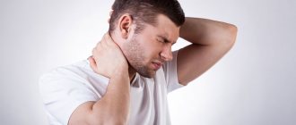
Manifestations of hemiplegia
In any case, the main manifestation of hemiplegia is the complete loss of voluntary movements on one side of the body. This is what distinguishes hemiplegia from hemiparesis - with it, movements may be difficult or weakened, but the very ability to perform them remains. In addition, along with disturbances of voluntary movements and muscle spasms, other symptoms are present - an increase in healthy reflexes and the appearance of pathological ones, for example. You can often notice a change in the color and temperature of the skin on the paralyzed side, as well as swelling. Sometimes with hemiplegia there are also accompanying symptoms, such as epilepsy. All this data together helps doctors establish the most accurate diagnosis and select the optimal treatment regimen for hemiplegia.
Diagnosis of alternating hemiplegia in childhood
The diagnosis of alternating hemiplegia of childhood is based on the identification of characteristic symptoms, a detailed medical history of the patient, a thorough clinical assessment and a variety of specialized tests. Let us consider the specific diagnostic criteria proposed for alternating hemiplegia of childhood. There are seven criteria:
- onset of symptoms before 18 months;
- repeated episodes of hemiplegia, sometimes affecting both sides of the body;
- quadriplegia, occurring alone or as part of a hemiplegic attack;
- relief of symptoms during sleep;
- additional paroxysmal episodes such as dystonia, abnormal eye movements, or autonomic dysfunction;
- evidence of developmental delay or neurological abnormalities such as choreoathetosis, ataxia or cognitive impairment;
- symptoms cannot be attributed to another cause.
Methods for treating hemiplegia
Treatment of hemiplegia always relies on three pillars: drug therapy, surgical treatment and rehabilitation. Each of them complements the other two and they all work together for a common result, so they should be approached with equal attention.
Medicines help reduce the level of spasm, which makes life and care for patients easier, and most importantly, gives scope for rehabilitation. In addition, there are attempts to use nootropic drugs to treat the cause of hemiplegia - brain damage, but it is important to note that these are methods without proven effectiveness.
Surgical interventions also serve the purpose of reducing the level of spasticity and provide more opportunities for subsequent rehabilitation - staged fibrotomy according to the V.B. method. Ulzibata, for example, will free chronically spasming muscles from contractures, which in such a situation inevitably arise and in themselves further limit movement. Without this, further rehabilitation for hemiplegia would be much less effective and probably much more painful. The latter in itself reduces the effectiveness of rehabilitation procedures.
After this, rehabilitation comes into play - there are many ways and techniques, and many of them can really make the lives of patients with hemiplegia much easier. These include physiotherapy, for example electrical muscle stimulation, which helps reduce spasticity, and various massage techniques, and physical therapy, including the use of special equipment.
One of the most important rehabilitation methods is to involve the patient in normal daily activities - this helps not only to reduce the symptoms of hemiplegia, but also teaches new skills and helps socialization.
Among the relatively new rehabilitation techniques is “mirror therapy,” which actually uses mirrors to help the patient concentrate on one side of the body. According to studies from 2018-2019, mirror therapy shows significantly better results than conventional rehabilitation, although the specific mechanisms of its action are not yet fully understood.
There is one more aspect that is not directly related to the treatment of hemiplegia, but is very important for maximizing the results of therapy. This is Wednesday. A barrier-free environment is important for both children and adult patients with hemiplegia, and inclusive education helps children socialize better and quickly learn new skills.
Treatment of double hemiplegia
As with other forms of cerebral palsy, the earlier treatment is started, the more favorable the prognosis will be. Therapy is selected individually for each patient, since the cause of the disease and age matter. Therefore, self-medication will not bring any results.
Doctors use the following methods:
- vitamin therapy;
- medications aimed at restoring motor activity;
- neuroprotectors;
- antioxidants;
- massage;
- exercise therapy;
- electrophoresis.
The metameric method of treatment has proven its effectiveness. This is a targeted effect on the affected areas, which restores trophic, sensory and motor functions. The essence of the method is to administer small medicinal doses by injection. The needle, entering under the skin, activates reflexes, and the drug enhances the effect. The drugs used consist of nerve cells and amino acids.
Prognosis for hemiplegia
With functional hemiplegia, the prognosis is as favorable as possible - such conditions usually disappear completely and without a trace.
With organic hemiplegia, everything is somewhat more complicated. On the one hand, given that hemiplegia does not progress on its own unless caused by a tumor, worsening of symptoms over the course of life cannot be expected. On the other hand, it is important to remember that both cerebral hemiplegia and central hemiplegia lead to a decrease in muscle activity on one side of the body, and because of this, additional symptoms may develop - deterioration in joint mobility, the appearance of bedsores, or the formation of blood clots.
Unfortunately, at the current stage of development of medicine, one cannot expect a complete and complete disappearance of the symptoms of organic hemiplegia. But, despite this, with timely and comprehensive treatment in patients with hemiplegia in general and specifically in cases of cerebral palsy in the form of spastic hemiplegia, very noticeable improvements are possible, both in terms of symptoms and in terms of quality of life and socialization.
We hope that now for you the expressions “Central non-spastic right-sided hemiplegia” or “Cerebral spastic left-sided hemiplegia” will not sound like a Chinese letter or a sentence!
Return to list
Consequences of double hemiplegia
Most children with this diagnosis cannot be taught. The reason is not only low intellectual abilities, but also a lack of sense of motivation. Much depends on the environment that surrounds the child and the efforts made. For example, despite severe musculoskeletal disorders, a patient suffering from double hemiplegia can learn to sit. Mental abnormalities do not allow such patients to fully adapt to society, but they can become partially socialized.
Forms of cerebral palsy and their clinical manifestations
In the first days and months of life, a child with cerebral palsy may not differ from his peers, and symptoms of the disease appear later. Their severity depends on the degree of brain damage, as well as the timeliness of diagnosis and treatment. The clinical picture of cerebral palsy may include the following disorders:
- increase or decrease in the tone of certain muscles;
- skeletal deformity;
- long-term preservation of reflexes, which normally disappear between the ages of 6 and 8 months;
- impaired reflexes, including swallowing;
- mental retardation, hearing and vision problems;
- convulsive syndrome.
Doctors at the Clinical Brain Institute argue that any changes in a child’s behavior should be a reason for additional examination. Currently, to determine an accurate diagnosis, a generally accepted classification is used, which identifies 5 main forms of cerebral palsy.
Spastic diplegia
This form of the disease is the most common and is recorded in more than half of patients with cerebral palsy. Its clinical signs can be clearly seen by the end of the first year of life. It was previously called Little's disease, named after the scientist who first began work on research into cerebral palsy. Most often, this spastic diplegia develops in premature babies and is manifested by a typical set of symptoms:
- paresis of the limbs, more pronounced in the legs;
- hypertonicity of the flexors and extensors, due to which the limbs are constantly in an unnatural forced position;
- foot deformities that increase as the child grows;
- difficulty concentrating, hearing and vision impairment, decreased mental development.
It is worth understanding that not every patient develops the symptom complex to its fullest extent. Experts say that timely and competent rehabilitation will allow significant work to be done on the child’s social adaptation. In addition, with spastic diplegia, convulsive syndrome is observed much less often than with other forms.
Double hemiplegia (spastic tetraplegia)
This form is considered the most severe, but it occurs in no more than 2.5% of patients. The disease develops as a result of prolonged hypoxia of the brain during fetal development, as well as due to severe damage to neurons due to infectious pathologies and poisoning. The term “tetraplegia” means a uniform impairment of the motor function of all limbs. If the symptom manifests itself when the syndrome is more severe on the hands, the form “double hemiplegia” is distinguished. The disease is detected already in the first month of a child’s life. It is characterized by the following symptoms:
- paresis of the upper and lower extremities, muscle hypertonicity, unnatural position of the extremities (arms are bent at the elbows and pressed to the body, legs can be bent at the joints or straightened);
- hearing and vision impairment, pseudobulbar disorders;
- cognitive disorders, difficulties with adaptation and self-care;
- secondary microcephaly is a decrease in brain volume.
The prognosis depends on the frequency and severity of seizures. In this form, they occur in most cases, and some patients experience epileptic seizures. Adaptation of such patients is difficult, but they also require long-term rehabilitation to improve their quality of life.
Hemiplegic form
The hemiplegic form is registered in more than 32% of patients with cerebral palsy. It manifests itself immediately after birth and is characterized by unilateral paresis of the limbs, most often the upper ones. Its causes can be ischemic infarction, hemorrhage in brain tissue, congenital abnormalities of the central nervous system. As the child grows up, he or she develops gait disturbances and foot deformities may occur. In addition, the hemiplegic form often develops with decreased vision and hearing, concentration and speech. The possibility of social adaptation is determined, first of all, by the level of the child’s intellectual abilities. This form is also characterized by individual epileptic seizures of varying severity.
Hyperkinetic (dyskinetic) form
Most often, the hyperkinetic form develops as a consequence of hemolytic jaundice, a disease of newborns. It is diagnosed in no more than 10% of patients and has a good prognosis. The clinical picture includes involuntary muscle contractions and various speech disorders. It may consist of a number of symptoms:
- involuntary contractions of the muscles of the limbs, facial and neck muscles - these signs are especially pronounced in a stressful environment;
- delayed speech development, speech articulation disorders;
- convulsions are a very rare occurrence in the dyskinetic form.
The patient's intellectual abilities most often remain unchanged. Competent and timely adaptation allows such children to attend regular educational institutions and continue to develop in various areas.
Atonic-astatic form
The ataxic form of cerebral palsy is manifested by decreased muscle tone. Its causes may be abnormalities in the development of the nervous system, cerebral ischemia, or birth trauma. Such patients have difficulties with motor activity - children begin to hold their heads, sit and crawl later than their peers. In addition, hearing and vision disturbances may occur. In most cases, the child’s intellectual abilities remain unchanged, which significantly increases the chances of adaptation and socialization.
Mixed forms
In most cases, a specific form of cerebral palsy can be identified. However, there remains the possibility of simultaneous damage to all brain systems: cerebellar, pyramidal and extrapyramidal. In this case, there is a combination of several symptom complexes of cerebral palsy. It is possible to develop simultaneously spastic and hyperkinetic, as well as spastic and hemiplegic forms.