Microcephaly is a congenital anomaly, which is characterized by a significant reduction in the size of the brain (and, as a result, the skull) as a result of disruption of the growth processes of brain tissue during the period of intrauterine development of the baby.
It can be an independent defect or a manifestation of any other genetic diseases (in this case we are talking about true microcephaly), and it can also develop as a consequence of various diseases the mother suffered during pregnancy (secondary, or false, microcephaly). However, in any case, this pathology is accompanied by a lag in the child’s mental development of varying degrees of severity and the presence of other neurological symptoms.
Statistical information
The incidence of the defect ranges from approximately 1:2000 to 1:10000 among all newborns. As an independent disease, true microcephaly is much less common and is detected in one out of 50 thousand children, both girls and boys. If this pathology occurs as a manifestation of another hereditary disease, then its frequency in the population is determined by the frequency of the underlying disease.
Prevention of microcephalic syndrome
There are several prevention methods that, if followed, can significantly reduce the risk of such a terrible disease as microcephaly in children. These include:
- A competent approach to pregnancy planning;
- Passing all necessary tests;
- Ultrasound examination of the fetus;
- Compliance with safety measures during pregnancy and childbirth.
If the disease has been identified, early termination of pregnancy may be recommended. Today, all newborns and their parents undergo genetic testing.
This allows you to do everything possible to prevent the potential risk of microcephaly in the future.
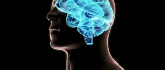
Anatomy of the brain
The brain is the main organ of the human nervous system; its formation and development begin in the earliest stages of the embryonic period.
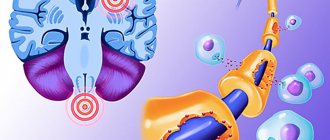
The first rudiments of nervous tissue are formed already on the 18th day of intrauterine development. They are a neural plate, which consists of a small number of neurons (nerve cells). As the embryo forms, their number increases, as a result of which the brain and spinal cord and other elements of the system are formed from the neural plate.
A neuron is a highly specialized cell that consists of a body and many processes through which it communicates with a huge number of other cells. Thanks to this structure, the main function of the nervous system is ensured, namely the reception and processing of information, including the regulation of the vital functions of the entire organism as a whole. Through some neurons (they are called afferent), information in the form of nerve impulses enters the brain, where it is processed and a response nerve impulse is formed, which is sent to organs and tissues through other neurons (they are called efferent).
The formation of nerve cells in the brain is completely completed only at the time of birth, on average reaching about 150 billion neurons. However, throughout a person’s life they are not able to reproduce (divide), and at the same time they gradually die due to so-called apoptosis (a genetically programmed process of death of malfunctioning or damaged cells).
The brain is anatomically divided into three sections.
- Large hemispheres of the brain (right and left). Being the higher parts of the brain, they consist of the cortex and subcortical structures (hypothalamus, thalamus, etc.). The cerebral cortex contains neurons that are responsible for the formation of personality, the processes of self-knowledge and learning (higher nervous activity of a person). Subcortical structures ensure the formation of instinctive and behavioral reactions (sexual, food, etc.). It is also worth noting that the cerebral cortex under normal conditions has an inhibitory effect on subcortical structures; in other words, a person’s consciousness can suppress his instinctive reactions.
- Brain stem. It includes the medulla oblongata, the pons, and the midbrain and diencephalon. They provide reflex activity of the human body, and also maintain communication between the brain and spinal cord. In addition, the trunk contains various nerve centers (for example, vasomotor, respiratory, etc.). They, in turn, ensure the performance of those physiological functions that are not controlled by consciousness (for example, a person breathes automatically, without thinking about it).
- The cerebellum is a small structural formation that is responsible for coordinating human movements, maintaining muscle tone and maintaining balance during complex movements (walking, running, etc.).
The human skull is divided into the brain and facial sections. The latter includes the bones of the face. The development of the skull comes from embryonic tissue called ectoderm. It surrounds the rudiments of the brain from the very beginning of their formation.
There are three stages in the formation of the skull bones:
- membranous - a layer of cells that surrounds the future brain, becomes soft membranous tissue, simultaneously increasing with the growth of brain tissue;
- cartilaginous - from about the sixth to seventh week of embryonic development, this tissue in the area of the base of the skull gradually turns into denser cartilaginous tissue;
- bone - already from the eighth to tenth week of the intrauterine period, ossification points are formed in various parts of the skull, which are zones of the beginning of the growth of bone tissue.
By the time of birth, some areas of membranous tissue (fontanelles) remain in the roof of the skull of a newborn child, which ensure bone mobility during childbirth or when the brain enlarges for any reason.
The large (anterior) fontanelle has a diamond shape and is located at the junction of the frontal and parietal bones. It completely ossifies and closes by the age of two years.
The small (posterior) fontanel has the shape of a triangle and is located in the occipital region at the junction of the parietal and occipital bones. As a rule, it becomes overgrown by the second month of life.
The lateral fontanels are paired and small in size, they are located on the side surface of the head, slightly in front and behind the ears, and are also completely overgrown by the second month of life.
The bones of the facial skull are formed from the so-called gill arches, which are derivatives of mesoderm (another embryonic tissue). Thus, the development of the brain and facial parts of the skull occurs independently of each other. In addition, the formation of the facial skull is practically independent of the development and growth of the brain; however, the size of the cerebral skull normally always corresponds to the volume of the cerebral hemispheres.
At the end of the 19th and beginning of the 20th centuries in Europe and North America, children with microcephaly were sold to the circus. There they took part in the so-called “freak show”.
The difference between baby teeth and molars
A child’s milk teeth differ from permanent teeth in their structure, shape and color. We strongly recommend that you look at numerous photos in which you can easily tell the difference. In addition, children's teeth have a thin layer of enamel, a small crown and dentin. In the photographs you can easily notice that such teeth have widely spaced roots that are located at an angle. This feature is explained by the need for the formation of permanent teeth. An X-ray of a child's jaw with baby teeth will help determine how much the new molars have formed. If you pull out children's teeth before the molars are formed, you are likely to encounter serious deficiencies. This usually causes a cosmetic defect in the form of a diastema, or a large gap between the teeth.
Babies grow 20 milk teeth, which are gradually replaced by molars (for more details, see the article: when do children change their milk teeth to molars?). Their rudiments are formed in the jaw at the stage of embryonic development of the child, so the skull of a newborn has a special structure. The chin is weakly expressed, the brain part is larger than the facial part. The lower jaw is located slightly further in relation to the upper. As temporary teeth appear, its position changes.
The formation of baby teeth begins at 5 weeks of pregnancy. From the 20th, the rudiments of permanent incisors and canines are formed. Premolars are formed after birth, and molars are formed closer to the year, usually at 10 months of life. The growth of the roots of permanent and temporary teeth lasts about 2 years, and begins only before their eruption.
Often, crooked molars grow in place of non-permanent teeth pulled out prematurely (see also: how molars grow in children: photo). They occupy an incorrect position in the dentition, which is not only a cosmetic defect, but also leads to dental problems. That is why, when the first signs of carious lesions are detected, doctors rush to fill temporary teeth.
The change in the skull with baby teeth takes place in 2 stages. Up to 3 years of age, the teeth are tightly packed, the enamel is not erased, and the lower jaw occupies a neutral position. The second period ends by age 7. It is characterized by the appearance of interdental spaces, changes in bite, and abrasion of the enamel.
The buds of permanent teeth put pressure on the roots of temporary teeth, which leads to their resorption (loss). When permanent teeth emerge, they have a size and shape that will last a lifetime. Eruption pathologies are often associated with inflammatory processes at the roots of temporary teeth. This can be seen on x-rays and corrected early.
Reasons for the development of the anomaly
Taking into account the causes and time of occurrence, primary microcephaly (true or hereditary) and secondary (syndromic) are distinguished.
Primary microcephaly in children is a manifestation of hereditary diseases; it accounts for about 7–34% of all types of this anomaly.
Primary microcephaly
The following diseases are independent forms.
Giacomini syndrome is a hereditary microcephaly, which is predominantly manifested by mental disorders in combination with the occurrence of convulsive conditions, as well as the progression of paralysis.
Payne's syndrome - this disease is also hereditary. It affects only boys and is manifested by constant convulsive conditions of the lower extremities, nervous disorders and abnormalities in the functioning of the heart.
Microcephaly can be a manifestation of the following pathologies:
- Down syndrome - the appearance of an extra chromosome in the XXI pair, clinically manifested by impaired mental development of the child, a decrease in the size of the brain and cerebellum, as well as other characteristic symptoms;
- Patau syndrome - the appearance of an extra chromosome in the XIII pair, characterized by low birth weight of the newborn, microcephaly and other anomalies of the skull and internal organs;
- Edwards syndrome - the appearance of an extra chromosome in the XVIII pair, while children have a small or long and narrow skull, low birth weight, underdeveloped brain and cerebellum, as well as developmental defects of the facial skeleton, legs (mainly feet) and internal organs;
- cat cry syndrome is a chromosome defect in the V pair, which is manifested by microcephaly, low body weight, retardation in physical and mental development, as well as a characteristic cry in a child, similar to the meowing of a cat (due to damage to the cartilage of the larynx);
- Prader-Willi syndrome - damage to chromosome XV, characterized by microcephaly, delayed psychomotor development and various anomalies of internal organs and skeletal bones;
- Miller-Dieker syndrome is a defect in the XVII chromosome, leading to disruption of the development of brain neurons in the embryo, as a result of which the brain of a newborn child is reduced and devoid of convolutions, in addition, there is severe muscle weakness, delayed physical and mental development and defects of internal organs;
- phenylketonuria is a hereditary disease characterized by impaired processing and use of one of the amino acids - phenylalanine; the resulting toxic substances have a damaging effect on the central nervous system of the pregnant woman, and also inhibit the development and growth of the fetal brain.
Secondary microcephaly develops due to the influence of unfavorable factors on the fetus in the embryonic period, when the formation and formation of all brain structures occurs. In this case, the genetic apparatus of the parents is not damaged.
Causes of secondary microcephaly
- viruses and infections (rubella, cytomegalovirus, measles, toxoplasmosis);
- alcohol abuse - ethanol almost freely penetrates from the mother’s blood through the placenta into the fetus’s body, the most severe manifestation is the so-called fetal alcohol syndrome, manifested by mental retardation and defects in the development of internal organs;
- drug abuse - they can also easily penetrate the placental barrier and adversely affect the fetus;
- taking certain medications that are contraindicated during pregnancy (for example, anticancer drugs such as colchicine or busulfan);
- intrauterine hypoxia - insufficient oxygen delivery to the tissues and organs of the fetus, which leads to a delay in its growth and development, as well as the occurrence of mental retardation and various anomalies;
- fasting - if the expectant mother fasts for a long time or does not take the required amount of protein food, then the fetal organs cannot form normally, which can be fraught with the development of congenital dystrophies and pathologies;
- mechanical injuries in the early stages of pregnancy can lead to disruption of the development of the brain and internal organs, often even incompatible with life;
- exposure to radiation - in the embryonic period, very intensive growth of all tissues occurs, therefore the damaging effects of radiation on the fetus lead to irreversible changes in the brain.
Folk remedies
Photo: avrorra.com
Traditional medicine is used as an additional measure in the treatment of microcephaly. For example, the well-known dark chocolate is not only a favorite treat for children and adults, but also, due to its richness in flavanols, has a beneficial effect on a person’s mental abilities and mood. The action is realized through the interaction of antioxidant molecules that stimulate brain perfusion.
The effects of ginseng have been known in Chinese medicine for a long time. This amazing plant affects almost all processes of brain activity. It is taken to improve short-term memory, improve attention, improve mood, and also reduce fatigue.
There is another unique plant - Rhodiola rosea. Thanks to research, it has become known that this plant can enhance performance by increasing the threshold of mental fatigue and fatigue caused by stress. In addition, actions such as strengthening associative thinking, improving short-term memory, and increasing the ability to concentrate on various objects and phenomena have been identified.
The diet should contain foods containing sufficient amounts of omega-3 fatty acids (walnuts, flax seeds, herbivore meat, legumes). In addition, you can buy fish oil capsules at the pharmacy that are rich in omega-3 fatty acids. This substance is considered absolutely deservedly useful, since increased mental activity is observed not only in people with reduced intellectual ability, but also numerous studies have led to the conclusion that healthy people also experience increased mental activity. You should also give preference to complex carbohydrates when choosing between simple and complex ones. The main source of complex carbohydrates is cereals. Moreover, it is worth noting that we are talking about whole grain products, and not about their processed products. That is, it is recommended to use oats, buckwheat, wheat, and brown rice. But oatmeal, buckwheat flakes, and semolina porridge can be excluded from the diet. Also, special attention is paid to the amount of vegetables eaten per day. Almost all vegetables are a source of complex carbohydrates. It should be remembered that to preserve the beneficial properties of vegetables, it is recommended to consume them raw.
The information is for reference only and is not a guide to action. Do not self-medicate. At the first symptoms of the disease, consult a doctor.
SEARCH FOR TREATMENT AROUND THE WORLD WITH YELLMED
Symptoms of microcephaly
In most cases, it appears immediately after the baby is born. If concomitant pathologies are compatible with life, then as the child grows older, the skull may undergo some changes and even increase slightly in size, but will always lag behind the age norm.
The usual appearance of such a newborn is characterized by the following features:
- noticeable predominance of the facial part of the skull over the brain;
- reduction in the vertical dimensions of the skull;
- prominent brow ridges;
- the presence of skin folds in the occipital region;
- the forehead is narrow and, in the case of a pronounced form, can be inclined at the back;
- wide and short nose;
- large, low-set ears.
The large fontanel and cranial sutures with this anomaly heal already in the first months of the baby’s life. In the future, children with microcephaly, as a rule, are lagging behind in weight and height, have a disproportionate physique, large sparse teeth and a narrow, high (Gothic) palate.
Neurological disorders can be represented by severe muscle weakness and loss of coordination, spastic paresis, convulsions and strabismus. Often such children may suffer from cerebral palsy (CP) and epilepsy. They begin to hold their heads up, crawl, sit and walk late. In addition, there is a noticeable delay in speech development, limited vocabulary, unclear articulation and impaired understanding of speech addressed to them.
With mild mental retardation, such patients can be quite trainable, capable of self-care, as well as performing simple tasks. But, in most cases, a microcephalic child requires care and supervision from adults.
Depending on their temperamental characteristics, these children can be classified into the so-called torpid or eretic group. Patients in the first case are characterized by lethargy, inactivity, and indifference to the environment; in the second case - hyperactivity, fussiness and unstable attention. The emotional sphere in patients with microcephaly is relatively preserved: children are good-natured and friendly, much less often they are emotionally unstable and have a tendency to affective outbursts.
Depending on the postnatal course of microcephaly, 4 types of its clinical manifestation are distinguished.
- Type I is characterized by the absence of an increase in symptoms of the disease. At the same time, the child retains completely normal motor activity, convulsions and spasms are not observed, and the IQ level is within 60;
- Type II is characterized by frequent manifestations and rapid progression of convulsive syndrome. The death of patients with this form of pathology, as a rule, occurs mainly from respiratory infections at the age of 10–12 years;
- Type III manifests itself with rapid progression; convulsive syndrome develops quite early, while its spastic component is sharply expressed. There is also a delay in psychomotor development;
- Type IV is an autosomal recessive pathology (unclassified microcephaly). It is characterized by the complete absence of convulsive and spastic syndrome.
Why was the child’s skull deformed?
Skull deformation in infants can occur not only at birth. Sometimes parents notice that during development, the child’s skull has changed unnaturally. What's happened?
Let's look at the most common cases:
Strongly elongated or sloping back of the head. In this case, the head may be uneven, flattened, and its size no longer corresponds to normal. What does this type of skull shape indicate? The problem most often is that the baby is in the same type of lying position for too long. Newborns have the peculiarity of tilting their head to a certain side. This leads to the development of skull deformation in children. The baby’s skull bones remain soft for a long time. This is provided by nature for a reason: this feature allows the brain to develop unhindered and protects the child himself from injury. Therefore, if he often turns his head in a certain direction, lies on one side, all this can affect the shape of his skull. Mothers always move the child from one position to another, placing him in different directions from the object of interest. Don't forget about the fontanel. This is an area on the head characterized by elastic soft tissues. While the fontanel is open and not closed, the shape of the child’s skull can sometimes change significantly. The head becomes lopsided or flat if the baby just lies in the same position for a long time
Therefore, parents should always pay attention to this fact so that in the future the adult child does not blame them for his disproportionate skull shape.
Consequences and quality of life
The quality of life of children with microcephaly is primarily determined by the severity of mental development disorders, which determines their future ability to self-care and learning.
Debility is a mild degree of mental retardation. In this case, children can be taught basic self-care skills, simple work, as well as speech, reading and writing. Such patients get along well in society, however, they cannot master the regular school curriculum, so they must go to special schools designed for children with mental disabilities. In this case, the prognosis for life is relatively favorable - these people can live up to 30 years, and in the most rare cases even into old age.
Imbecility is severe mental retardation. In such children, the ability to self-care is sometimes preserved, but often they still require constant care. Their intellectual abilities are extremely underdeveloped, and they are quite difficult to train. In this case, the prognosis for life is also less favorable - in quite rare cases, such patients even survive to adulthood. The cause of death, as a rule, is infectious diseases of the upper respiratory tract (pneumonia) or malformations of other internal organs, which are very often detected in children with microcephaly.
Idiocy - severe mental retardation. Such children are not able to care for themselves and cannot be taught. Their survival directly depends on the care of those around them. In this case, the prognosis is unfavorable - death occurs already in the first years of the child’s life due to disruption of the functioning of internal organs, concomitant developmental defects or infectious complications.
Deformation of the skull in a newborn
It is the mobility of the bones that allows them to connect, protecting the baby’s head from injury and helping to move along the birth canal. The shape of a newborn baby’s skull depends on the structure of the mother’s birth canal and can be:
- brachycephalic - in caesarean children, that is, round, with clearly visible frontal tubercles;
- elongated oval, that is, dolichocephalic - in children born naturally.
Due to the difficulties of passing along the mother's path, the baby may be born with asymmetry of the head, sometimes with a cephalohematoma. Deformation of the skull in infants is, to a greater extent, caused by this reason.
Within a few months, the shape of the newborn’s skull returns to normal.
Immediately after birth, the newborn’s skull is elongated in the anterior and posterior regions, after a few months it increases in the transverse region, and the shape of the head takes on its usual appearance. The normal size of a newborn's head is about 36 cm.
It is not dangerous if the size is larger. Sometimes a baby inherits this feature from its parents; with age, everything returns to normal. Indicators below the norm are found in premature babies, in children with compression, and Down syndrome.
Deviations from the norm
The main reason for deviations from the norm is birth trauma and congenital diseases. Injury can occur if the fetal head does not fit the size of the woman's tract or she suffers from diabetes. Many consequences can now be cured and the head can be straightened by the year.
For your information! Any deviations from the norm frighten adults; they do not know what to do. Doctors reassure: as long as the fontanel is not ossified, all the flaws can level out on their own.
Pathologies of the skull
It is much more difficult to cure congenital diseases, for example, hydrocephalus or hydrocephalus, which is a serious and dangerous disease. A symptom of another rare pathology, microcephaly, is a too small head, which indicates underdevelopment of the brain.
Such problems can be detected during pregnancy, with an x-ray, and then treatment can begin. There is also a pathological deformation of the head in a child:
- sloping and asymmetrical back of the head – plagiocephaly;
- acrocephaly or elongated form;
- curvature of the frontal or occipital areas - scaphocephaly.
Asymmetry of the skull in children can lead to neurological pathologies and retardation in mental or physical development.
The size of a newborn's head must correspond to certain parameters
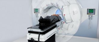
Diagnostics
Prenatal diagnosis of microcephaly includes ultrasound examination (ultrasound). During this procedure, the doctor compares the data obtained on the size of the fetus's head and torso with normal values. The most reliable information can only be obtained if you know exactly the duration of pregnancy.
Determining mutations of genes and chromosomes is the so-called invasive diagnostics. This method is carried out with a puncture of the fetal bladder, because the material for research is fetal villi, amniotic fluid, and also particles of epithelium.
Prenatal screening also includes a biochemical blood test. In addition, the pregnant woman is asked to fill out a special form, which, among other questions, contains a column about the timing of pregnancy. All information received and test results are processed in a special computer program that shows the likelihood of developing brain microcephaly.
A newborn baby is examined by a neonatologist in the delivery room in the very first minutes of his life through an external examination. If the diagnosis is confirmed, then an additional comprehensive examination is required.
Children with microcephaly must be consulted by a geneticist to identify concomitant hereditary diseases. In order to determine the degree and prognosis, it is also important to conduct a full instrumental examination: ultrasound of the brain (neurosonography), electroencephalography (EEG), radiography of the skull, as well as computed and magnetic resonance imaging (CT and MRI).
In addition, in order to identify defects in the development of other organs and systems, the child should receive consultations from specialized specialists: otolaryngologist (ENT), ophthalmologist, cardiologist, neurologist, neurosurgeon, orthopedist.
How to determine the presence of a disease?
The main sign of microcephaly in a child is a decrease in the size of the skull at birth. This sign persists throughout the baby’s life and becomes more obvious with age.
In most cases, in children with this diagnosis, the fontanelles close at an accelerated pace (in the first month of life).
Gradually, the main symptom of microcephaly is joined by additional signs due to the progression of the pathological process.
You can detect microcephaly in a child in the following ways:
- conducting a comprehensive examination in a medical institution;
- laboratory examination of amniotic fluid;
- fetal cord blood analysis;
- confirmation or exclusion of the diagnosis by laboratory tests.
Treatment
Medical care for microcephaly comes down mainly to symptomatic support for patients, since it is impossible to restore normal brain activity, but there are ways to correct it. Treatment implies an integrated approach aimed at the physical and intellectual development of the child with the goal of maximizing his adaptation in society. It is carried out in three directions.
- Drug therapy. It is used to stimulate metabolic processes in the brain. For this, nootropic, sedative, anticonvulsant and dehydration drugs, as well as B vitamins, are prescribed.
- Physiotherapeutic procedures, physical therapy and massage.
- Therapeutic measures aimed at correcting the mental development of the child. In the event that mental retardation does not reach a severe degree (idiocy), then it is quite possible for such a patient to be taught basic self-care skills, performing simple work, and sometimes even speech and writing. For this purpose, long-term classes with specialists, special training programs and other necessary activities are held.
There is a so-called conductive pedagogy. Its essence is to create conditions that encourage the child to mental, motor and emotional activity, which creates the prerequisites for the further development of his intellect and psyche. During this treatment, the patient learns both simple and complex movements, learns to think and make decisions. At the beginning, the child develops motor behavioral stereotypes, which, as a result of long and hard work with teachers, gradually become meaningful and automated, that is, he does not just learn and repeat certain movements, but realizes the purpose for which he does this.
For each patient, an individual training program is drawn up, which in turn includes therapeutic exercises and physical education, exercises with various sports equipment, classes with a speech therapist, audiologist, psychotherapist and other specialists. This technique allows one to achieve fairly good results in children with various forms of mental retardation, including microcephaly.
Treatment of children with the primary form of pathology is sometimes successful. However, it should be remembered that even with timely and well-chosen therapy, the baby will never be completely healthy, but he will be able to lead a more simplified social life.
Psychosocial adaptation
In the psychological and social adaptation of children with microcephaly, the main role is given to parents.
The child must take all prescribed medications in a timely manner, attend classes with speech therapists and other teachers, engage in physical therapy and receive a full-fledged massage.
Any interruptions in therapy can cause an exacerbation of the disease and accelerate its progression.
Recommendations for parents of children with this diagnosis:
- Raising children with microcephaly is accompanied by increased responsibility on the part of adults (parents must be aware of the scale of the problem).
- Psychological assistance should be provided not only to the child, but also to his parents.
- Slowing down the progression of the disease and the emergence of positive dynamics is only possible with constant training with the child and following the recommendations of doctors.
- Thanks to special classes, you can increase a child’s self-esteem and develop his individuality (such classes are in most cases conducted in a group form, children learn to communicate with each other, acquire basic skills and more easily adapt to external conditions).
- Children with a mild form of intellectual disability can attend remedial classes, but classes with a remedial teacher are mandatory.