Good afternoon, dear readers. Today we will continue to study the spinal cord with an emphasis on its internal structure. In the last article we got acquainted with the external structure of this organ, and today we will look inside. Today we will also study spinal roots and spinal nerves.
As you remember from the illustrations from the previous article, on each section of the spinal cord we saw a strange figure that looked like a butterfly. This happened even in the most primitive schemes, such as this one:
We see approximately the same picture in photographs of spinal cord preparations:
As you can see, in each illustration there is a certain dark figure that resembles a butterfly. This dark substance is nothing more than the bodies of neurons. Anatomists call this butterfly gray matter (substantia grisea). There is white fabric around the “butterfly”. This tissue is called white matter (substantia alba). You can easily remember the Latin translation of the word “white” if you remember the name of the main good wizard from Harry Potter - Albus Dumbledore.
Let's return to the spinal cord. White matter, unlike gray matter, is formed by the processes of neurons, not their cell bodies.
Reflex arcs
Before we look at the spinal cord in more detail, we need to talk a little about reflex arcs. As you know, the main part of our relationship with the outside world is built on the basis of reflexes - behavioral stereotypes with which we react to any stimulus.
Reflexes are divided into innate (for example, constriction of the pupil from bright light) and acquired (for example, a trained fighter’s avoidance of a punch to the head). Reflexes are also divided into two-neuron (monosynaptic) and polyneuron (polysynaptic).
The simplest reflex arcs have a two-neuron structure. Such, for example, is the well-known knee reflex, in which a light blow to the tendon of the quadriceps femoris muscle under the patella causes the leg to straighten at the knee joint.
By the way, the junction of two neurons is called a synapse. The synapse is quite complex, I plan to make a separate article about the physiology of synapses, this is a very important topic.
In fact, most reflex arcs are polyneuronal - that is, they contain three or more neurons. Imagine that you accidentally touched a hot iron with your finger and immediately pulled your hand away. This reflex arc will work with the participation of a third neuron, which is called an interneuron. An interneuron is an intermediate link between a sensory and a motor neuron.
When we talk about more complex, multi-neuron reflex arcs, we mean a reflex arc in which one sensory neuron, one motor neuron, and many intercalary ones. True, this does not apply to such complex processes as, for example, planning, building logical connections or thoughtful analysis of any events. Here we are talking about the connections of neurons in the cerebral cortex and this is a slightly different story.
And we return to our spinal cord horns.
Symptoms
The main manifestations of spinal injury are pain and a burning sensation, swelling, and redness at the site of injury. A hematoma or even an open wound may also be observed. But the clinical picture largely depends on the level of damage and its severity. Thus, cervical spine injuries are characterized by:
- impaired sensitivity and mobility of the hands up to complete paralysis (tetraplegia);
- difficulty turning your head;
- respiratory failure, shortness of breath, respiratory arrest.
Injuries to the cervical spine are the most dangerous in terms of disability and death.
Damage at the level of the thoracic and lumbar vertebrae may be accompanied by:
- decreased or loss of sensation in the legs;
- disorders of urination and defecation;
- inability to raise a straight leg while lying on your back;
- back muscle tension.
Thus, a high risk of injury is indicated by the appearance of disturbances in sensitivity and motor functions.
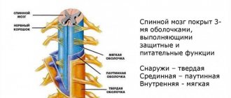
Gray matter of the spinal cord
So, we found out that in the spinal cord there is gray matter - the bodies of neurons and white matter - the processes of neurons. The gray matter forms a pattern that resembles a butterfly. In this “butterfly” we can see quite clear outgrowths that form horns if we are talking about a section of the spinal cord, and gray columns (columnae grisea) if we look at the spinal cord as a whole.
In a cross section we can see the anterior horns (cornu anterius), posterior horns (cornu posterius) and lateral horns (cornum lateralis). Do not forget that the front horns are directed towards the stomach, and the rear horns are directed towards the back. However, I encourage you to use anatomical terminology, and when talking about directions, use the term not “anterior” but “ventral” and not “posterior” but “dorsal”. Personally, I remember the term “ventral” from its association with the Latin term for “stomach,” which is “ventriculus.” The stomach is located in the abdominal cavity, and this is a rather convenient memory.
In this illustration we see the posterior horns (blue), the anterior horns (red) and the lateral horns (yellow).
Anterior horns of the spinal cord
The anterior horns are formed by the cell bodies of motor neurons. This means that the processes of such neurons will be directed from the spinal cord to the skeletal muscles. The excitation from such a neuron will be transmitted to a skeletal muscle - for example, to the biceps of the arm - and cause it to contract to bend the arm at the elbow joint.
The fundamental point here is precisely the direction from the spinal cord to the working organ, in this case, to the muscle. Let's draw an arrow from the spinal cord to emphasize the direction of the nerve impulse. Remember this thing, it is very important.
Anatomists call this direction efferent, and the opposite direction, which corresponds to sensory neurons, called afferent. To avoid confusion, I came up with an association - the term “efferent” begins with the letter “E”, as well as the transcription of the English word “exit”, which means “exit”.
Posterior horns of the spinal cord
The dorsal horns are formed by the bodies of interneurons. Most reflex arcs have three neurons—a sensory neuron, an interneuron, and a motor neuron. The sensory neuron acts first. For example, you took a hot mug of tea in your hand. Long processes of sensory neurons reach the skin of the palm and fingers with which we touched the hot mug. The temperature of this mug is lower than the hot iron from the previous example, and you did not drop it immediately. You realized the mug was hot, but it was tolerably hot, so you were able to wait a second while your hand set it back on the table.
What happened? The processes of the sensitive neuron (we do not yet know where they are) transmitted the signal to the spinal cord. Next, the signal from the spinal cord went to the brain, after which the temperature was analyzed and the trajectory of the movement of your hand with the mug to the table was calculated. This information left the brain and again went down to, as we now know, the neurons of the dorsal horns.
So, in this story, all neurons except the first (sensitive) and posterior (motor) are intercalary. That is, the interneuron does not receive information from its receptor process (as a sensitive one) and does not send information to the working organ (as a motor one). An interneuron is an intermediate neuron between sensory and motor neurons.
Let's add the body of the interneuron to our diagram to remember where the interneurons are located.
In total, it is the bodies of interneurons that are located in the dorsal horns of the spinal cord.
By the way, pay attention to a fundamental thing. The processes of interneurons cannot be located inside the gray matter, right? Of course, they can’t, because gray matter is the bodies of neurons, not processes. This is why the processes of interneurons extend beyond the gray matter and then enter it again.
Lateral horns of the spinal cord
The lateral horns of the spinal cord are not found everywhere. You can find them from the 8th cervical to the 1st lumbar segments of the spinal cord. I will definitely tell you what segments are and return to the lateral horns, but for now it is important for us to understand that the lateral horns occupy only the conditional middle of the spinal cord, and they are absent very high or very low.
The lateral horns also contain the bodies of interneurons, but only those that belong to the autonomic nervous system. Autonomic neurons innervate smooth muscles, that is, muscles of internal organs and blood vessels. The fundamental difference with the autonomic nervous system is that it cannot be controlled by our will - we may want to bend our arm and flex it, but we cannot want to clench our stomach and put that desire into action by force of will.
So, the lateral horns are formed by the bodies of autonomic neurons, and autonomic intercalary ones. I have drawn one autonomic neuron, the process of which, as you can see, extends beyond the spinal cord - and this is a very significant feature, in fact. In the somatic nervous system, interneurons are nestled inside the gray matter, and that's all. But in the vegetative processes of the intercalary neurons, they extend beyond the spinal cord. We will look at where exactly they come from in a separate article about the autonomic nervous system.
Where are the cell bodies of sensory neurons located?
Now, in theory, you should have a question. We analyzed the localization of the bodies of motor neurons, the bodies of interneurons, and even the bodies of vegetative interneurons. Something's missing, isn't it? Of course, there are not enough cell bodies of sensory neurons. Where are they located if we have already dismantled all the gray matter of the spinal cord?
The bodies of sensory neurons are located in the spinal ganglion. To understand where this is located, we need to disassemble the structure of the spinal roots, we will do this a little later. Now, to avoid confusion, it is important for us to note that the cell bodies of sensory neurons are located outside the spinal cord.
With the help of these arrows, I emphasize that the nerve impulse travels along the sensory fibers to the spinal cord, that is, in the afferent direction.
Let me remind you that motor neurons transmit information in the direction from the spinal cord to target organs; in the case of the somatic nervous system, these target organs are skeletal muscles.
Causes
Spinal injury is usually caused by the application of an external force. Most often it occurs as a result of:
- falls from a height, which is called catatrauma – 50%;
- road traffic accidents – 30%;
- diving head down in shallow water – 10%.
This is often accompanied by damage to the spinal cord, i.e., a spinal cord injury initially occurs. In such situations, the spinal cord suffers due to instant or gradually increasing compression by bone structures, displacement of the vertebrae in an unstable segment of the spine. It can also be stretched and ruptured, which leads to disability.
The spinal cord is a sensitive anatomical structure that can also be damaged, even if initially only the spine was damaged at the time of injury. In such situations, spinal cord injury is secondary and becomes a consequence of impaired microcirculation, severe swelling of the surrounding soft tissues, hypoxia (lack of oxygen), disturbances in electrolyte metabolism and the action of a number of other factors.
However, the situation is extremely serious in both cases, regardless of whether primary or secondary spinal cord injury occurs.
Less commonly, the cause of spinal injury, namely vertebral compression fracture, is osteoporosis. This disease is most common in older people, although it can also occur at a young age without long-term use of corticosteroids and some other drugs. It is characterized by a decrease in bone density, which leads to the fact that the vertebral bodies become more fragile and can flatten, forming a wedge-shaped deformity, even as a result of a sudden movement, coughing, not to mention a fall or blow.
Other causes of spinal injury without significant external influence may be tumors of the spine itself or the formation of metastases in it.
Spinal cord segments
We finally come to a rather important question. Remember in the last article I published this illustration?
Here we see the spinal cord in the form of a smooth, elongated thing, similar to a rolled up umbrella. But in many textbooks and atlases you could see illustrations of the spinal cord, similar to an insect with a lot of legs, such as here (this is, of course, Sinelnikov):
What are these strange appendages? These are the roots of the spinal cord that form the spinal nerves. Let's figure out what it is. But before we begin, you must be sure that you have thoroughly mastered the previous material, because without it it will be impossible to understand anything.
So, the spinal cord actually has paired “processes,” which are first called roots, then spinal nerves, and then branches of the spinal nerves. Quite difficult, isn't it? Everything will be clear soon, but for now let’s just agree that the spinal cord has certain processes.
Anterior roots of the spinal cord
Let's remember the illustration we worked with in the previous section.
of somatic motor neurons and autonomic emerge from the anterior, that is, ventral horns of the spinal cord . Each neuron process is very small, however, in the spinal cord there are a lot of neurons and a lot of processes, so we can see clusters of these processes with the naked eye. The processes of somatic motor cells, as well as intercalary vegetative ones, form noticeable formations called the anterior roots of the spinal cord (radix anterior).
A quick note - in my diagram the neuron bodies look very large - four neuron bodies occupy the entire anterior horn. In reality, of course, the proportions are completely different - hundreds of thousands of neurons fit in the anterior horns, but I wouldn’t be able to draw them all, so you see these four neurons.
Now let’s mark the same area, that is, the anterior root, in the illustration from Sinelnikov’s atlas:
I like it when Latin expressions are chosen precisely and correspond to the shape of the object being called. This is why I was unhappy in the article about hand bones. But everything is fine here - these bunches of shoots really look like the roots of plants that gather into single ones... and I’ll tell you what they gather into when we look at the dorsal roots.
Posterior roots
Here we will need our illustration again. As you remember, the dorsal horns of the spinal cord contain the bodies of sensory neurons. The bodies of the sensory neurons themselves are located in some strange place outside the spinal cord. Only I corrected the processes of the sensory neurons - in previous illustrations they looked too long in contrast to the very short processes of the motor neurons. I also added arrows to show the direction of the nerve impulse
As in the case of motor neurons, the processes of sensory neurons are grouped into clearly visible bundles. These bundles are called the posterior roots (radix posterior). Only, unlike the anterior motor nerve fibers that exit the spinal cord, the dorsal roots enter the spinal cord. This is said because of the direction of the nerve impulse, which enters the spinal cord along the anterior roots, and exits it to the periphery along the posterior roots.
Speaking of the dorsal roots, we can see an unusual rounded formation. This is the spinal ganglion (ganglion sensorium nervi spinalis). Please do not confuse the dorsal ganglion with the spiral ganglion, which lies in the cochlea and is involved in the perception of sound. The spinal ganglion is a small round structure within which the bodies of sensory neurons are located. Let's label the dorsal root and dorsal root ganglion first in our illustration:
And, of course, in the illustration from Sinelnikov (the colors are similar):
But that is not all. The spinal ganglion also contains sensory neurons of the autonomic reflex arc. Let's mark them in orange in our drawing:
Spinal nerves
So, we have two pairs of roots - two front and two back. In my illustration I show all sorts of anatomical things on only one side of the spinal cord, but you need to understand that they are symmetrical.
As you remember, the dorsal roots enter the spinal cord, and the anterior ones exit it. Near the spinal cord, the dorsal and anterior roots are significantly removed from each other. However, slightly distal to the spinal cord, the roots begin to approach each other, and eventually unite into a noticeable but very short trunk. This trunk is called the spinal nerve (nervus spinalis). Let's draw it.
There are a few problems with my drawing - it's too far gone to need redrawing, so I'll just describe those problems. First, I had to significantly increase the length of both spines in order to draw the spinal nerve and not move the titles from previous sections. Second, the dorsal ganglion is located too proximal to the spinal cord. In reality, it is, of course, much more distal and very close to the spinal nerve.
Again, the spinal nerve is a very short nerve trunk. Many people believe that the spinal nerves are something large, entwining the entire body. Of course, this is a mistake, because the spinal nerve cannot be longer than 1 cm, as a rule, even less.
However, the spinal nerve is divided into several branches, which in turn actually entwine the torso, limbs and even internal organs. We will talk about this a little further, but now we must project a section of the spinal column along which we can find the exit point of the spinal nerve.
Such a site is the intervertebral foramen (foramen intervertebrale), which is formed by the superior vertebral notch (incisura vertebralis superior) of the underlying vertebra and the lower vertebral notch (incisura vertebralis inferior) of the overlying vertebra. I have highlighted the intervertebral foramina in red:
Let's look at a suitable illustration from Sinelnikov's atlas, which very clearly shows the relationship between the spinal nerve and the intervertebral foramen.
The spinal nerve is designated here as number 6. As you can see, this is a very short section of nerve fibers that extends just a few millimeters from the intervertebral foramen and is immediately divided into several branches. Let's mark the spinal nerves in red:
Branches of the spinal nerve
And now we can trace all the anatomical structures that extend from the spinal nerve. To do this, we will use our old illustration and continue to complete it.
Shell (return) branch
So, first of all, a small, thin branch departs from the spinal nerve, which, immediately after being disconnected from the nerve trunk, turns back to the spinal cord to innervate the dura mater. This branch is called the shell branch (ramus meningius), another name is the recurrent branch (ramus recurrens). It consists of sensitive somatic and autonomic motor fibers, which I reflected in the diagram.
Only you may have a completely fair question - why do somatic sensory nerves go to the spinal cord, and not autonomic sensory ones? In fact, I could not find such reflex arcs in any of my four atlases. I will be sure to clarify this issue and correct it in a later version of this article.
Autonomic motor fibers are necessary here in order to control the contraction and relaxation of the vascular wall. As we remember, the dura mater of the spinal cord and, especially, the epidural space are connected with a large number of vessels.
One more point - in Sinelnikov’s drawing we cannot see the recurrent branch, because, apparently, it is too small, and this drawing was created to show the relationship of the spinal nerve, root, ganglion and intervertebral foramen.
White connecting branch
The next branch that arises from the short spinal nerve is the autonomic branch. This is a small branch called the white connecting branch (ramus communicans albus). The white connecting branch is formed by processes of intercalary autonomic neurons, which are located in the lateral horns of the spinal cord. The white connecting branch has this name because the processes of the neurons that form it are covered with a protective material, myelin, consisting of lipids and proteins.
Let's draw a white connecting branch in our illustration:
Sympathetic trunk
As you know, I always pay attention to names. If you also have this feature, you probably wondered - why is the white connecting branch a connecting branch? What does it connect to what?
We know that the white communicating branch arises from the spinal nerve, that is, it connects the spinal nerve to something. This “something” is the sympathetic trunk, an important component of the autonomic nervous system.
The autonomic nervous system, as you know, is divided into sympathetic and parasympathetic. The mediator of the sympathetic system is adrenaline, so the effects produced by the influence of the sympathetic system can be easily remembered by the effect of adrenaline on the body. Imagine that some predator is chasing you, and your adrenaline is released. Adrenaline dilates your pupils (mydriasis), increases your breathing and your pulse, while blocking gastrointestinal motility so you don't have to go to the toilet during the chase. All this is the influence of the sympathetic system.
Accordingly, parasympathetic influences lead to the opposite effect - constriction of the pupils (miosis), increased gastrointestinal motility, decreased pulse and pressure.
We will not study the anatomy of the parasympathetic nervous system in this article, but we will definitely touch on it during our study of the vagus nerve (nervus vagus, 10th pair of cranial nerves).
But the sympathetic nervous system will be the subject of our study, more precisely, its part, which is called the sympathetic trunk.
The sympathetic trunk (truncus sympathicus) is a paired formation that looks like a Christmas tree garland, only located vertically. Both sympathetic trunks - right and left - are located a short distance from the spinal cord.
This is a blurry and rather small illustration, but both sympathetic trunks are very clearly represented here:
As you can see, both trunks connect approximately in the area of the sacrococcygeal joint.
When I said that the sympathetic trunk is like a Christmas tree garland, I meant that it consists of round nodes, which are called the nodes of the sympathetic trunk (ganglia trunci sympathici). These nodes are connected to each other by thin nerve bundles called internodal branches (rami interganglionares).
In our illustration, I have highlighted the nodes of the sympathetic trunk in blue, and the internodal branches in green.
So we can go back to our diagram of the spinal cord because we found out that the white communicating branch connects the ganglion of the sympathetic trunk and the spinal nerve. Let's draw this:
Gray connecting branch
We have just studied the white communicating branch, which arises from the spinal nerve and connects it to the sympathetic ganglion. However, another autonomic branch immediately departs from the sympathetic ganglion, which again joins the spinal nerve. This is the gray connecting branch (ramus communicans griseus).
In a separate lesson, I will talk about how this branch is formed and, in general, about the physiology of the autonomic system. For now, we need to understand that the gray branches are processes of neurons whose bodies are located in the ganglion of the sympathetic trunk. These fibers are called postganglionic fibers because they emerge from the ganglion rather than enter it.
Posterior branch
We have finished studying the thin branches that arise from the spinal nerve, and now we are moving on to the larger ones. By the way, the recurrent and autonomic branches are so small that they are often not depicted on diagrams of the spinal nerves. For example, they are not in the already familiar illustration from Sinelnikov’s atlas:
However, we see two large branches, about which we cannot say that they arise from the spinal nerve. It rather bifurcates into these large branches, doesn't it? One of these branches in the illustration seems to continue the course of the spinal nerve, while the other deviates backward. The branch that deviates backward is called the posterior branch (ramus posterior).
The posterior rami of the spinal nerves, true to their name, travel posteriorly in a dorsal direction to innervate the skin of the back and the deep muscles of the back. By the way, this is one of the most difficult topics in myology, and it would not be amiss to repeat these muscles again.
Let's mark this branch in blue in Sinelnikov's illustration:
And we’ll also draw it in our illustration:
As you can see from the colors, the posterior branch includes fibers of the autonomic nervous system, motor somatic fibers and sensory somatic fibers.
First aid for spinal injury and choice of treatment tactics
It is important not to move or lift the patient after receiving a blow or other traumatic impact and immediately call an ambulance. Under no circumstances should the victim be allowed to sit or stand, but it is worth giving pain medication and attempting to calm him down. If there is no breathing or heartbeat, resuscitation should be performed. Specialists who arrive at the scene carefully immobilize the patient at the scene by securely fixing him on a rigid shield using a rigid head holder, and then transport him to a medical facility.
Any patient who is suspected of having a spinal injury is immediately treated with a spinal injury protocol until a full investigation has been completed and it is proven that no injury has occurred.
If the presence of an injury is confirmed, its type and characteristics are clarified, it becomes clearly clear what treatment is indicated in a particular case. Conservative therapy is carried out only in the mildest cases in the absence of signs of neurological deficit, when only a violation of the anatomy of bone structures is observed without signs and risks of complications, in particular compression of the spinal cord. It is usually indicated for mild compression fractures, dislocations, fractures of processes, arches, and vertebral bodies without the formation of fragments.
Conservative treatment consists of:
- bed rest or immobilization of the affected part of the spine;
- drug therapy appropriate to the situation;
- traction therapy (spinal traction using a special apparatus);
- physiotherapy;
- manual therapy.
If necessary, vertebral reduction is initially performed.
But in the vast majority of cases, surgery is indicated for spinal injuries. Emergency surgery is required when:
- the presence and especially intensification of signs of neurological deficit;
- severe deformation of the spinal canal due to bone fragments, dislocated vertebrae or significant curvature;
- the presence of a large hematoma formed as a result of injury to a herniated intervertebral disc, injury to the yellow ligament, the presence of a foreign body that threatens compression of the spinal cord;
- the presence of isolated hematomyelia, i.e. hemorrhage in the spinal cord;
- compression of a large blood vessel supplying the spinal cord;
- pronounced compression of the spinal roots;
- instability of spinal motion segments when creating a threat of their displacement and compression of the spinal cord;
- the presence of foreign bodies in the spine;
- liquorrhea, i.e. leakage of cerebrospinal fluid through defects at the base of the skull;
- injuries received from gunshot or stab wounds.
But there are also contraindications for performing the operation even if there are compelling indications for it. In such situations, spinal surgery is postponed until the patient's condition is stabilized. This is about:
- traumatic or hemorrhagic shock with unstable hemodynamics;
- the presence of severe damage to internal organs, leading to internal bleeding, associated with the risk of developing peritonitis;
- extremely severe traumatic brain injuries with signs of intracranial hematoma formation.
However, it is also possible to carry out the operation as planned. They are required when conservative therapy is ineffective, but they allow you to carefully prepare the patient for the upcoming surgical intervention and select a treatment tactic that suits him.
Surgery for spinal injury
The goals of surgical treatment are:
- rapid and complete elimination of the factor leading to compression of the spinal cord and other neurovascular formations;
- restoration of the correct axis of the spine;
- fixation and stabilization of the spine, which will allow the patient to be activated as early as possible and avoid deformation in the future.
If there are signs of spinal cord damage, it is recommended to perform surgery as early as possible, since up to 70% of cases of the development of all irreversible changes occur in the first hours after injury. But there are quite a few types of operations that can be indicated for spinal injuries. The choice of a specific one is made by the neurosurgeon in each case individually.
Halotraction
Halotraction is an operation indicated for complicated or unstable injuries of the cervical spine. It involves dynamic reposition and reliable fixation of the vertebrae while maintaining the patient’s physiological activity using a special halo apparatus.
It is a device, part of which reliably connects the damaged vertebrae to the base of the skull, and the other is located outside the body. Its design features make it possible to gradually perform traction, i.e. stretching of damaged spinal motion segments until complete reposition of the vertebrae is achieved.
On average, the halo apparatus is removed after 2.5-3 months. After this, the use of a head holder is prescribed for 3-4 weeks.
Laminectomy
Laminectomy is a decompression operation widely used for various spinal injuries. It allows you to eliminate compression of the spinal cord and its roots by various anatomical structures by removing the vertebral arches or only their processes, or to provide access to the spinal cord to remove foreign bodies or perform other manipulations on it.
Often the situation requires a discectomy, i.e., removal of the intervertebral disc, and in some cases the surgeon is forced to remove the entire damaged vertebra completely, i.e., perform a corpectomy.
Laminectomy requires further installation of stabilizing systems in order to achieve spinal fusion.
Discectomy
Discectomy is a decompression operation indicated for traumatic injuries of the intervertebral disc that have led to compression of the spinal roots or spinal cord. It involves removing the damaged intervertebral disc and can be combined with laminectomy.
The operation is performed through an anterolateral approach (for injuries to the cervical spine) or posterior (for damage to the thoracic or lumbosacral discs). It is performed under general anesthesia, and the removed disc is replaced with a bone graft, synthetic implant, or spinal fusion is achieved.
In cases uncomplicated by serious spinal cord injuries, microdiscectomy may be performed. This operation involves making a much smaller incision and may involve removing only a formed hernia while preserving the disc. Endoscopic surgery can also be an alternative, but spinal injuries are rarely limited to isolated disc damage, so discectomy is often only one of the stages of surgical intervention.
Spinal fusion
Spondylodesis is an operation whose main goal is the reliable fusion of several spinal motion segments with each other, which leads to their complete immobilization, and therefore eliminating the risk of displacement and subsequent injury to the spinal cord. For this purpose, a fragment of the patient’s own bone (autograft) or artificial bone can be used. But more often special titanium structures are used, i.e., transpedicular fixation of the vertebrae is performed, since this technique is associated with fewer risks and is more reliable.
Transpedicular fixation involves a strong connection of adjacent vertebrae using special screws and rods passed through their heads. Titanium screws are screwed into the intersection of the transverse process of the vertebra with the superior articular process. In this case, it is necessary to fix at least 3 vertebrae, even if only one of them is affected, since otherwise the proper degree of stabilization of the spine will not be achieved.
The transpedicular fixation method is the main way to achieve stabilization of the spinal column in various spinal injuries.
Spinal fusion can be performed in isolation or as one of the stages of surgical treatment of spinal injury, for example, after decompression of the spinal cord or its roots. The operation may also involve removal of the intervertebral disc, which is especially important in case of traumatic damage. In this case, a modern, precisely sized cage is installed in place of the removed disc.
If initial decompression of spinal structures is necessary, this task can be solved through an anterior or posterior approach. Most often, the operation is performed through a posterior approach, since this is associated with lower intraoperative risks and is less likely to lead to postoperative complications.
Vertebroplasty and kyphoplasty
Vertebroplasty and kyphoplasty are two similar techniques that can be used for vertebral compression fractures. Their essence is to restore the strength of a broken vertebral body using specially developed, quickly hardening bone cement.
Both operations are minimally invasive and involve the introduction of a thin cannula into the body of a broken vertebra, through which its integrity will be restored by injecting freshly mixed bone cement into it. It will fill all natural bone pores and provide high strength to the vertebra within 10 minutes, since this is exactly the time it takes for it to completely harden.
But when choosing kyphoplasty, a special balloon is first inserted into the vertebral body through the same cannula and filled with liquid. As a result, it swells and helps restore the anatomy of the fractured vertebral body. After this, the balloon is deflated and removed, and the resulting space and the entire vertebral body are filled with bone cement. Once it has hardened, the cannula is removed and the remaining puncture is covered with a sterile bandage.
The features of these two percutaneous surgery techniques determine the specifics of their application. Thus, vertebroplasty is indicated for mild compression fractures, when the height of the vertebral body decreases by less than 70%. At the same time, kyphoplasty has wider possibilities, so it can be used for severe compression fractures with a decrease in vertebral height by more than 70%.
Thus, no one is immune from spinal injury. But if it happens, it is important not to try to cope with the situation on your own, but to immediately call an ambulance, ensuring the victim is completely immobile. The prognosis of a spinal injury largely depends on both its type and the speed of receiving qualified medical care. In mild, uncomplicated cases, complete recovery usually occurs, but if the spinal cord is damaged, there is a high risk of complications, including loss of control over urination and defecation, and disability.